





 Video Library


Please set your exam date
Preterm Labor
Study Questions
Practice Exercise 1
A nurse is caring for a client at 29 weeks of gestation who reports uterine tightening. Which of the following findings would confirm a diagnosis of preterm labor rather than false labor?
Explanation
Preterm labor is characterized by regular uterine contractions occurring before 37 weeks of gestation that lead to progressive effacement and dilation. This pathological process involves myometrial activation and decidual activation triggered by inflammatory or mechanical stressors.
Rationale for correct answer:
3. Regular uterine contractions occurring at least every 5 minutes that lead to progressive cervical effacement and dilation between 20 and 37 weeks of gestation confirm preterm labor. The hallmark of true labor is cervical change driven by coordinated myometrial activity.
Rationale for incorrect answers:
1. Contractions that resolve with hydration and rest represent false labor, often termed Braxton Hicks contractions. True preterm labor contractions persist and intensify despite supportive measures like oral rehydration and lateral recumbent positioning, which reduce uterine irritability caused by dehydration.
2. Irregular contractions occurring at wide intervals without back discomfort are characteristic of false labor. True labor contractions are regular, increase in frequency and intensity, and often cause discomfort that radiates from the back to the fundus due to pacemaker activity in the cornua.
4. Mild cramping that subsides after voiding and resting in the lateral position indicates transient uterine irritability rather than labor. Emptying a distended bladder removes mechanical pressure on the uterus, which resolves spurious contractions without causing cervical remodeling.
Test-taking strategy:
- Analyze the scenario/question: The clinical scenario requires the nurse to identify the clinical findings that differentiate true preterm labor from false labor in a client at 29 weeks of gestation.
- Decision Framework: Obstetrical Knowledge of Labor Assessment.
- Apply knowledge of cervical change as the definitive diagnostic criterion for labor. True labor involves regular uterine contractions that result in progressive effacement and dilation of the cervix, whereas false labor contractions do not produce cervical changes and typically resolve with hydration, rest, or position changes.
- Rule out Choice 1 because contractions that resolve with hydration and rest are characteristic of false labor or uterine irritability rather than active labor.
- Rule out Choice 2 because irregular, widely spaced contractions without radiation of pain do not indicate progressive cervical changes or true labor.
- Rule in Choice 3 because regular contractions accompanied by progressive cervical effacement and dilation between 20 and 37 weeks confirm preterm labor.
- Rule out Choice 4 because mild cramping that subsides after voiding represents transient irritability from bladder distension, not pathological labor.
Take home points
- Preterm labor is clinically defined as regular uterine contractions occurring between 20 and 37 weeks of gestation that are accompanied by progressive cervical effacement and dilation.
- False labor, or Braxton Hicks contractions, is characterized by irregular contractions that do not increase in frequency or intensity and do not cause cervical changes.
- True labor contractions typically begin in the lower back and radiate to the abdomen, becoming stronger and more frequent with ambulation, and do not resolve with rest or hydration.
- Initial management of suspected preterm uterine irritability includes hydration, bladder emptying, and lateral recumbent positioning to rule out spurious contractions before diagnosing preterm labor.
A client at 26 weeks of gestation asks the nurse to explain the difference between "extremely preterm" and "very preterm" birth. Which of the following responses by the nurse is most accurate?
Explanation
Preterm birth subcategories are defined by gestational age thresholds, where extremely preterm is < 28 weeks and very preterm is 28 to < 32 weeks, which directly correlates with neonatal mortality rates, pulmonary surfactant deficiency, and long-term neurodevelopmental deficits.
Rationale for correct answer:
1. This choice is correct because standard clinical guidelines define extremely preterm birth as occurring before 28 weeks of gestation. In contrast, very preterm birth is defined as occurring from 28 weeks to just under 32 weeks of gestation.
Rationale for incorrect answers:
2. This choice is incorrect because birth before 32 weeks is too broad, and the range of 32 to 34 weeks actually represents moderate preterm birth. These definitions do not align with standard neonatal classification systems.
3. This choice is incorrect because these 2 terms represent distinct clinical categories with different prognoses. They do not share 1 category of less than 30 weeks, which violates standard gestational age classifications.
4. This choice is incorrect because preterm classifications are based strictly on gestational age. They are not defined by clinical status at birth, such as the need for resuscitation or the ability to sustain independent respiration.
Test-taking strategy:
- Analyze the scenario/question: The question asks for the correct clinical definitions of "extremely preterm" and "very preterm" birth to explain to a client at 26 weeks of gestation.
- Apply knowledge of Obstetrical Knowledge and neonatal classification systems. Preterm birth is categorized into subcategories based on gestational age to guide clinical expectations, risk assessment, and neonatal intensive care unit preparation. Understanding these precise gestational thresholds is essential for accurate patient education and interdisciplinary communication.
- Rule in Choice 1 because it accurately reflects the World Health Organization definitions, which designate extremely preterm as < 28 weeks and very preterm as 28 to < 32 weeks.
- Rule out Choice 2 because it incorrectly defines the gestational ranges, mislabeling moderate preterm as very preterm and failing to recognize the correct thresholds for early gestation.
- Rule out Choice 3 because it falsely groups these 2 distinct clinical categories into 1 range, which obscures the unique prognostic differences associated with each sub-classification.
- Rule out Choice 4 because neonatal classification systems are based on objective gestational age rather than subjective clinical presentation or immediate respiratory support requirements at delivery.
Take home points
- Preterm birth is defined as any birth occurring before 37 completed weeks of gestation.
- Extremely preterm birth refers specifically to deliveries that occur before 28 weeks of gestation.
- Very preterm birth is defined as deliveries occurring from 28 weeks up to but not including 32 weeks of gestation.
- Moderate to late preterm birth encompasses deliveries from 32 weeks up to 37 weeks of gestation.
A nurse is reviewing the chart of a client at 31 weeks of gestation with suspected preterm labor. Which finding on transvaginal ultrasound would be most concerning for impending preterm birth?
Explanation
A cervical length less than 2.5 cm and cervical funneling before 34 weeks indicate cervical insufficiency. This structural weakness significantly increases the risk of spontaneous preterm birth and premature rupture of membranes.
Rationale for correct answer:
2. A cervical length of less than 2.5 cm at 31 weeks of gestation indicates significant cervical shortening, which is a strong predictor of preterm delivery. The presence of funneling at the internal os further demonstrates structural incompetence and imminent labor.
Rationale for incorrect answers:
1. A cervical length of 4.2 cm is well within the normal physiological range of 3.0 to 5.0 cm during the third trimester. A closed internal os indicates structural integrity, representing a very low risk of spontaneous preterm birth and confirming cervical competence.
3. A measurement of 3.5 cm represents a normal, reassuring cervical length that indicates adequate structural support. The absence of funneling confirms that the internal os remains stable, indicating a low probability of premature rupture of membranes or imminent delivery.
4. A cervical length of 3.0 cm is at the lower limit of normal but remains clinically reassuring. A thick, closed cervix indicates that no effacement or dilation has initiated, suggesting a low risk of cervical insufficiency and preterm labor.
Test-taking strategy:
- Analyze the scenario/question: The clinical scenario requires identifying the transvaginal ultrasound finding that indicates the highest risk of impending preterm birth in a client at 31 weeks of gestation.
- Use the Obstetrical Knowledge framework to evaluate cervical parameters.
- Apply knowledge of cervical length assessment and sonographic markers of preterm labor. Transvaginal ultrasound is the gold standard for measuring cervical length, where a length of < 2.5 cm or the presence of funneling indicates a high risk of preterm birth.
- Rule out Choice 1 because a cervical length of 4.2 cm with a closed os represents normal anatomy and indicates low risk of preterm delivery.
- Rule in Choice 2 because a cervical length of 1.8 cm (which is < 2.5 cm) combined with funneling indicates cervical effacement and high risk of imminent birth.
- Rule out Choice 3 because a length of 3.5 cm without funneling is a reassuring finding that indicates cervical stability.
- Rule out Choice 4 because a length of 3.0 cm with a thick, closed cervix represents a reassuring measurement that does not suggest active labor.
Take home points
- Transvaginal ultrasound is the gold standard imaging modality for assessing cervical length and predicting preterm birth risk in symptomatic clients.
- A cervical length of less than 2.5 cm before 34 weeks of gestation is a major risk factor for spontaneous preterm delivery.
- Cervical funneling represents the protrusion of the amniotic sac into the internal cervical os and indicates progressive cervical incompetence.
- Normal cervical length during the third trimester typically ranges between 3.0 and 5.0 cm, which indicates a low risk of imminent delivery.
A primary health care provider documents that a client is experiencing "threatened preterm labor." Which of the following statements by the nurse best reflects understanding of this diagnosis?
Explanation
Threatened preterm labor involves uterine contractions between 20 and 37 weeks of gestation without cervical dilation or effacement. Management focuses on monitoring to prevent preterm birth and administering corticosteroids for fetal lung maturity if progression occurs.
Rationale for correct answer:
2. Threatened preterm labor is characterized by regular uterine contractions occurring between 20 and 37 weeks of gestation without progressive cervical change. This clinical state requires close monitoring to distinguish it from true preterm labor and prevent premature delivery.
Rationale for incorrect answers:
1. A cervical dilation of 4 cm indicates active preterm labor rather than threatened preterm labor. True preterm labor involves progressive cervical effacement and dilation, which typically leads to imminent delivery within a short timeframe, requiring immediate tocolytic therapy.
3. Ruptured membranes prior to 37 weeks of gestation define preterm premature rupture of membranes, not threatened preterm labor. This condition significantly increases the risk of intra-amniotic infection and requires distinct management strategies to prevent maternal and fetal sepsis.
4. The completion of corticosteroids and tocolytics represents a therapeutic intervention rather than a diagnostic definition. These treatments are administered to accelerate fetal lung maturity and delay delivery, but they do not define the clinical diagnosis itself.
Test-taking strategy:
- Analyze the scenario/question: The question asks for the statement that best reflects an understanding of "threatened preterm labor."
- Decision framework: Obstetrical Knowledge of labor progression and diagnostic criteria.
- Apply knowledge of gestational assessment and labor definitions. Threatened preterm labor involves uterine activity without the cervical effacement or dilation that characterizes active labor. Distinguishing between threatened and active labor is crucial for determining the necessity of aggressive interventions like tocolysis or corticosteroid administration.
- Rule out Choice 1 because a dilation of 4 cm indicates active labor rather than a threatened state, meaning delivery is highly likely.
- Rule in Choice 2 because threatened preterm labor is clinically defined by contractions without documented cervical effacement or dilation.
- Rule out Choice 3 because membrane rupture indicates premature rupture of membranes, which is a distinct clinical entity with different risks.
- Rule out Choice 4 because completing medical interventions describes a treatment protocol rather than the diagnostic criteria of the condition.
Take home points
- Threatened preterm labor is defined by regular uterine contractions between 20 and 37 weeks of gestation without progressive cervical change.
- Active preterm labor is diagnosed when uterine contractions are accompanied by progressive cervical dilation of 3 cm or more or effacement of 80 percent or more.
- Preterm premature rupture of membranes is the rupture of chorioamniotic membranes prior to 37 weeks of gestation and before the onset of labor.
- Management of threatened preterm labor focuses on monitoring uterine activity and cervical status to avoid unnecessary interventions while preparing for potential preterm birth.
A nurse is teaching a prenatal class about factors that increase the global burden of preterm birth. Which of the following statements should the nurse include? Select all that apply.
Explanation
Preterm birth, defined as delivery before 37 completed weeks of gestation, is driven by uterine overdistension, cervical insufficiency, or decidual senescence. It manifests with regular contractions and cervical effacement, leading to high neonatal morbidity globally.
Rationale for correct answers:
1. Preterm birth is the leading cause of neonatal mortality worldwide, accounting for a significant proportion of deaths in children under 5 years of age. This global burden highlights the critical need for effective preventative strategies in obstetric care.
2. Limited access to prenatal care prevents early identification and management of risk factors, which significantly increases the risk of preterm delivery. Adequate prenatal monitoring is essential for optimizing maternal-fetal outcomes and reducing complications.
3. An interpregnancy interval of less than 6 months depletes maternal nutritional stores and prevents uterine recovery, which increases the risk of preterm labor. This short interval is a major contributor to recurrent prematurity globally.
Rationale for incorrect answers:
4. Preterm birth rates are not equally distributed but show significant socioeconomic disparities, with higher rates in low-resource settings. Disadvantaged populations face increased exposure to chronic stress, malnutrition, and infections, which drive premature births.
5. Multifetal pregnancies actually increase the risk of preterm labor due to uterine overdistension and cervical insufficiency. Overdistension triggers early uterine contractions, making multiple gestations a primary driver of iatrogenic prematurity and spontaneous preterm birth.
Test-taking strategy:
- Analyze the scenario/question: The clinical scenario requires identifying the global risk factors and epidemiological facts associated with preterm birth to educate prenatal clients.
- The decision framework used is Obstetrical Knowledge of maternal-fetal risk factors and global health statistics. Apply knowledge of gestational physiology, socioeconomic determinants of health, and interpregnancy intervals to evaluate how these factors influence the incidence of preterm labor. This requires understanding that uterine capacity, maternal health optimization, and healthcare access directly impact gestational length.
- Rule in Choice 1 because preterm birth is globally recognized as the primary cause of neonatal mortality and under-5 deaths.
- Rule in Choice 2 because inadequate prenatal care prevents the mitigation of modifiable gestational risks.
- Rule in Choice 3 because an interpregnancy interval of less than 6 months prevents uterine involution and nutritional replenishment.
- Rule out Choice 4 because preterm birth rates exhibit profound epidemiological disparities across different socioeconomic strata.
- Rule out Choice 5 because multifetal gestations cause uterine overdistension, which triggers early labor.
Take home points
- Preterm birth remains the leading cause of neonatal mortality and morbidity worldwide, necessitating global public health interventions.
- Inadequate access to prenatal care is a major modifiable risk factor that prevents early detection of complications like cervical shortening or infections.
- An interpregnancy interval of less than 6 months significantly increases the risk of preterm birth due to incomplete maternal tissue healing and nutrient depletion.
- Multifetal gestations and lower socioeconomic status are strongly associated with higher rates of preterm labor due to physiological and environmental stressors.
A nurse is caring for a client at 33 weeks of gestation with a history of a prior spontaneous preterm birth at 30 weeks. Which of the following statements by the nurse demonstrates accurate understanding of this client's risk?
Explanation
Spontaneous preterm birth, occurring at <37 weeks of gestation, is primarily driven by pathological inflammation, cervical insufficiency, or uterine overdistension. It presents with uterine contractions and cervical dilation, often requiring progesterone therapy or cervical cerclage to prevent recurrence.
Rationale for correct answer:
2. A history of spontaneous preterm birth is the strongest clinical predictor for recurrence. This history increases the risk of subsequent preterm delivery by 1.5 to 2.5 times, requiring close monitoring and potential progesterone supplementation to mitigate myometrial activation.
Rationale for incorrect answers:
1. This statement is incorrect because a prior preterm birth is the most significant risk factor for recurrence. Ignoring this history neglects critical preventive strategies, such as cervical length screening, which are essential to prevent cervical insufficiency and subsequent preterm labor.
3. This statement is incorrect because even 1 prior spontaneous preterm birth significantly elevates the risk of recurrence. While multiple preterm births further compound the risk, clinical protocols for risk stratification and preventive interventions are initiated after just 1 occurrence.
4. This statement is incorrect because preterm births occurring before 34 weeks, especially early preterm births like at 30 weeks, carry an even higher risk of recurrence. The gestational age of the prior delivery is inversely proportional to the recurrence risk of preterm delivery.
Test-taking strategy:
- Analyze the scenario/question: The clinical scenario describes a pregnant client at 33 weeks of gestation with a history of a prior spontaneous preterm birth at 30 weeks. The decision framework used is Obstetrical Knowledge regarding risk factors for preterm labor. Apply knowledge of risk factors for preterm birth, specifically focusing on how a history of spontaneous preterm birth affects the risk profile of subsequent pregnancies. A prior spontaneous preterm birth is the most significant risk factor for recurrence, and the risk is inversely related to the gestational age of the previous delivery.
- Rule out Choice 1 because it falsely claims that prior preterm birth has no impact on the current pregnancy, ignoring established epidemiological data on recurrence rates.
- Rule in Choice 2 because it correctly identifies that a prior spontaneous preterm birth significantly increases the risk of recurrence, which is vital for clinical surveillance and preventive therapies.
- Rule out Choice 3 because it incorrectly limits the risk to clients with multiple prior preterm births, whereas 1 event is sufficient to warrant high-risk classification.
- Rule out Choice 4 because it incorrectly states that the risk is only relevant after 34 weeks, whereas earlier preterm births actually carry a higher recurrence risk.
Take home points
- Spontaneous preterm birth is defined as delivery occurring between 20 and 36 weeks and 6 days of gestation.
- A history of a prior spontaneous preterm birth is the strongest clinical risk factor for a subsequent preterm delivery.
- The risk of recurrence is inversely proportional to the gestational age of the prior preterm delivery, with earlier deliveries carrying higher risk.
- Preventive strategies for clients with a history of preterm birth include serial cervical length measurements and progesterone supplementation.
A nurse is assessing a client at 28 weeks of gestation who reports contraction frequency. Which pattern would meet the diagnostic threshold for preterm labor?
Explanation
Preterm labor involves regular uterine contractions occurring before 37 weeks of gestation, leading to progressive cervical effacement and dilation. It is diagnosed when contraction frequency reaches >= 4 in 20 minutes or >= 8 in 60 minutes, accompanied by effacement >= 80% or dilation >= 2 cm.
Rationale for correct answer:
2. This pattern meets the diagnostic threshold because preterm labor is characterized by regular uterine contractions occurring at a frequency of 4 in 20 minutes or 8 in 60 minutes. These contractions must be palpable and accompanied by progressive cervical change to confirm the diagnosis.
Rationale for incorrect answers:
1. This frequency is insufficient to meet the diagnostic criteria for preterm labor. 2 contractions in 60 minutes represent a hypoactive uterine pattern that does not generate enough intrauterine pressure to induce myometrial remodeling or subsequent effacement of the cervix.
3. Irregular contractions of mild intensity do not represent true labor. Preterm labor requires regular, coordinated contractions that lead to cervical change, whereas irregular contractions are typical of physiological uterine activity, such as Braxton-Hicks contractions, which do not cause cervical dilation.
4. Contractions that resolve with position changes or hydration are indicative of false labor. True preterm labor contractions are persistent, regular, and do not subside with rest, indicating active pacemaker activity within the myometrium that requires clinical intervention.
Test-taking strategy:
- Analyze the scenario/question: The clinical scenario asks the nurse to identify which contraction pattern meets the diagnostic threshold for preterm labor in a client at 28 weeks of gestation.
- Decision Framework: This question requires the application of Obstetrical Knowledge, specifically the diagnostic criteria for preterm labor.
- Apply knowledge of preterm labor diagnostics, which require regular uterine contractions occurring between 20 and 37 weeks of gestation. The standard clinical threshold for contraction frequency is defined as 4 contractions in 20 minutes or 8 contractions in 60 minutes, accompanied by progressive cervical effacement or dilation.
- Rule out Choice 1 because 2 contractions in 60 minutes is far below the required frequency threshold for active labor.
- Rule in Choice 2 because 8 contractions in 60 minutes meets the established diagnostic criteria for regular uterine activity in preterm labor.
- Rule out Choice 3 because irregular contractions of mild intensity represent spurious labor rather than true preterm labor.
- Rule out Choice 4 because contractions that resolve with position changes are characteristic of transient Braxton-Hicks contractions.
Take home points
- Preterm labor is defined as regular uterine contractions occurring between 20 and 37 weeks of gestation accompanied by cervical change.
- The diagnostic threshold for contraction frequency in preterm labor is at least 4 contractions in 20 minutes or 8 contractions in 60 minutes.
- Braxton-Hicks contractions are typically irregular, mild, and resolve with rest, hydration, or changes in position.
- True preterm labor contractions are regular, palpable, and lead to progressive cervical effacement and dilation.
A client at 30 weeks of gestation asks why late preterm birth, occurring between 34 and 36-6/7 weeks, is still considered a risk category. Which response by the nurse is most appropriate?
Explanation
Late preterm infants often suffer from surfactant insufficiency and poor thermoregulation capabilities. These neonates frequently present with hypoglycemia and hyperbilirubinemia, necessitating close monitoring of blood glucose levels and serum bilirubin concentrations.
Rationale for correct answer:
2. Late preterm infants exhibit physiological immaturity despite their size, often manifesting as surfactant deficiency or poor coordination of suck-swallow-breathe reflexes. These neonates face a significantly higher risk of morbidity compared to term infants due to these developmental gaps.
Rationale for incorrect answers:
1. This statement is clinically inaccurate because late preterm neonates demonstrate higher rates of hypoglycemia and hyperbilirubinemia. These infants lack the physiological reserves of term infants, leading to increased hospital readmission rates and potential long-term neurodevelopmental complications during the neonatal period.
3. This assertion is false because late preterm birth is defined by gestational age, not by the necessity of surgical procedures. Many infants in this category are born vaginally and require only supportive nursing care, though they remain at higher risk for metabolic instability.
4. This classification is incorrect as extremely preterm infants are defined as those born before 28 weeks of gestation. Late preterm infants typically do not require prolonged mechanical ventilation, as their pulmonary development is significantly more advanced than that of extremely preterm neonates.
Test-taking strategy:
- Analyze the scenario/question: The nurse must identify the clinical rationale for classifying late preterm infants (34 to 36-6/7 weeks) as a high-risk group. The decision framework relies on neonatal physiological development and evidence-based obstetric outcomes. Apply knowledge of fetal maturation milestones to differentiate between term and preterm outcomes. Understanding that late preterm infants possess immature organ systems helps identify why they are prone to metabolic and respiratory instability.
- Rule out Choice 1 because late preterm infants have higher rates of complications than term infants.
- Rule in Choice 2 because it accurately describes the physiological challenges faced by late preterm neonates.
- Rule out Choice 3 because the definition is based on gestational age rather than surgical necessity.
- Rule out Choice 4 because it misidentifies the classification of extremely preterm infants.
Take home points
- Late preterm infants are born between 34 and 36-6/7 weeks of gestation.
- These neonates are at increased risk for respiratory distress syndrome and feeding difficulties.
- Metabolic instability including hypoglycemia and jaundice is more common in late preterm infants.
- Late preterm birth is associated with higher rates of neonatal morbidity compared to term births.
Practice Exercise 2
A nurse is reviewing risk factors with a client at 24 weeks of gestation who has a history of a cone biopsy for cervical dysplasia. Which of the following statements by the nurse best reflects the relevance of this history?
Explanation
Cervical insufficiency is characterized by painless cervical dilation leading to recurrent second-trimester pregnancy loss. It is often caused by iatrogenic trauma, such as cone biopsy, resulting in collagen disruption and premature effacement.
Rationale for correct answer:
2. A cone biopsy involves the surgical excision of a portion of the cervix, which can lead to structural cervical insufficiency. This procedure reduces the total volume of cervical tissue, potentially compromising the mechanical integrity required to maintain pregnancy until term.
Rationale for incorrect answers:
1. This statement is clinically inaccurate because surgical procedures involving the cervix are documented risk factors for obstetric complications. The removal of tissue can cause cervical scarring or shortening, which significantly increases the likelihood of preterm labor during gestation.
3. This assertion is false because the anatomical changes resulting from a cone biopsy are permanent regardless of when the procedure occurred. The resulting cervical incompetence persists as a long-term structural deficit that elevates the risk of spontaneous abortion or preterm birth.
4. This claim is incorrect because the primary obstetric risk associated with cervical excisional procedures is related to structural weakness rather than vascularity. While hemorrhage is a surgical risk, the long-term sequelae involve cervical shortening and an increased risk of preterm delivery rather than postpartum bleeding.
Test-taking strategy:
- Analyze the scenario/question: The clinical scenario involves a pregnant patient at 24 weeks gestation with a history of cervical surgery. The objective is to identify the correct physiological correlation between prior cervical trauma and current obstetric risk.
- Use the clinical framework of obstetric risk assessment and anatomical pathology.
- Apply knowledge of cervical physiology to understand how surgical excision impacts structural support. The cervix must maintain a closed internal os against the increasing pressure of the gravid uterus throughout the second and third trimesters.
- Rule out Choice 1 because it ignores established obstetric evidence regarding cervical trauma.
- Rule out Choice 3 because the anatomical impact of the procedure is permanent regardless of timing.
- Rule out Choice 4 because the primary concern is structural integrity rather than hemorrhagic complications.
- Rule in Choice 2 because it correctly identifies the mechanical risk of insufficiency.
Take home points
- Cervical cone biopsy is a recognized risk factor for preterm labor due to the reduction of cervical tissue volume.
- Structural changes to the cervix can lead to cervical insufficiency, which is the inability of the cervix to remain closed during pregnancy.
- Obstetric history including surgical procedures on the cervix necessitates increased surveillance for signs of preterm labor.
- Mechanical weakness of the cervix is a direct consequence of excisional procedures that remove significant portions of the cervical stroma.
A nurse is providing prenatal education about behavioral risk factors for preterm labor. Which of the following statements should the nurse include? Select all that apply.
Explanation
Preterm labor is defined as regular uterine contractions occurring before 37 weeks gestation. It is often triggered by placental insufficiency, uterine distension, or maternal infection, leading to cervical effacement and dilation.
Rationale for correct answers:
1. Cigarette smoking introduces nicotine and carbon monoxide into the maternal circulation, which induces significant placental vasoconstriction. This reduction in uteroplacental perfusion creates a hypoxic environment that triggers the release of inflammatory mediators, ultimately increasing the preterm labor risk.
2. Cocaine acts as a potent sympathomimetic agent that causes acute hypertension and uterine artery vasospasm. This physiological stress frequently leads to placental abruption, a catastrophic obstetric complication that necessitates immediate delivery, thereby directly contributing to the incidence of preterm labor.
4. Psychosocial stress and the absence of a robust support network are recognized as significant determinants of health in pregnancy. Chronic stress elevates maternal cortisol levels, which can stimulate the hypothalamic-pituitary-adrenal axis and promote the early onset of preterm birth.
Rationale for incorrect answers:
3. Occupational hazards, including prolonged standing and heavy lifting, are clinically associated with increased risks of adverse pregnancy outcomes. These physical demands can increase intra-abdominal pressure and uterine activity, potentially leading to cervical insufficiency or premature uterine contractions in vulnerable patients.
5. Alcohol is a known teratogen that exerts toxic effects on the developing fetus and maternal physiology. Contrary to the statement, alcohol consumption is associated with various obstetric complications, including intrauterine growth restriction and an increased risk of spontaneous abortion or preterm birth.
Test-taking strategy:
- Analyze the scenario/question: The objective is to identify evidence-based behavioral risk factors for preterm labor.
- Decision framework: Use the biopsychosocial model to categorize maternal behaviors that impact fetal and placental health.
- Apply knowledge of: Maternal physiology and the pathophysiology of placental insufficiency. Understanding how exogenous substances and environmental stressors disrupt normal gestation is critical for identifying modifiable risk factors.
- Rule out Choice 3 because occupational physical strain is a documented risk factor for gestational complications.
- Rule out Choice 5 because alcohol is a teratogen that negatively impacts pregnancy outcomes.
- Rule in Choice 1 because nicotine-induced vasoconstriction compromises fetal oxygenation.
- Rule in Choice 2 because cocaine-induced abruption is a major cause of indicated preterm delivery.
- Rule in Choice 4 because psychosocial factors influence maternal endocrine responses during pregnancy.
Take home points
- Cigarette smoking reduces placental blood flow through vasoconstriction and increases the risk of preterm delivery.
- Cocaine use is a major risk factor for placental abruption and subsequent preterm labor due to sympathomimetic effects.
- High levels of psychosocial stress and lack of social support are linked to increased rates of preterm birth.
- Occupational physical demands and alcohol consumption are contraindicated during pregnancy due to their association with adverse outcomes.
A nurse is caring for a client at 27 weeks of gestation diagnosed with chorioamnionitis. Which finding would the nurse expect to identify as part of the classic triad associated with this condition?
Explanation
Chorioamnionitis is an intra-amniotic infection characterized by maternal fever, fetal tachycardia, and uterine tenderness. It results from ascending bacteria, often requiring prompt antibiotic administration and delivery to prevent sepsis.
Rationale for correct answer:
2. The classic clinical presentation of chorioamnionitis, or intra-amniotic infection, involves a systemic inflammatory response. Maternal fever is a hallmark sign, often accompanied by fetal tachycardia resulting from fetal distress and uterine tenderness due to the inflammatory process affecting the myometrium.
Rationale for incorrect answers:
1. Maternal hypothermia is not a diagnostic criterion for chorioamnionitis, as the condition typically induces a febrile response. Furthermore, uterine relaxation is clinically inconsistent with the uterine irritability and tenderness commonly observed during an active intra-amniotic infection process.
3. Maternal hypertension is not associated with chorioamnionitis and may suggest preeclampsia. Fetal bradycardia is a late, ominous sign of fetal hypoxia rather than an early indicator, and painless contractions are characteristic of Braxton Hicks rather than infectious labor.
4. Maternal hypoglycemia is not a clinical feature of chorioamnionitis. While cervical shortening is a component of preterm labor, it is not part of the specific diagnostic triad used to identify this inflammatory condition of the amniotic membranes.
Test-taking strategy:
- Analyze the scenario/question: The nurse must identify the classic clinical triad for chorioamnionitis at 27 weeks gestation.
- Decision framework: Clinical diagnostic criteria for intra-amniotic infection.
- Apply knowledge of: The systemic inflammatory response syndrome (SIRS) criteria in obstetrics. This involves recognizing that maternal fever, fetal tachycardia, and uterine tenderness are the primary indicators of infection.
- Rule out Choice 1 because hypothermia and uterine relaxation are not consistent with the pathophysiology of an acute infectious inflammatory process.
- Rule out Choice 3 because hypertension and painless contractions are not diagnostic of intra-amniotic infection or chorioamnionitis syndrome.
- Rule out Choice 4 because hypoglycemia is not a clinical marker for this condition and cervical shortening is a non-specific sign of preterm labor progression.
Take home points
- Chorioamnionitis is an intra-amniotic infection typically characterized by the Gibbs criteria.
- Maternal fever is defined as a temperature greater than or equal to 38.0 degrees Celsius.
- Fetal tachycardia is defined as a baseline heart rate greater than 160 beats per minute.
- Uterine tenderness and maternal leukocytosis are additional clinical indicators of this condition.
A nurse is explaining the pathophysiology of preterm labor to a nursing student. Which statement by the student indicates a need for further teaching regarding the decidual hemorrhage pathway?
Explanation
Decidual hemorrhage involves the extravasation of blood into the decidua, triggering the coagulation cascade. The resulting thrombin generation induces myometrial contractions, often leading to placental abruption and preterm labor.
Rationale for correct answer:
3. This statement is incorrect and indicates a need for further teaching because decidual hemorrhage is fundamentally linked to placental abruption. The disruption of the maternal-fetal interface leads to thrombin generation, which is a hallmark of the pathological process associated with placental abruption and subsequent preterm labor.
Rationale for incorrect answers:
1. This statement is accurate regarding the pathophysiology of preterm labor. Decidual hemorrhage involves the disruption of the maternal-fetal interface, which triggers the coagulation cascade and the subsequent release of thrombin at the site of the decidual-placental interface.
2. This statement is accurate because thrombin functions as a potent uterotonic agent. It binds to protease-activated receptors on myometrial cells, increasing intracellular calcium levels and promoting myometrial contractility, which contributes to the initiation of labor contractions.
4. This statement is accurate because the activation of the coagulation cascade and the release of thrombin directly stimulate the uterine muscle. This biochemical pathway serves as a recognized mechanism by which decidual bleeding can precipitate the onset of preterm labor.
Test-taking strategy:
- Analyze the scenario/question: The student must identify the incorrect statement regarding the pathophysiology of preterm labor linked to decidual hemorrhage. The question requires distinguishing between established clinical mechanisms and false associations.
- Use the clinical framework of pathophysiology and etiology to evaluate the statements. Apply knowledge of the coagulation cascade and its role in uterine physiology. Recognize that thrombin is a known mediator of uterine activity and that decidual hemorrhage is clinically synonymous with placental abruption.
- Rule out Choice 1 because it correctly identifies the release of thrombin at the interface.
- Rule out Choice 2 because it accurately describes the uterotonic properties of thrombin on myometrial tissue.
- Rule out Choice 4 because it correctly links the biochemical pathway to the clinical outcome of preterm labor.
- Rule in Choice 3 because it falsely claims the pathway is unrelated to placental abruption, contradicting established obstetric pathology.
Take home points
- Decidual hemorrhage triggers the coagulation cascade leading to the release of thrombin at the placental interface.
- Thrombin acts as a direct uterotonic agent by binding to protease-activated receptors on myometrial cells.
- The presence of decidual hemorrhage is a primary mechanism associated with placental abruption and preterm delivery.
- Understanding these biochemical pathways is essential for managing patients at risk for spontaneous preterm labor.
A nurse is assessing a client at 26 weeks of gestation with twins. Which pathophysiological mechanism explains this client's increased risk for preterm labor?
Explanation
The myometrium undergoes significant structural remodeling during pregnancy, transitioning from a quiescent state to a contractile phenotype. Mechanical distension triggers the expression of connexin-43, facilitating intercellular communication and synchronized myometrial contractions.
Rationale for correct answer:
2. Uterine overdistension is a primary mechanical trigger for preterm labor in multifetal pregnancies. The physical stretching of the myometrium induces the expression of contraction-associated proteins, which facilitate the development of gap junction channels, thereby enhancing electrical coupling and myometrial synchronicity for labor.
Rationale for incorrect answers:
1. Multifetal gestations are associated with significantly elevated levels of corticotropin-releasing hormone. This placental hormone stimulates the production of fetal cortisol, which promotes the maturation of the fetal lung and triggers a positive feedback loop that increases myometrial sensitivity to uterotonics.
3. Increased placental mass in twin pregnancies leads to a higher production of prostaglandins. These lipid compounds are essential for cervical ripening and the initiation of uterine contractions, meaning that prostaglandin synthesis is actually upregulated rather than reduced in cases of multifetal gestation.
4. Pregnancy is a hypercoagulable state characterized by increased levels of clotting factors. Thrombin production is typically elevated, not decreased, and it acts as a potent stimulant for uterine contractions by activating protease-activated receptors on the myometrium, contributing to the pathophysiology of preterm labor.
Test-taking strategy:
- Analyze the scenario/question: The clinical scenario involves a patient at 26 weeks gestation with a multifetal pregnancy, which is a known risk factor for preterm labor. The question requires identifying the specific physiological mechanism linking uterine volume to the onset of labor.
- Decision framework: Use the physiological principles of uterine distension and the transition from uterine quiescence to active labor.
- Apply knowledge of: The mechanical stretch theory suggests that excessive uterine volume triggers molecular changes in the myometrium. This process involves the upregulation of proteins that facilitate intercellular communication and synchronized contractions.
- Rule out Choice 1 because multifetal gestations are characterized by elevated, not decreased, corticotropin-releasing hormone levels which accelerate parturition through endocrine pathways.
- Rule out Choice 3 because increased placental mass leads to higher prostaglandin levels, which promote cervical ripening and uterine contractility.
- Rule out Choice 4 because thrombin levels are typically increased in pregnancy, and it acts as a pro-inflammatory mediator that enhances myometrial activity.
- Rule in Choice 2 because mechanical stretch is the established etiological factor for increased myometrial gap junction formation.
Take home points
- Multifetal gestation significantly increases the risk of preterm labor due to mechanical uterine overdistension.
- Uterine stretching induces the expression of contraction-associated proteins and gap junctions in the myometrium.
- Elevated placental corticotropin-releasing hormone levels in twins contribute to the activation of the fetal-placental unit.
- Prostaglandins and thrombin are mediators that promote uterine activity and are generally increased in multifetal pregnancies.
A nurse reviews a transvaginal ultrasound report noting funneling of the internal cervical os at 25 weeks of gestation. Which of the following best describes this finding?
Explanation
Cervical insufficiency involves the painless dilation of the cervix, often leading to second-trimester pregnancy loss. It is characterized by a cervical length < 25 mm and protrusion of the membranes. Management includes cerclage placement or progesterone supplementation to prevent preterm birth.
Rationale for correct answer:
1. This finding represents the classic sonographic appearance of cervical insufficiency, where the internal os undergoes premature effacement. The amniotic membranes bulge into the endocervical canal, creating a V-shaped or U-shaped appearance, which indicates significant mechanical instability of the cervix.
Rationale for incorrect answers:
2. Complete closure of the internal and external os describes a normal, competent cervix during the second trimester. This state is characterized by a closed internal os and a length typically > 25 mm, which contradicts the presence of pathological funneling.
3. Thickening of the cervical stroma is the opposite of the effacement process observed in cervical insufficiency. In this clinical scenario, the cervical length is decreasing rather than increasing, indicating a loss of structural integrity and collagen cross-linking within the cervix.
4. Absence of cervical change implies a stable, non-dilated cervix that maintains its normal length. Funneling is a dynamic, abnormal process that signifies active cervical shortening and a high risk for preterm delivery before the third trimester.
Test-taking strategy:
- Analyze the scenario/question: The clinical presentation involves a transvaginal ultrasound finding of cervical funneling at 25 weeks gestation, which is a diagnostic marker for cervical insufficiency.
- Decision framework: Use the clinical definition of cervical incompetence and sonographic markers of preterm labor risk.
- Apply knowledge of: Cervical anatomy and the pathophysiology of premature cervical ripening. The internal os is the primary site of structural failure in patients with cervical insufficiency, leading to the protrusion of membranes.
- Rule out Choice 2 because it describes a healthy, closed cervix rather than the pathological changes associated with funneling.
- Rule out Choice 3 because cervical stroma thinning, not thickening, is the hallmark of cervical effacement and impending preterm birth.
- Rule out Choice 4 because funneling is a distinct, measurable sonographic change that indicates a deviation from the baseline, stable cervical morphology.
- Rule in Choice 1 because it accurately describes the protrusion of the amniotic sac into the cervical canal, which is the definition of funneling.
Take home points
- Cervical funneling is defined as the protrusion of amniotic membranes into the internal cervical os.
- Transvaginal ultrasound is the gold standard for assessing cervical length and detecting early signs of insufficiency.
- Cervical insufficiency is a major risk factor for mid-trimester pregnancy loss and preterm delivery.
- Clinical management may involve cerclage placement if significant shortening or funneling is identified before 24 weeks.
A nurse is caring for a client with elevated fetal fibronectin detected at 24 weeks of gestation. Which of the following best explains the clinical significance of this finding?
Explanation
Fetal fibronectin is a glycoprotein found at the choriodecidual junction. Its presence in cervicovaginal secretions indicates mechanical or inflammatory disruption, significantly increasing the statistical probability of preterm labor occurring.
Rationale for correct answer:
2. Fetal fibronectin is an extracellular matrix glycoprotein that acts as a biological glue between the fetal membranes and the uterine decidua. Its detection in cervicovaginal secretions between 22 and 34 weeks gestation signifies a pathological disruption of the choriodecidual interface, which correlates with an elevated risk of preterm labor.
Rationale for incorrect answers:
1. Fetal fibronectin is not a marker for pulmonary maturity, which is assessed via the lecithin-to-sphingomyelin ratio or phosphatidylglycerol levels. Corticosteroids are indicated for fetal lung maturation when preterm birth is imminent, but this specific protein does not reflect alveolar development.
3. While inflammation can lead to the release of fetal fibronectin, the test itself is not diagnostic for intra-amniotic infection. Chorioamnionitis is typically confirmed through clinical criteria, amniotic fluid analysis, or histopathological examination of the placenta rather than this specific biochemical marker alone.
4. Detection of fetal fibronectin in the cervicovaginal fluid during the second or early third trimester is considered an abnormal finding. It is associated with a higher predictive value for preterm delivery, meaning it does not represent normal placental function or a lack of risk.
Test-taking strategy:
- Analyze the scenario/question: The clinical scenario involves a positive fetal fibronectin test at 24 weeks gestation. The decision framework relies on obstetric pathophysiology and the interpretation of diagnostic screening tools in high-risk pregnancies. Apply knowledge of the anatomical location of fetal fibronectin, which is the interface between the chorion and the decidua. Understanding that its presence in the vagina indicates mechanical or inflammatory separation of these layers is essential for identifying the correct clinical implication.
- Rule out Choice 1 because it confuses biochemical markers for pulmonary maturity with markers for membrane integrity.
- Rule out Choice 3 because fetal fibronectin lacks the specificity required to diagnose an active intra-amniotic infection.
- Rule out Choice 4 because a positive result is clinically significant and indicates an increased risk for preterm birth.
- Rule in Choice 2 because it accurately describes the anatomical disruption associated with the presence of this glycoprotein.
Take home points
- Fetal fibronectin is a protein that maintains the adherence of the fetal membranes to the uterine wall.
- Detection of this protein in cervicovaginal fluid between 22 and 34 weeks gestation is a marker for increased preterm birth risk.
- The test has a high negative predictive value, meaning a negative result is very reliable for predicting the absence of imminent labor.
- Positive results should prompt clinical vigilance, including the consideration of antenatal corticosteroids and transfer to a facility capable of neonatal intensive care.
Practice Exercise 3
A nurse is assessing a client at 28 weeks of gestation who reports pelvic pressure and low backache. Which additional finding would prompt the nurse to suspect preterm labor rather than normal pregnancy discomfort?
Explanation
Preterm labor involves myometrial contractions leading to cervical effacement and dilation before 37 weeks. Diagnosis requires regular contractions occurring at least every 10 minutes, often accompanied by fetal fibronectin presence or ultrasonographic evidence of shortened cervical length.
Rationale for correct answer:
3. This finding indicates uterine contractions that are frequent and persistent, which are hallmark signs of preterm labor. When contractions occur at a rate of more than 4 to 6 per hour, they suggest cervical change is likely occurring, necessitating immediate clinical intervention.
Rationale for incorrect answers:
1. Symptoms that resolve with rest are typically attributed to physiologic changes of pregnancy, such as round ligament pain or venous congestion. This pattern suggests the discomfort is not related to myometrial activity indicative of true labor progression.
2. Intermittent symptoms occurring only once or twice daily are generally considered benign manifestations of the enlarging uterus and fetal movement. These findings do not meet the clinical threshold for preterm labor diagnosis, which requires regular, progressive uterine activity.
4. Discomfort relieved by bladder emptying often points to suprapubic pressure caused by a distended bladder rather than uterine contractions. This clinical presentation is common in pregnancy and is not associated with the pathologic process of preterm labor.
Test-taking strategy:
- Analyze the scenario/question: The nurse must differentiate between benign pregnancy discomforts and the onset of preterm labor, which is defined as regular uterine contractions occurring before 37 weeks gestation.
- Decision framework: Clinical assessment of uterine activity and cervical status.
- Apply knowledge of: The physiological differences between Braxton Hicks contractions and true labor. True labor is characterized by increasing frequency, intensity, and duration that does not subside with rest or hydration.
- Rule out Choice 1 because rest-induced resolution indicates transient discomfort rather than progressive labor.
- Rule out Choice 2 because infrequent, sporadic symptoms lack the rhythmic nature required for cervical dilation.
- Rule out Choice 4 because bladder-related relief confirms an extrinsic cause rather than intrinsic uterine pathology.
Take home points
- Preterm labor is defined as regular uterine contractions occurring between 20 and 37 weeks of gestation.
- Frequent contractions exceeding 4 to 6 per hour are a clinical red flag for preterm labor.
- Braxton Hicks contractions are typically irregular, painless, and resolve with rest or position changes.
- Persistent pelvic pressure and backache that do not improve with rest require immediate cervical assessment.
A nurse is preparing to collect a fetal fibronectin sample from a client at 26 weeks of gestation. Which action by the nurse is most appropriate to ensure test accuracy?
Explanation
Fetal fibronectin is a glycoprotein found at the maternal-fetal interface. Its presence in cervicovaginal secretions between 24 and 34 weeks indicates a high risk for preterm labor and membrane rupture.
Rationale for correct answer:
2. The fetal fibronectin test detects the presence of a glycoprotein that acts as a biological glue between the fetal sac and the uterine lining. To ensure diagnostic accuracy, the specimen must be obtained before any physical manipulation of the cervicovaginal area occurs.
Rationale for incorrect answers:
1. Performing a digital cervical examination immediately prior to collection introduces mechanical disruption of the cervicovaginal secretions. This physical contact can cause a false positive result by contaminating the sample with blood or lubricants, thereby compromising the clinical validity of the assay.
3. There is no physiological requirement for the client to maintain a state of fasting before this specific diagnostic procedure. The presence of fetal fibronectin is independent of metabolic status or gastrointestinal intake, making this instruction an unnecessary clinical intervention that does not improve test sensitivity.
4. Sexual intercourse within 24 hours of the procedure is a strict contraindication because seminal fluid and mechanical friction can lead to inaccurate results. Collecting the sample within 2 hours of intercourse significantly increases the risk of a false positive due to specimen contamination and local irritation.
Test-taking strategy:
- Analyze the scenario/question: The nurse must identify the correct protocol for fetal fibronectin collection to avoid false results in a preterm pregnancy.
- The decision framework used is the Clinical Protocol and Diagnostic Integrity framework.
- Apply knowledge of the biochemical properties of fetal fibronectin, which is a protein found at the maternal-fetal interface. Understanding that mechanical or chemical interference leads to false positives is essential for accurate screening.
- Rule out Choice 1 because digital exams cause iatrogenic interference with the cervicovaginal fluid.
- Rule in Choice 2 because it maintains the integrity of the specimen collection process.
- Rule out Choice 3 because fasting is irrelevant to protein detection in the vaginal vault.
- Rule out Choice 4 because recent coitus introduces exogenous substances that invalidate the test.
Take home points
- Fetal fibronectin is a glycoprotein that maintains the adherence of the chorion to the decidua.
- The test is used to predict the risk of preterm labor between 24 and 34 weeks of gestation.
- Any physical manipulation of the cervix or vagina must be avoided for 24 hours prior to sampling.
- Contamination from lubricants, blood, or semen will result in a false positive test result.
A client at 25 weeks of gestation reports a sudden gush of clear fluid from the vagina. Which nursing action takes priority?
Explanation
Preterm premature rupture of membranes involves the premature loss of amniotic fluid, increasing risks for chorioamnionitis and fetal distress. Clinical management requires sterile visualization to confirm rupture and prevent ascending infection.
Rationale for correct answer:
2. The sterile speculum examination is the gold standard for evaluating preterm premature rupture of membranes. It allows for direct visualization of the cervical os and the pooling of amniotic fluid in the posterior vaginal fornix, confirming membrane integrity without introducing pathogens.
Rationale for incorrect answers:
1. Digital cervical examinations are strictly contraindicated in cases of suspected rupture of membranes due to the significant risk of introducing ascending infection into the uterine cavity. This maneuver increases the likelihood of chorioamnionitis and subsequent neonatal sepsis.
3. Instructing the client to ambulate is clinically inappropriate as it may promote further loss of amniotic fluid or increase the risk of umbilical cord prolapse. Maintaining bed rest is essential to minimize intrauterine pressure and preserve the remaining amniotic fluid volume.
4. Delaying evaluation is dangerous because preterm premature rupture of membranes necessitates immediate assessment for infection, fetal distress, and labor onset. Prompt clinical intervention is required to manage potential preterm complications and optimize maternal and fetal outcomes.
Test-taking strategy:
- Analyze the scenario/question: The clinical presentation of a sudden gush of fluid at 25 weeks gestation is highly suggestive of preterm premature rupture of membranes (PPROM). The priority is to confirm the diagnosis while minimizing the risk of infection and cord prolapse.
- Decision framework: Safety and Infection Prevention (Aseptic Technique).
- Apply knowledge of: Obstetric protocols regarding membrane rupture. Digital exams are avoided to prevent bacterial translocation, while sterile speculum exams provide diagnostic confirmation via ferning or nitrazine testing.
- Rule out Choice 1 because digital exams increase sepsis risk and iatrogenic complications.
- Rule in Choice 2 because it utilizes sterile technique to confirm amniotic fluid presence.
- Rule out Choice 3 because ambulation increases cord prolapse risk and fetal compromise.
- Rule out Choice 4 because delayed care risks maternal infection and neonatal morbidity.
Take home points
- Preterm premature rupture of membranes is defined as membrane rupture occurring before 37 weeks of gestation.
- Digital vaginal examinations are contraindicated in suspected rupture of membranes to prevent the introduction of bacteria into the uterus.
- Sterile speculum examination is the preferred diagnostic method to visualize pooling, perform nitrazine testing, and observe ferning patterns.
- Management focuses on monitoring for signs of chorioamnionitis, fetal heart rate abnormalities, and the administration of prophylactic antibiotics and corticosteroids.
A nurse is reviewing diagnostic criteria for preterm labor with a nursing student. Which statement indicates accurate understanding?
Explanation
Preterm labor is characterized by regular uterine contractions and progressive cervical change occurring before 37 weeks of gestation. Clinical management often involves tocolytic therapy and corticosteroid administration for fetal lung maturation.
Rationale for correct answer:
2. The clinical diagnosis of preterm labor necessitates the presence of regular uterine contractions occurring between 20 and 37 weeks of gestation, accompanied by documented progressive cervical change, such as effacement or dilation, or a cervical dilation of >= 3 cm. This diagnostic standard ensures that false labor is distinguished from true preterm labor.
Rationale for incorrect answers:
1. A single cervical examination is insufficient to establish a diagnosis of preterm labor because it provides only a static anatomical snapshot. Without evidence of change over time, one cannot confirm the active labor process required for clinical intervention.
3. Contractions alone are indicative of uterine irritability or Braxton Hicks contractions rather than true labor. The absence of cervical structural modification means the patient is not in labor, as the myometrial activity has not yet resulted in cervical ripening or dilation.
4. Cervical effacement is a critical component of the Bishop score and is essential for assessing the progression of labor. Ignoring this physiological parameter would lead to significant diagnostic errors, as effacement represents the mechanical thinning of the cervix necessary for fetal descent.
Test-taking strategy:
- Analyze the scenario/question: The question requires identifying the clinical criteria for diagnosing preterm labor, which is defined as regular uterine contractions associated with cervical change before 37 weeks gestation.
- Decision framework: Clinical diagnostic criteria for obstetric emergencies.
- Apply knowledge of: Obstetric protocols regarding preterm labor diagnosis. This involves distinguishing between uterine irritability and true labor by evaluating cervical status and contraction frequency.
- Rule out Choice 1 because a single exam lacks the longitudinal data required to confirm cervical progression.
- Rule in Choice 2 because it correctly identifies the essential criteria of contractions and cervical change.
- Rule out Choice 3 because contractions without cervical change are classified as threatened preterm labor or uterine irritability.
- Rule out Choice 4 because cervical effacement is a primary indicator of labor progression.
Take home points
- Preterm labor is defined as regular uterine contractions occurring between 20 and 37 weeks gestation.
- Diagnosis requires documented progressive cervical change or a cervical dilation of at least 3 cm.
- Contractions alone do not constitute labor if the cervix remains stable and unchanged over time.
- Cervical effacement and dilation are both critical clinical markers used to determine the onset of active labor.
A nurse is caring for a client at 29 weeks of gestation with suspected rupture of membranes. Which of the following findings would support this diagnosis? Select all that apply.
Explanation
Spontaneous rupture of membranes involves the loss of amniotic fluid due to a breach in the chorioamnionic sac. This condition increases the risk of chorioamnionitis, preterm delivery, and fetal distress. Clinicians monitor for oligohydramnios and infection.
Rationale for correct answers:
1. The nitrazine test relies on the alkaline nature of amniotic fluid, which typically has a pH between 7.0 and 7.5. When the paper turns blue, it indicates a pH > 6.5, confirming the presence of amniotic fluid.
2. The fern test utilizes the high concentration of sodium chloride and protein in amniotic fluid. When dried on a slide, these substances crystallize into a characteristic arborization pattern, which is a definitive diagnostic marker for membrane rupture.
4. During a sterile speculum examination, the visualization of fluid pooling in the posterior vaginal fornix is a classic clinical sign. This accumulation of amniotic fluid confirms the loss of membrane integrity and the presence of leakage.
Rationale for incorrect answers:
3. Amniotic fluid is alkaline, whereas normal vaginal secretions are acidic, typically ranging from 3.8 to 4.5. A result showing acidic pH on nitrazine paper would indicate intact membranes and exclude the diagnosis of rupture of membranes.
5. The absence of fluid during a sterile speculum examination suggests that the membranes are likely intact. If there is no visible pooling or leakage, the clinical suspicion for spontaneous rupture of membranes is significantly decreased in this patient.
Test-taking strategy:
- Analyze the scenario/question: The clinical objective is to identify diagnostic criteria for preterm premature rupture of membranes (PPROM) at 29 weeks gestation.
- Use the clinical diagnostic triad: pH testing, ferning, and visualization.
- Apply knowledge of physiological changes in vaginal pH and amniotic fluid composition. The alkaline shift is the primary indicator of membrane rupture.
- Rule out Choice 3 because amniotic fluid is alkaline, not acidic, making this result inconsistent with the diagnosis of membrane rupture.
- Rule out Choice 5 because the absence of fluid is a negative finding that contradicts the presence of rupture, which requires objective evidence of fluid loss.
- Rule in Choice 1 because the alkaline shift is a standard diagnostic indicator for amniotic fluid presence.
- Rule in Choice 2 because the crystallization pattern is highly specific for amniotic fluid.
Take home points
- Amniotic fluid is alkaline with a pH typically between 7.0 and 7.5.
- The fern test identifies sodium chloride crystallization patterns under microscopic examination.
- Nitrazine paper turns blue when exposed to fluids with a pH greater than 6.5.
- Sterile speculum examination is the gold standard for visualizing fluid pooling in the vaginal vault.
A nurse obtains a negative fetal fibronectin result for a client at 28 weeks of gestation with mild contractions. Which of the following statements best reflects the clinical significance of this result?
Explanation
The fetal fibronectin assay detects the presence of extracellular matrix proteins in cervicovaginal secretions. Its absence indicates an intact choriodecidual interface, suggesting that spontaneous preterm labor is unlikely within a 14-day interval.
Rationale for correct answer:
2. The fetal fibronectin test serves as a biochemical marker for the integrity of the choriodecidual interface. A negative result demonstrates a high negative predictive value, indicating that the risk of spontaneous preterm birth within the next 14 days is minimal in symptomatic patients.
Rationale for incorrect answers:
1. A negative fetal fibronectin result does not provide an absolute guarantee against preterm birth. While it significantly reduces the likelihood of imminent delivery, it cannot exclude the possibility of spontaneous labor or other obstetric complications that might necessitate clinical intervention later.
3. Immediate hospitalization and the administration of tocolytic agents are not indicated based solely on a negative fetal fibronectin result. This diagnostic tool is specifically utilized to avoid unnecessary treatment and reduce the burden of inpatient care for patients at low risk.
4. The fetal fibronectin test is clinically indicated for symptomatic patients between 24 and 34 weeks of gestation. Performing this test after 34 weeks is generally considered clinically irrelevant because the physiological presence of fibronectin increases naturally, leading to a high rate of false positives.
Test-taking strategy:
- Analyze the scenario/question: The clinical scenario involves a patient at 28 weeks gestation presenting with uterine activity. The primary objective is to interpret the diagnostic utility of a negative fetal fibronectin assay in the context of preterm labor assessment.
- Decision framework: Evidence-based obstetric triage and diagnostic utility.
- Apply knowledge of: The biochemical properties of fetal fibronectin as a glycoprotein found at the choriodecidual junction. Understand that its presence indicates disruption of the placental-decidual interface, while its absence serves as a reliable indicator of membrane integrity.
- Rule out Choice 1 because medical diagnostics rarely provide absolute certainty, and preterm birth can be triggered by multifactorial processes beyond the scope of a single biomarker test.
- Rule in Choice 2 because the high negative predictive value is the primary clinical utility of this test, allowing for the safe deferral of aggressive interventions.
- Rule out Choice 3 because the test is designed to prevent iatrogenic complications and unnecessary resource utilization in patients who are not in active labor.
- Rule out Choice 4 because the test is specifically validated for the 24 to 34-week window, and its utility is well-established within this specific gestational timeframe.
Take home points
- Fetal fibronectin is a glycoprotein that acts as a biological glue between the fetal membranes and the decidua.
- The test is indicated for symptomatic patients between 24 and 34 weeks of gestation to assess the risk of preterm delivery.
- A negative result has a high negative predictive value, meaning there is a low probability of delivery within 14 days.
- The test should not be used after 34 weeks because the physiological breakdown of the choriodecidual interface leads to frequent false positive results.
Practice Exercise 4
A nurse is caring for a client at 30 weeks of gestation receiving terbutaline for tocolysis. Which finding requires immediate notification of the primary health care provider?
Explanation
Terbutaline-induced pulmonary edema is a life-threatening complication resulting from fluid retention and increased cardiac output. Clinicians must monitor for tachypnea, bilateral crackles, and decreased oxygen saturation, which indicate cardiopulmonary decompensation requiring immediate cessation of the tocolytic agent.
Rationale for correct answer:
3. Terbutaline is a beta-2 adrenergic agonist that can cause significant cardiovascular and pulmonary side effects. The development of bilateral crackles and dyspnea indicates potential pulmonary edema, a serious complication of fluid retention and cardiac stress, necessitating immediate intervention by the provider.
Rationale for incorrect answers:
1. A maternal heart rate of 110 beats/min is a common, expected side effect of beta-adrenergic stimulation. While tachycardia is a known pharmacologic response, it typically does not require immediate cessation of therapy unless it exceeds 120 beats/min or causes hemodynamic instability.
2. Mild jitteriness and tremors are frequently reported adverse effects associated with the systemic activation of beta-adrenergic receptors. These symptoms are generally considered tolerable side effects of tocolytic therapy and do not represent an acute pathological crisis requiring immediate provider notification.
4. A maternal blood glucose level of 110 mg/dL is a common finding during terbutaline administration due to glycogenolysis. This mild hyperglycemic effect is a known metabolic consequence of the drug and is generally managed through routine monitoring rather than urgent clinical intervention.
Test-taking strategy:
- Analyze the scenario/question: The nurse must identify a life-threatening complication associated with the administration of a beta-2 agonist for preterm labor management.
- Decision framework: Use the ABC (Airway, Breathing, Circulation) priority framework to assess for signs of systemic toxicity or organ failure.
- Apply knowledge of: Terbutaline pharmacodynamics, specifically its potential to cause fluid overload and pulmonary compromise. Recognizing that pulmonary edema is a rare but fatal adverse event is critical for patient safety.
- Rule out Choice 1 because tachycardia is a common expected effect of beta-agonists.
- Rule out Choice 2 because tremors are a benign side effect of sympathetic stimulation.
- Rule out Choice 4 because transient metabolic changes are anticipated during therapy.
- Rule in Choice 3 because respiratory distress indicates a medical emergency.
Take home points
- Terbutaline is a beta-2 adrenergic agonist used for short-term tocolysis in preterm labor.
- Common side effects include maternal tachycardia, tremors, and mild hyperglycemia.
- Pulmonary edema is a rare but serious complication characterized by crackles and dyspnea.
- Immediate notification of the provider is required for signs of respiratory compromise or cardiac failure.
A nurse is administering betamethasone to a client at 28 weeks of gestation. Which of the following statements by the nurse best reflects the purpose of this medication?
Explanation
Antenatal corticosteroids promote fetal lung maturation by increasing the synthesis of surfactant proteins. This intervention reduces the incidence of neonatal respiratory distress syndrome by improving alveolar compliance and gas exchange.
Rationale for correct answer:
2. This choice correctly identifies the primary pharmacological objective of antenatal corticosteroid administration. Betamethasone induces the synthesis of surfactant within the fetal type II pneumocytes, which significantly decreases the incidence of respiratory distress syndrome in preterm neonates.
Rationale for incorrect answers:
1. Betamethasone is a glucocorticoid, not a tocolytic agent. It does not possess the pharmacological capacity to inhibit myometrial contractility or terminate preterm labor. Its administration is intended to improve neonatal outcomes rather than achieve uterine quiescence or cessation of labor.
3. Betamethasone is an immunosuppressive agent that may theoretically exacerbate an active infectious process. It is not an antibiotic or antimicrobial therapy and has no role in treating chorioamnionitis or other underlying pathogenic causes of preterm labor or premature rupture of membranes.
4. Betamethasone does not influence the coagulation cascade or uterine involution processes. It is not indicated for the prevention of postpartum hemorrhage, which is typically managed through uterotonics like oxytocin or mechanical interventions to ensure adequate hemostasis following the delivery of the placenta.
Test-taking strategy:
- Analyze the scenario/question: The clinical scenario involves the administration of a specific corticosteroid, betamethasone, at 28 weeks gestation. The objective is to identify the therapeutic intent of this intervention in the context of preterm labor management.
- Decision framework: Pharmacological mechanism of action and clinical indication.
- Apply knowledge of: Fetal development and the physiological role of glucocorticoids in accelerating organ maturation. Specifically, understand that betamethasone crosses the placenta to stimulate fetal lung maturation.
- Rule out Choice 1 because corticosteroids lack tocolytic properties and do not stop contractions.
- Rule out Choice 3 because the drug is not an antibiotic and does not treat infection.
- Rule out Choice 4 because the medication does not affect coagulation or prevent hemorrhage.
- Rule in Choice 2 because it accurately describes the maturation of fetal alveoli.
Take home points
- Betamethasone is an antenatal corticosteroid administered to pregnant patients between 24 and 34 weeks of gestation.
- The primary mechanism involves the induction of fetal lung surfactant production to improve neonatal pulmonary compliance.
- Administration of this medication significantly reduces the risk of respiratory distress syndrome, intraventricular hemorrhage, and necrotizing enterocolitis.
- It is not a tocolytic and does not treat maternal infection or prevent postpartum hemorrhage.
A nurse is monitoring a client receiving magnesium sulfate for fetal neuroprotection at 30 weeks of gestation. Which findings indicate magnesium toxicity? Select all that apply.
Explanation
Magnesium toxicity occurs when serum levels exceed 8 to 10 mEq/L, leading to neuromuscular blockade, respiratory depression, and cardiac arrest. Clinical management requires immediate cessation of the magnesium infusion and administration of intravenous calcium gluconate to stabilize the cardiac membrane and restore respiratory function.
Rationale for correct answers:
1. Magnesium sulfate acts as a neuromuscular blocking agent by inhibiting the release of acetylcholine at the motor end plate. The loss of deep tendon reflexes is a classic clinical early sign of hypermagnesemia, typically occurring when serum levels exceed 7 to 10 mEq/L, necessitating immediate intervention.
2. Respiratory depression is a critical indicator of magnesium toxicity because high concentrations of magnesium ions interfere with the transmission of nerve impulses to the diaphragm. A respiratory rate below 12 breaths/min indicates impending failure and requires the nurse to stop the infusion and administer calcium gluconate.
4. Therapeutic serum magnesium levels for neuroprotection or seizure prophylaxis generally range between 4 and 7 mEq/L. A serum level of 9 mEq/L exceeds the therapeutic window, placing the patient at high risk for cardiac arrhythmias and severe respiratory compromise.
Rationale for incorrect answers:
3. Urine output is a vital parameter for monitoring renal excretion of magnesium, as the kidneys are the primary route of elimination. A rate of 45 mL/hr is within the normal range, indicating adequate renal perfusion and sufficient clearance to prevent toxic accumulation.
5. Deep tendon reflexes rated as 2+ are considered within the normal physiological range for a healthy adult. The presence of these reflexes suggests that the neuromuscular junction is functioning normally and that the current serum magnesium concentration has not reached pathological levels of neuromuscular blockade.
Test-taking strategy:
- Analyze the scenario/question: The nurse must identify clinical manifestations of magnesium sulfate toxicity in a preterm patient.
- The decision framework used is the Safety and Physiological Integrity model, focusing on identifying life-threatening adverse effects of high-alert medications.
- Apply knowledge of the therapeutic index of magnesium sulfate. Recognize that magnesium acts as a central nervous system depressant and peripheral neuromuscular blocker.
- Rule out Choice 3 because adequate urine output ensures the drug is being excreted, preventing systemic toxicity.
- Rule out Choice 5 because normal reflexes indicate the absence of neuromuscular inhibition.
- Rule in Choice 1 because the loss of patellar reflexes is the primary clinical marker.
- Rule in Choice 2 because respiratory depression is a lethal complication.
- Rule in Choice 4 because the serum level is above the therapeutic range.
Take home points
- Magnesium sulfate is a high-alert medication used for fetal neuroprotection and eclampsia prophylaxis.
- The therapeutic range for serum magnesium is typically 4 to 7 mEq/L.
- Loss of deep tendon reflexes is the earliest clinical sign of magnesium toxicity.
- Calcium gluconate is the specific antidote used to reverse magnesium-induced respiratory depression.
A nurse prepares to administer calcium gluconate to a client receiving magnesium sulfate. Which assessment finding warrants this intervention?
Explanation
Magnesium toxicity occurs when serum concentrations exceed therapeutic limits, often secondary to renal insufficiency. Clinical manifestations include profound hyporeflexia, skeletal muscle weakness, bradypnea, and cardiac conduction abnormalities culminating in asystole.
Rationale for correct answer:
2. The administration of parenteral magnesium sulfate can precipitate severe hypermagnesemia, leading to neuromuscular blockade and central nervous system depression. A respiratory rate of 8 breaths/min indicates impending respiratory failure, and absent deep tendon reflexes signify profound magnesium toxicity requiring immediate antagonism with calcium gluconate.
Rationale for incorrect answers:
1. Deep tendon reflexes rated as 2+ bilaterally represent a normal physiological response on the standard neurological grading scale. This demonstrates adequate neuromuscular function and indicates that serum magnesium concentrations remain within a safe therapeutic range, negating the need for calcium gluconate administration or any pharmacological intervention.
3. A measured urine output of 50 mL/hr over the past hour exceeds the minimum physiological threshold of 30 mL/hr required for adequate renal clearance. Because magnesium is excreted primarily by the kidneys, this normal value ensures renal excretion and prevents drug accumulation or toxicity.
4. A serum magnesium level of 5 mEq/L falls well within the standard therapeutic target range of 4 to 7 mEq/L utilized during the management of preeclampsia. This concentration effectively prevents seizures without causing neurological depression or threatening cardiorespiratory stability.
Test-taking strategy:
- Analyze the scenario/question: The nurse must identify the specific clinical manifestation that mandates the administration of calcium gluconate as an antidote during magnesium sulfate therapy.
- Apply knowledge of pharmacology and critical care nursing to differentiate between normal therapeutic parameters and signs of drug toxicity.
- Utilize the pharmacological safety framework to evaluate adverse effects associated with high-alert medications like magnesium sulfate.
- Rule out Choice 1 because 2+ reflexes are normal and do not indicate toxicity.
- Rule out Choice 3 because adequate urine output ensures proper drug elimination.
- Rule out Choice 4 because the laboratory value is within the therapeutic range.
- Rule in Choice 2 because bradypnea and absent reflexes signify critical toxicity requiring antidote administration.
Take home points
- Magnesium sulfate is a high-alert medication used primarily for seizure prophylaxis in preeclampsia and requires strict physiological monitoring.
- The standard therapeutic serum range for magnesium sulfate is 4 to 7 mEq/L, and levels above this threshold increase the risk of adverse events.
- Loss of deep tendon reflexes and a respiratory rate below 12 breaths/min are hallmark signs of magnesium toxicity and impending respiratory arrest.
- Calcium gluconate acts as the specific pharmacological antagonist to magnesium and must be readily available at the bedside during therapy.
A nurse is caring for a client at 32 weeks of gestation with confirmed preterm premature rupture of membranes and unknown group B streptococcus status. Which action is most appropriate?
Explanation
Management of preterm premature rupture of membranes requires prompt administration of antibiotics to prevent ascending intrauterine infections. This critical intervention significantly decreases the incidence of neonatal sepsis while successfully promoting pregnancy latency to improve overall fetal maturity outcomes.
Rationale for correct answer:
2. The administration of broad-spectrum antibiotic therapy in preterm premature rupture of membranes promotes latency and reduces neonatal morbidity. Current guidelines mandate initiating intrapartum antibiotic prophylaxis for unknown group B streptococcus status to prevent sepsis.
Rationale for incorrect answers:
1. Withholding antimicrobial therapy until definitive culture results return exposes the preterm fetus to unacceptable risks of ascending intrauterine infection. Clinical protocols dictate immediate intervention to suppress pathogens and prevent severe maternal chorioamnionitis.
3. Antifungal medications are entirely inappropriate as a primary therapeutic intervention because the clinical presentation indicates a high risk for bacterial infection rather than a fungal etiology. Standard protocols target typical organisms associated with membranes rupture complications.
4. Delaying antimicrobial administration until the onset of active labor fails to provide necessary prophylactic coverage during the critical latency period. Early intervention is vital for preventing neonatal bacteremia and subsequent multi-system morbidity.
Test-taking strategy:
- Analyze the scenario/question: The nurse must evaluate the standard of care for a client at 32 weeks of gestation presenting with preterm premature rupture of membranes and an unknown group B streptococcus status.
- Decision framework: Obstetric and neonatal infection prevention guidelines.
- Apply knowledge of infectious disease management in obstetric clients experiencing preterm premature rupture of membranes. The nurse must prioritize preventing ascending reproductive tract infections and mitigating neonatal sepsis risks through prompt antimicrobial therapy.
- Rule out Choice 1 because withholding therapy increases the risk of ascending infection.
- Rule out Choice 3 because antifungals do not address the primary bacterial pathogens.
- Rule out Choice 4 because delaying treatment until labor elevates neonatal morbidity.
- Rule in Choice 2 because prompt antibiotics promote latency and prevent sepsis.
Take home points
- Preterm premature rupture of membranes requires immediate clinical evaluation and timely prophylactic interventions to optimize maternal and fetal outcomes.
- Unknown group B streptococcus colonization status in the setting of membrane rupture mandates empirical intrapartum antibiotic prophylaxis to prevent neonatal sepsis.
- Administering appropriate antimicrobial therapy effectively prolongs the latency period and reduces the incidence of chorioamnionitis.
- Delaying necessary antibiotic administration exposes the developing fetus to severe risks of ascending bacterial pathogens and associated morbidity.
A nurse is educating a client at 29 weeks of gestation on modified activity restriction for preterm labor. Which statement by the client indicates a need for further teaching?
Explanation
Preterm labor is characterized by regular uterine contractions accompanied by cervical changes occurring before 37 weeks of gestation. Etiologies include decidual hemorrhage, uterine overdistension, and intrauterine infection. Management involves corticosteroid administration for fetal lung maturation and tocolytic agents to delay delivery.
Rationale for correct answer:
3. The client statement that strict bed rest for the remainder of her pregnancy will definitely prevent preterm birth indicates a need for further teaching because robust clinical trials show bed rest lacks efficacy in preventing preterm birth and causes maternal muscle atrophy and significant thromboembolic risk.
Rationale for incorrect answers:
1. Performing range-of-motion exercises while on bed rest is a correct clinical recommendation to maintain joint mobility and promote venous return, thereby mitigating the hazards of prolonged immobilization such as venous stasis and progressive musculoskeletal deconditioning during the management of preterm labor.
2. Reporting any calf pain or swelling immediately is a correct statement reflecting vigilant monitoring for deep vein thrombosis, a critical hypercoagulable complication associated with prolonged inactivity, elevated estrogen states, and altered hemodynamic parameters during advanced gestation.
4. Avoiding sexual intercourse as part of pelvic rest is a correct statement because seminal prostaglandins and mechanical cervical stimulation can trigger uterine contractions, making pelvic rest a standard non-pharmacological adjunct to tocolytic therapy in preventing cervical dilation.
Test-taking strategy:
- Analyze the scenario/question: The nurse must identify which client statement demonstrates a misunderstanding regarding modified activity restriction and bed rest for preterm labor at 29 weeks of gestation.
- Decision framework: Obstetric Knowledge and Evidence-Based Practice.
- Apply knowledge of antepartum complications and activity restrictions. Understand that current obstetric guidelines do not support bed rest due to lack of efficacy and severe adverse effects.
- Rule out Choice 1 because isometric and range-of-motion exercises are medically advised to prevent complications during immobility.
- Rule out Choice 2 because monitoring for unilateral lower extremity pain is essential to detect early signs of thromboembolism.
- Rule in Choice 3 because bed rest is ineffective for stopping preterm labor and carries severe maternal morbidity risks rather than proven gestational benefits.
- Rule out Choice 4 because pelvic rest including sexual abstinence is routinely prescribed to minimize uterine irritability and prevent ascending infection.
Take home points
- Bed rest lacks clinical efficacy in preventing preterm birth and fails to improve neonatal outcomes.
- Prolonged immobilization increases the risk of maternal venous thromboembolism, muscle atrophy, and bone demineralization.
- Safe management during modified activity restriction includes maintaining hydration, performing range-of-motion exercises, and monitoring for deep vein thrombosis.
- Pelvic rest entails avoiding sexual intercourse to minimize mechanical and biochemical stimulation of the cervix.
Practice Exercise 5
A nurse is caring for a client at 28 weeks of gestation admitted with preterm labor. Which nursing action is the priority upon admission?
Explanation
Preterm labor is defined as regular uterine contractions resulting in cervical change prior to 37 weeks of gestation, often managed with tocolytics and antenatal corticosteroids. Complications include respiratory distress syndrome and intraventricular hemorrhage, managed by maintaining optimal uteroplacental perfusion and preventing neonatal morbidity.
Rationale for correct answer:
2. Continuous electronic fetal monitoring is the primary clinical action for a patient presenting with preterm labor at 28 weeks of gestation. This intervention allows the medical team to accurately assess uterine activity and evaluate fetal well-being in real time, ensuring that appropriate interventions are initiated promptly upon admission.
Rationale for incorrect answers:
1. Positioning the client supine is strictly contraindicated because it compresses the inferior vena cava, leading to severe maternal hypotension and subsequent uteroplacental insufficiency. The nurse must maintain left lateral tilt to optimize hemodynamic stability and placental perfusion.
3. Encouraging ambulation is inappropriate for a patient experiencing active preterm labor because physical activity can stimulate further cervical change and exacerbate uterine contractions. Bed rest and tocolytic administration are typically required to manage and halt the progression of labor.
4. Delaying intravenous access is unsafe practice because an acute obstetric emergency requires immediate vascular access for fluid resuscitation and medication administration. Establishing a secure intravenous line is critical for the rapid delivery of corticosteroids or tocolytics as ordered.
Test-taking strategy:
- Analyze the scenario/question: The nurse must prioritize immediate clinical interventions for a pregnant client at 28 weeks of gestation admitted with preterm labor. The decision framework relies on maternal-fetal triage priorities, emphasizing fetal assessment and stabilization over non-urgent procedures. Apply knowledge of obstetric pathophysiology to identify that fetal assessment supersedes ambulation or delayed vascular access.
- Rule out Choice 1 because the supine position causes inferior vena cava compression, which precipitates hypotension and reduces uteroplacental blood flow.
- Rule in Choice 2 because continuous electronic fetal monitoring is essential to evaluate baseline fetal heart rate, variability, and uterine contraction patterns immediately upon presentation.
- Rule out Choice 3 because ambulation stimulates uterine activity and accelerates cervical dilation, which is detrimental in active preterm labor.
- Rule out Choice 4 because delaying intravenous access compromises patient safety by impeding the rapid administration of emergency medications and fluid resuscitation.
Take home points
- Preterm labor management requires immediate assessment of fetal well-being and uterine activity using continuous electronic fetal monitoring.
- The supine position must be avoided during pregnancy to prevent inferior vena cava compression and maternal hypotension.
- Ambulation is contraindicated in active preterm labor as it can accelerate cervical dilation and delivery.
- Prompt establishment of intravenous access is vital for emergent medication administration and fluid management in obstetric complications.
A nurse positions a client with preterm labor in the left lateral position. Which of the following best explains the rationale for this intervention?
Explanation
Aortocaval compression occurs when the gravid uterus occludes maternal central vessels in the supine position, leading to supine hypotension syndrome, reduced cardiac output, fetal distress, and decreased placental perfusion. Interventions like lateral positioning restore venous return.
Rationale for correct answer:
2. The left lateral decubitus position successfully displaces the gravid uterus off the maternal great vessels, specifically mitigating the mechanical pressure exerted on the inferior vena cava. This hemodynamic adjustment significantly enhances maternal venous return, thereby optimizing cardiac output, augmenting uteroplacental blood flow, and ensuring adequate placental perfusion for the fetus.
Rationale for incorrect answers:
1. Aspiration risk is primarily associated with impaired laryngeal reflexes, general anesthesia, or neurological deficits, not preterm labor. Positioning a client laterally does prevent supine hypotension syndrome, but it has no physiological impact on gastroesophageal reflux or preventing pulmonary aspiration during sleep in this clinical context.
3. Uterine contractility in preterm labor is governed by complex endocrine and paracrine pathways involving oxytocin receptors and prostaglandins. Simply altering maternal posture via lateral positioning does not exert a direct tocolytic effect to decrease the intrinsic frequency of uterine contractions mechanically.
4. Vaginal examinations require specific gynecological positioning such as the lithotomy posture or a modified dorsal recumbent stance with adequate visualization and draping. The lateral position actually obscures the perineum, thereby hindering rather than facilitating clinical pelvic examinations by the primary care provider.
Test-taking strategy:
- Analyze the scenario/question: The clinician must evaluate the physiological mechanisms underlying standard nursing interventions implemented during obstetric complications like preterm labor.
- Apply knowledge of maternal-fetal physiology and hemodynamics during pregnancy, recognizing that maternal positioning directly impacts cardiovascular status and fetal well-being.
- Rule out Choice 1 because aspiration is unrelated to obstetric positioning for preterm labor and concerns airway protection.
- Rule in Choice 2 because shifting the uterus relieves vena cava compression and maximizes cardiac output.
- Rule out Choice 3 because posture changes lack the pharmacological mechanism required to stop uterine contractions.
- Rule out Choice 4 because anatomical access for examinations is optimized in dorsal positions, not lateral ones.
Take home points
- Preterm labor management often involves optimizing maternal hemodynamics to support fetal oxygenation and well-being.
- The left lateral decubitus position is the gold standard for relieving maternal aortocaval compression during pregnancy.
- Relieving inferior vena cava compression enhances maternal venous return and prevents supine hypotension syndrome.
- Positional interventions alone do not halt active preterm labor and must be paired with appropriate medical management.
A nurse is caring for a client at 27 weeks of gestation in preterm labor who expresses fear about her infant's survival. Which nursing response is most therapeutic?
Explanation
Preterm labor is characterized by regular uterine contractions accompanied by cervical changes occurring before 37 weeks of gestation. Etiologies include intrauterine infection, uteroplacental ischemia, and uterine overdistension. Clinical management involves tocolytic therapy, corticosteroid administration for fetal lung maturation, and comprehensive multidisciplinary support to mitigate severe neonatal complications such as intraventricular hemorrhage and bronchopulmonary dysplasia, which heavily impact overall infant survival.
Rationale for correct answer:
2. Choice 2 is correct because the nurse provides essential empathic support while validating the client's emotional distress. Addressing the fear directly and offering structured education regarding neonatal outcomes and support services promotes optimal psychosocial adaptation during a high-risk antepartum hospitalization.
Rationale for incorrect answers:
1. Choice 1 is incorrect because providing false reassurance minimizes the client's genuine anxiety and contradicts established neonatal morbidity statistics associated with delivery at 27 weeks of gestation, where significant risks like respiratory distress syndrome remain prevalent.
3. Choice 3 is incorrect because instructing the client to remain calm invalidates her affective state and inappropriately attributes the exacerbation of preterm uterine contractions entirely to maternal anxiety, ignoring complex underlying pathological triggers.
4. Choice 4 is incorrect because deflecting the client's emotional and informational needs to the provider represents a therapeutic barrier and poor communication strategy, failing to utilize the nurse's role in supportive counseling.
Test-taking strategy:
- Analyze the scenario/question: The nurse must evaluate communication techniques when interacting with an anxious client experiencing preterm labor at 27 weeks of gestation.
- Decision framework used: Patient-Centered Care and Therapeutic Communication Principles.
- Apply knowledge of psychosocial integrity during high-risk obstetrical complications to select responses that validate feelings, provide realistic information, and avoid false reassurance or dismissal.
- Rule out Choice 1 because giving false reassurance diminishes the client's legitimate fears and misrepresents actual infant morbidity statistics.
- Rule out Choice 3 because blaming maternal stress for worsening contractions ignores multifaceted pathophysiology and induces guilt.
- Rule out Choice 4 because refusing to discuss outcomes creates a therapeutic barrier and abandons the client's immediate psychosocial needs.
- Rule in Choice 2 because it acknowledges emotions and offers structured education alongside available support.
Take home points
- Therapeutic communication in high-risk obstetrics requires acknowledging patient fears without resorting to false reassurance or dismissive statements.
- Preterm labor at 27 weeks carries significant neonatal risks that necessitate compassionate, realistic, and supportive nursing interventions.
- Instructing patients to simply remain calm during preterm labor invalidates their emotional distress and fails to address underlying physiological causes.
- Nurses play a vital role in providing immediate emotional support and initiating discussions about expected neonatal outcomes and available resources.
A nurse is providing discharge teaching to a stabilized client following an episode of preterm labor at 30 weeks of gestation. Which statements should the nurse include? Select all that apply.
Explanation
Preterm labor is defined as regular uterine contractions resulting in cervical changes before 37 weeks of gestation, often triggered by decidual hemorrhage, intrauterine infection, or uteroplacental ischemia. Management requires tocolytic therapy and corticosteroid administration to enhance fetal lung maturity.
Rationale for correct answers:
1. Uterine irritability and preterm contractions exceeding 4 contractions per hour indicate potential myometrial activation and cervical change, necessitating immediate clinical evaluation to prevent recurrent preterm labor and delivery.
2. A sudden gush of fluid strongly suggests preterm premature rupture of the membranes, which poses severe infectious and fetal risks requiring urgent inpatient assessment and intervention.
4. The presence of vaginal bleeding can signify placental abruption or cervical pathology, making immediate reporting critical for maternal and neonatal safety during gestation.
Rationale for incorrect answers:
3. Increasing pelvic pressure that progressively worsens is not a normal physiologic adaptation and often represents downward fetal descent or progressive cervical effacement, which must be reported rather than ignored.
5. Discharging a patient without a strict outpatient surveillance plan is clinically negligent, as recurrent preterm labor requires systematic obstetrical follow-up and serial ultrasound cervical length monitoring to ensure well-being.
Test-taking strategy:
- Analyze the scenario/question: The nurse must identify all accurate discharge instructions for a client stabilized after preterm labor at 30 weeks of gestation by evaluating maternal warning signs of recurrent labor and adverse obstetrical outcomes.
- Obstetrical Knowledge and maternal-fetal triage framework.
- Apply knowledge of preterm labor recurrence risks, recognizing that patients require explicit parameters regarding uterine activity, membrane integrity, and vaginal discharge.
- Rule out Choice 3 because increasing pelvic pressure is a pathologic sign of cervical change, not a normal variant.
- Rule out Choice 5 because mandatory clinical follow-up is essential for ongoing surveillance of high-risk pregnancies.
- Rule in Choice 1 because tracking contraction frequency is vital for identifying uterine hyperactivity.
- Rule in Choice 2 and 4 because fluid leakage and hemorrhage are dangerous complications.
Take home points
- Preterm labor survivors require explicit parameters for reporting uterine contractions exceeding 4 per hour to prevent delivery.
- Any sudden gush of fluid from the vagina must be reported immediately to evaluate for membrane rupture.
- Vaginal bleeding during gestation always warrants urgent medical assessment to rule out placental pathology.
- Routine follow-up appointments are mandatory after a preterm labor episode to monitor maternal and fetal status.
A nurse is preparing a client at 29 weeks of gestation and her family for the possibility of imminent preterm delivery. Which action best supports the family's psychosocial needs?
Explanation
Preterm labor involves cervical dilation and effacement occurring between 20 and 37 weeks of gestation, frequently triggered by intrauterine inflammation, infection, or uteroplacental insufficiency. Clinical manifestations include regular uterine contractions, pelvic pressure, and vaginal bleeding, requiring tocolytic therapy, corticosteroids for fetal lung maturation, and comprehensive multidisciplinary management to optimize neonatal outcomes.
Rationale for correct answer:
2. Offering a tour or detailed informational materials regarding the neonatal intensive care unit actively reduces parental apprehension through empowerment and familiarity. This evidence-based nursing intervention promotes family-centered care by mitigating the psychological trauma associated with unexpected preterm birth and complex neonatal technological environments.
Rationale for incorrect answers:
1. Discouraging inquiries regarding the neonatal intensive care unit fosters information deprivation, which exacerbates maternal anxiety and maladaptive coping mechanisms. Comprehensive psychological support mandates open communication channels to address anticipatory grief and facilitate effective adaptation during high-risk antepartum complications.
3. Postponing developmental discussions until after delivery increases acute stress responses and parental helplessness by inducing sudden informational shock. Establishing early collaborative dialogue allows families adequate time for psychological processing of potential neonatal morbidities and intensive care requirements.
4. Referring emotional concerns exclusively to pastoral care circumvents the comprehensive scope of holistic nursing practice and therapeutic communication. Nurses must directly validate parental feelings and coordinate interprofessional support teams rather than completely delegating essential psychosocial interventions.
Test-taking strategy:
- Analyze the scenario/question: The nurse is caring for a client at 29 weeks of gestation facing imminent preterm delivery, requiring an evaluation of appropriate psychosocial interventions for the family.
- Decision framework: Family-Centered Nursing Care and Psychosocial Support Principles.
- Apply knowledge of antepartum crisis management, recognizing that empowering families through transparent education significantly decreases anxiety and improves long-term coping outcomes.
- Rule out Choice 1 because suppressing inquiries intensifies parental distress and prevents healthy coping mechanisms.
- Rule out Choice 3 because delaying vital information causes acute informational shock and hinders psychological preparation.
- Rule out Choice 4 because transferring all emotional care externally neglects holistic nursing responsibilities.
- Rule in Choice 2 because providing proactive unit orientation and welcoming questions directly alleviates fear and fosters trust in the healthcare team during crises.
Take home points
- Preterm delivery at 29 weeks of gestation triggers significant acute anxiety and requires targeted family-centered psychosocial nursing interventions.
- Providing early orientation and informational resources regarding the neonatal intensive care unit empowers parents and reduces psychological trauma.
- Suppressing parental inquiries or delaying critical discussions worsens emotional distress and maladaptive coping strategies during high-risk situations.
- Holistic nursing care mandates direct therapeutic communication and emotional support rather than total delegation of psychosocial concerns.
Comprehensive Questions
A nurse is caring for a client at 27 weeks of gestation who reports low backache and pelvic pressure occurring six times per hour. Which action should the nurse take first?
Explanation
Preterm labor involves uterine contractions occurring before 37 weeks of gestation, leading to progressive cervical dilation. Common symptoms include pelvic pressure, menstrual-like cramps, and backache. Management often requires tocolytic therapy and corticosteroids to enhance fetal lung maturity.
Rationale for correct answer:
2. The client presents at 27 weeks of gestation with symptoms indicative of preterm labor, characterized by regular uterine contractions occurring at a frequency of 6 times per hour accompanied by pelvic pressure and low backache. Immediate assessment using an external fetal monitor is crucial to evaluate uterine activity and fetal well-being before initiating any further therapeutic interventions.
Rationale for incorrect answers:
1. Dismissing the reported symptoms as normal pregnancy discomforts is clinically dangerous because regular uterine contractions before 37 weeks gestation strongly suggest preterm labor requiring immediate obstetrical evaluation and continuous monitoring rather than false reassurance of the patient.
3. Instructing the client to increase oral fluid intake and rest is a subsequent nursing intervention often used for mild dehydration-related uterine irritability, but it is not the first action when objective signs of established preterm contractions are clinically documented.
4. Scheduling a follow-up appointment in 1 week delays essential medical evaluation and potential tocolytic administration, entirely failing to address the acute risks associated with active cervical change and progressing preterm labor requiring urgent intervention.
Test-taking strategy:
- Analyze the scenario/question: The nurse must evaluate a pregnant client at 27 weeks gestation reporting low backache and pelvic pressure occurring six times per hour, which constitutes regular uterine activity.
- Decision framework used: Obstetrical Knowledge and the Nursing Process (Assessment before intervention).
- Apply knowledge of preterm labor criteria, noting that contractions occurring at frequency > 4 per hour before 37 weeks require immediate electronic fetal monitoring.
- Rule out Choice 1 because providing false reassurance ignores critical warning signs of premature uterine activity.
- Rule out Choice 3 because hydration and rest are secondary interventions that follow baseline fetal and uterine assessment.
- Rule out Choice 4 because delaying assessment endangers the fetus and ignores the acute presentation of preterm labor.
- Rule in Choice 2 because assessing the uterine contractions and fetal heart rate is the immediate priority action for this clinical presentation.
Take home points
- Preterm labor is defined as regular uterine contractions occurring before 37 completed weeks of gestation accompanied by objective cervical changes or persistent pelvic pressure.
- Assessment always precedes intervention in the nursing process, requiring electronic fetal monitoring before administering hydration, tocolytics, or other comfort measures.
- Dismissing maternal reports of low backache and pelvic pressure can lead to delayed diagnoses and catastrophic neonatal outcomes associated with extreme prematurity.
- Recognizing contraction frequency greater than four per hour in a preterm gestation warrants immediate clinical escalation and potential intervention to prolong gestation.
A nurse is reviewing the history of a client at 22 weeks of gestation. Which finding is most significant for increased risk of preterm labor?
Explanation
Preterm labor is defined as regular uterine contractions resulting in cervical change prior to 37 weeks of gestation, triggered by mechanisms such as intrauterine infection, uteroplacental ischemia, and progesterone withdrawal. Clinical management involves tocolytic therapy and corticosteroids to improve neonatal respiratory distress.
Rationale for correct answer:
2. The strongest single clinical predictor of preterm labor is a history of a prior spontaneous preterm birth at 31 weeks of gestation, as documented in obstetric literature. Previous cervical incompetence, uterine pathology, or subclinical intrauterine infection can precipitate recurrent uterine contractions and early cervical remodeling.
Rationale for incorrect answers:
1. A maternal age of 29 years represents a low-risk cohort for obstetric complications compared to extremes of reproductive age under 17 or over 35 years. Nulliparity alone without additional uterine anomalies or cervical insufficiency does not significantly elevate the risk of preterm labor.
3. A maternal body mass index of 24 kg/m² falls within the normal weight classification range of 18.5 to 24.9 kg/m² according to standard guidelines. Normal gestational weight gain and BMI are associated with optimal fetal outcomes rather than adverse events like preterm labor.
4. An interprepregnancy interval of 24 months is considered optimal to allow for complete maternal physiological recovery and uterine involution. Intervals shorter than 18 months or longer than 59 months are typically correlated with adverse outcomes such as spontaneous prematurity.
Test-taking strategy:
- Analyze the scenario/question: The nurse must evaluate multiple clinical variables in a pregnant client at 22 weeks of gestation to identify the most potent risk factor for preterm labor.
- Decision framework used: Obstetrical Risk Assessment.
- Apply knowledge of high-risk obstetrics to weigh historical factors. A prior obstetric complication, particularly an early delivery, outweighs demographic norms or normal physiological measurements.
- Rule out Choice 1 because normal reproductive age and nulliparity lack the predictive weight of prior surgical or obstetrical history.
- Rule in Choice 2 because a documented prior preterm birth is the single greatest risk factor for subsequent premature delivery.
- Rule out Choice 3 because a normal body mass index is protective against metabolic and mechanical stressors associated with prematurity.
- Rule out Choice 4 because a 24-month interprepregnancy interval is well within the recommended window to prevent fetal morbidity.
Take home points
- A history of prior spontaneous preterm birth is the single most significant clinical predictor for recurrent preterm labor in subsequent pregnancies.
- Maternal age between 20 and 35 years is generally associated with the lowest baseline obstetrical risk profiles for prematurity.
- Normal body mass index ranges are associated with optimal placental perfusion and physiological adaptations during gestation.
- Optimal interprepregnancy intervals of 18 to 59 months minimize maternal nutrient depletion and reduce risks of adverse neonatal outcomes.
A nurse is caring for a client at 25 weeks of gestation with twins. Which pathophysiological process explains this client's elevated preterm labor risk?
Explanation
Multifetal gestations frequently precipitate preterm birth due to excessive mechanical uterine overdistension. This pathological stretch activates myometrial stretch receptors, upregulating contraction proteins and gap junctions that initiate premature and synchronized parturition cascades.
Rationale for correct answer:
2. The physiological mechanism underlying elevated preterm labor risk in multiple gestation involves excessive uterine overdistension resulting from the expanding products of conception. This physical stretch triggers the upregulation of myometrial contraction-associated proteins, prominently enhancing gap junction formation to synchronize coordinated myometrial contractions.
Rationale for incorrect answers:
1. This option is incorrect because successful parturition and labor initiation actually require a significant upregulation of intercellular channels. Decreased myometrial gap junction formation would impair intercellular communication, prevent synchronous contractions, and inhibit myometrial contractility rather than elevate the risk of preterm labor.
3. This option is incorrect because multiple gestations typically exhibit accelerated placental and fetal maturation pathways. Consequently, there is an elevated rather than reduced corticotropin-releasing hormone secretion, which stimulates the fetal adrenal axis to promote placental clock activation and early labor.
4. This option is incorrect because a larger placental mass in twin gestations actually increases the total biosynthetic output of inflammatory and uterotonic mediators. This results in elevated prostaglandin synthesis that drives cervical ripening and myometrial activation instead of reducing them.
Test-taking strategy:
- Analyze the scenario/question: The clinician must evaluate the physiological and molecular mechanisms connecting multifetal gestation to an increased probability of early parturition at 25 weeks of gestation.
- Decision framework used: Obstetrical pathophysiology and molecular endocrinology of parturition.
- Apply knowledge of uterocervical mechanics by evaluating how mechanical stretch and endocrine shifts dictate the timing of delivery.
- Rule out Choice 1 because impaired intercellular communication prevents coordinated smooth muscle contractions.
- Rule out Choice 3 because accelerated endocrine maturation increases rather than decreases hormonal triggers.
- Rule out Choice 4 because augmented placental mass enhances local inflammatory prostaglandin synthesis.
- Rule in Choice 2 because excessive mechanical uterine stretch activates myometrial gap junctions.
Take home points
- Multifetal gestations inherently elevate the physiological risk of preterm labor due to mechanical and endocrine pathways.
- Excessive physical stretch of the uterine muscle upregulates contraction-associated proteins and intercellular communication channels.
- Uterine overdistension promotes the formation of gap junctions that synchronize powerful myometrial contractions.
- Accelerated endocrine signaling and heightened inflammatory mediator production further contribute to early parturition in twin pregnancies.
A nurse reviews a transvaginal ultrasound showing a cervical length of 1.9 cm at 23 weeks of gestation. Which of the following is the priority nursing action?
Explanation
Cervical insufficiency is characterized by painless structural dilation and shortening of the uterine cervix during the second trimester, often leading to recurrent spontaneous abortion or extreme preterm delivery. Diagnostic criteria typically involve a sonographic cervical length less than 2.5 cm before 24 weeks, managed therapeutically via vaginal progesterone supplementation or surgical cerclage placement.
Rationale for correct answer:
2. The transvaginal ultrasound reveals a cervical length of 1.9 cm at 23 weeks of gestation, which represents a critical cervical insufficiency state requiring immediate clinical escalation. Because this biometric measurement falls below the pathological threshold of 2.5 cm before 24 weeks, the nurse must notify the primary health care provider urgently to initiate interventions such as progesterone or cerclage placement.
Rationale for incorrect answers:
1. Reassuring the client is clinically contraindicated because a cervical length measuring 1.9 cm before 24 weeks is highly abnormal and strongly predictive of preterm labor. Failing to recognize this pathological shortening exposes the developing fetus to extreme morbidity and mortality risks from prematurity.
3. Scheduling a repeat ultrasound in 6 weeks is an egregious error of omission given the advanced gestational age and critical measurement. Waiting 6 weeks would allow the pregnancy to progress well past the window for effective surgical intervention, thereby guaranteeing catastrophic gestational loss.
4. Documenting the finding with no further action violates standard obstetric safety protocols for managing abnormal sonographic biomarkers. Ignoring this critical sonographic anomaly prevents timely administration of life-saving tocolytic therapy or cervical reinforcement procedures.
Test-taking strategy:
- Analyze the scenario/question: The clinical scenario presents a pregnant client at 23 weeks gestation with a transvaginal ultrasound demonstrating a significantly shortened cervical length of 1.9 cm, demanding immediate nursing evaluation and response.
- Decision framework: Obstetrical Knowledge and Clinical Escalation.
- Apply knowledge of normal versus pathological cervical biometry during the second trimester. A cervical length < 2.5 cm prior to 24 weeks indicates high risk for spontaneous preterm birth and mandates immediate provider notification for potential interventions such as vaginal progesterone or rescue cerclage.
- Rule out Choice 1 because telling a patient this is normal is clinically false and dangerous, ignoring severe preterm risks.
- Rule out Choice 3 because waiting 6 weeks delays crucial treatment beyond the viable therapeutic window.
- Rule out Choice 4 because passive documentation without notification constitutes gross clinical negligence.
- Rule in Choice 2 because prompt provider communication is vital for patient safety and timely interventions.
Take home points
- Transvaginal ultrasound assessment of cervical length is a critical screening tool used during the second trimester to evaluate for the risk of spontaneous preterm birth.
- A cervical length measuring less than 2.5 cm prior to 24 weeks of gestation is considered abnormal and diagnostic of cervical insufficiency.
- Immediate interprofessional communication with the primary health care provider is required upon identifying abnormal ultrasound biometry to allow for timely rescue interventions.
- Passive documentation or delayed follow-up of pathological sonographic findings in obstetric care can result in severe neonatal morbidity and mortality associated with extreme prematurity.
A nurse collects a fetal fibronectin sample from a client at 27 weeks of gestation. Which finding would require the nurse to recollect the sample at a later time?
Explanation
Fetal fibronectin is an extracellular matrix glycoprotein normally found in cervicovaginal secretions up to 22 weeks and again at term, but its presence between 24 and 34 weeks strongly correlates with preterm labor. The diagnostic assay measures this biomarker to predict spontaneous delivery within 7 to 14 days, particularly when patients present with uterine contractions and progressive cervical changes. Clinical interpretation requires strict adherence to collection protocols because vaginal bleeding or recent coitus introduces exogenous proteins, resulting in misleading false positive diagnostic outcomes.
Rationale for correct answer:
2. The presence of seminal fluid or recent coitus severely confounds the assay, causing a false positive result due to chemical cross reactivity. Therefore, sexual intercourse within 24 hours of the test mandates a recollection at a later scheduled time.
Rationale for incorrect answers:
1. Performing a cervicovaginal secretion collection prior to any digital vaginal examination is standard clinical protocol. This prevents mechanical disruption of the extracellular matrix and avoids causing iatrogenic cervical trauma that might otherwise invalidate the specific fetal fibronectin assay results.
3. Maintaining a nothing by mouth status for 4 hours prior to a cervicovaginal collection is completely irrelevant to the biochemical stability of the sample. Fetal fibronectin is an extracellular glycoprotein found at the uteroplacental interface, and systemic gastrointestinal intake has no physiological impact on vaginal secretions.
4. Utilizing a sterile speculum without lubricant is the mandated methodology for visualizing the posterior fornix and safely obtaining the sample. Introduction of chemical lubricants would cause sample contamination, but standard speculum use ensures specimen integrity and accurate protein detection.
Test-taking strategy:
- Analyze the scenario/question: The nurse must identify the pre-analytical confounding factor that invalidates a fetal fibronectin assay, applying standard obstetrical knowledge regarding specimen collection protocols, timing, and patient preparation constraints between 24 and 35 weeks of gestation.
- Apply knowledge of pre-analytical variables that compromise diagnostic accuracy, specifically focusing on factors that introduce exogenous proteins or cause mechanical cervical inflammation.
- Rule out Choice 1 because performing the swab prior to digital exams prevents artifactual contamination and is the correct collection sequence.
- Rule out Choice 3 because maternal nutritional status has zero effect on cervicovaginal fluid composition or glycoprotein stability.
- Rule out Choice 4 because using a sterile speculum is required for visualization and does not necessitate sample recollection.
- Rule in Choice 2 because recent sexual activity introduces seminal fluid, causing assay interference and requiring delayed testing.
Take home points
- Fetal fibronectin acts as an adhesive glycoprotein glue between the chorion and decidua basalis, serving as a biochemical marker for impending preterm labor.
- Pre-analytical errors in fetal fibronectin collection commonly arise from recent sexual intercourse, vaginal bleeding, or digital examinations performed within 24 hours.
- Seminal fluid and lubricants can cause false positive immunoassay results, leading to unnecessary clinical interventions and hospital admissions.
- The specimen must be collected from the posterior vaginal fornix using a sterile polyester swab before any substances or instruments contact the cervix.
A nurse is caring for a client at 29 weeks of gestation receiving indomethacin for tocolysis. Which assessment finding requires immediate notification of the primary health care provider?
Explanation
Indomethacin therapy requires rigorous monitoring because prostaglandin inhibition directly reduces fetal urine production, causing severe oligohydramnios and potential renal failure. Furthermore, prolonged fetal exposure can induce premature constriction of the ductus arteriosus, leading to pulmonary hypertension and right ventricular failure in utero. Clinicians must strictly limit administration to less than 48 hours and avoid use beyond 32 weeks of gestation to mitigate these cardiopulmonary risks.
Rationale for correct answer:
2. Indomethacin is a nonsteroidal anti-inflammatory drug that inhibits prostaglandin synthesis, which decreases fetal renal blood flow and leads to fetal oligohydramnios. Prolonged use exceeding 48 hours or administration beyond 32 weeks can cause premature closure of the ductus arteriosus.
Rationale for incorrect answers:
1. A maternal heart rate of 88 beats per minute falls within the normal baseline range of 60 to 100 beats per minute, indicating cardiovascular stability. Indomethacin does not typically cause maternal tachycardia, making this finding entirely benign and reassuring for hemodynamic status.
3. Mild nausea is a common gastrointestinal side effect associated with oral nonsteroidal anti-inflammatory drugs due to mucosal irritation and prostaglandin inhibition. While uncomfortable, this symptom is minor and does not warrant immediate discontinuation or emergent notification.
4. A fetal heart rate baseline of 140 beats per minute is completely within the normal expected range of 110 to 160 beats per minute. This demonstrates excellent fetal well-being and reassuring uteroplacental gas exchange without any signs of hypoxic compromise.
Test-taking strategy:
• Analyze the scenario/question: The nurse must evaluate assessment findings for a client at 29 weeks of gestation receiving indomethacin for tocolysis to identify the critical adverse effect requiring immediate provider notification, utilizing the pharmacological principles of tocolytic medications, specifically nonsteroidal anti-inflammatory drugs.
• Decision framework: Obstetrical Knowledge and Pharmacological Safety.
• Apply knowledge of tocolytic therapy mechanisms: Indomethacin crosses the placenta and inhibits fetal prostaglandin synthesis, which decreases renal perfusion and amniotic fluid production, and can cause premature constriction of the ductus arteriosus.
• Rule out Choice 1 because maternal heart rate of 88 beats per minute is within the normal resting physiological limits.
• Rule in Choice 2 because decreased amniotic fluid volume indicates fetal renal impairment and oligohydramnios.
• Rule out Choice 3 because mild nausea represents a common gastrointestinal side effect that is not immediately dangerous.
• Rule out Choice 4 because a fetal heart rate of 140 beats per minute is a completely normal baseline demonstrating fetal preservation.
Take home points
• Indomethacin is a potent tocolytic agent used to suppress preterm labor by inhibiting prostaglandin synthesis through nonselective cyclooxygenase blockade.
• Fetal side effects of indomethacin administration include oligohydramnios and premature closure of the fetal ductus arteriosus, particularly after 32 weeks of gestation.
• Ultrasound monitoring of amniotic fluid index and fetal echocardiography for ductal constriction are essential clinical safety measures during prolonged indomethacin therapy.
• Normal baseline parameters such as maternal heart rate and fetal heart rate do not require urgent intervention unless accompanied by specific signs of distress or toxicity.
A nurse is administering the second dose of betamethasone to a client at 29 weeks of gestation. Which statement by the nurse best describes the expected timing of maximal fetal benefit?
Explanation
Antenatal corticosteroid therapy accelerates fetal maturation by upregulating surfactant synthesis in immature type 2 pneumocytes. This pharmacological intervention significantly decreases the incidence of respiratory distress syndrome, intraventricular hemorrhage, and neonatal mortality when administered between 24 and 34 weeks of gestation prior to anticipated preterm delivery.
Rationale for correct answer:
2. The administration of antenatal corticosteroids like betamethasone accelerates fetal maturation by stimulating surfactant production in type 2 pneumocytes. Pharmacokinetic profiles indicate that the optimal window for reducing neonatal respiratory distress syndrome occurs within the specified therapeutic timeframe following the initial injection.
Rationale for incorrect answers:
1. Therapeutic efficacy requires sufficient time for biochemical pathways to synthesize pulmonary surfactant. Suggesting immediate improvement disregards the necessary pharmacodynamic latency required for structural changes and protein synthesis in pulmonary tissue.
3. Clinical guidelines demonstrate that the protective window against neonatal morbidities begins shortly after the course is completed and wanes significantly after 7 days, making a two-week delay medically inaccurate for acute interventions.
4. Antenatal corticosteroids are specifically indicated for preterm deliveries expected before 34 weeks of gestation to decrease mortality, rendering the claim of zero benefit entirely contradictory to obstetric standards.
Test-taking strategy:
- Analyze the scenario/question: The nurse must identify the pharmacokinetic and pharmacodynamic parameters of betamethasone administration in preterm labor.
- Decision framework used: Obstetrical Knowledge and Pharmacology.
- Apply knowledge of antenatal corticosteroid administration, specifically focusing on the timing of fetal lung maturation, surfactant induction, and the therapeutic window designed to reduce neonatal morbidity and mortality.
- Rule out Choice 1 because cellular mechanisms require metabolic time to synthesize surfactant.
- Rule in Choice 2 because clinical trials establish peak efficacy within the designated multi-day window post-initiation.
- Rule out Choice 3 because therapeutic benefits decline rather than emerge weeks later.
- Rule out Choice 4 because the primary target population involves imminent preterm deliveries before 34 weeks.
Take home points
- Antenatal corticosteroids such as betamethasone are administered to accelerate fetal lung maturation and reduce neonatal respiratory morbidity.
- The standard dosing regimen consists of two intramuscular injections given 24 hours apart.
- Maximal fetal benefit is achieved approximately 24 hours after initiating the first dose and persists for up to 7 days.
- The primary clinical goal of this pharmacological intervention is to decrease the incidence of respiratory distress syndrome, intraventricular hemorrhage, and necrotizing enterocolitis in preterm neonates.
A nurse is caring for a client receiving magnesium sulfate for fetal neuroprotection at 29 weeks of gestation. Which assessment finding requires the nurse to stop the infusion and notify the primary health care provider?
Explanation
Hypermagnesemia causes neuromuscular blockade by inhibiting acetylcholine release at the motor endplate. Toxicity manifests as hyporeflexia, bradypnea below 12 breaths/min, and eventual cardiovascular collapse if the antidote calcium gluconate is not administered.
Rationale for correct answer:
2. A respiratory rate of 10 breaths/min indicates significant respiratory depression, while absent deep tendon reflexes signify neuromuscular blockade. These clinical manifestations are pathognomonic for magnesium toxicity, necessitating immediate cessation of the infusion to prevent cardiac arrest.
Rationale for incorrect answers:
1. Deep tendon reflexes rated as 2+ represent a normal physiological response. In the context of magnesium administration, maintaining active reflexes suggests that the serum concentration has not reached levels that cause pathological neuromuscular inhibition.
3. A urine output of 40 mL/hr exceeds the minimum threshold of 30 mL/hr required for adequate magnesium excretion. Since magnesium is primarily cleared by the kidneys, this rate prevents the accumulation of the drug to toxic levels.
4. A serum magnesium level of 5.5 mEq/L falls within the established therapeutic range of 4 to 7 mEq/L. This concentration provides effective neuroprotection or seizure prophylaxis without inducing life-threatening complications associated with hypermagnesemia.
Test-taking strategy:
- Analyze the scenario/question: The nurse must identify signs of magnesium sulfate toxicity in a pregnant client.
- Decision framework: The framework used is the Physiological Integrity category, specifically focusing on pharmacological and parenteral therapies.
- Apply knowledge of magnesium sulfate monitoring: Monitoring protocols focus on the triad of respiratory rate, deep tendon reflexes, and renal function. The nurse must recognize that magnesium acts as a central nervous system depressant that competes with calcium at the neuromuscular junction. Monitoring for signs of toxicity is paramount because the therapeutic window is narrow and excessive levels can lead to respiratory failure or cardiac arrest.
- Rule out Choice 1 because a 2+ reflex is a standard finding.
- Rule in Choice 2 because it demonstrates hypoventilation and areflexia.
- Rule out Choice 3 because the output is sufficient for clearance.
- Rule out Choice 4 because the value is non-toxic and therapeutic.
Take home points
- Magnesium sulfate is administered for fetal neuroprotection to reduce the risk of cerebral palsy in preterm infants.
- Therapeutic serum levels for magnesium sulfate typically range from 4 to 7 mEq/L or 5 to 8 mg/dL.
- Clinical signs of toxicity include a respiratory rate below 12 breaths per minute and the loss of patellar reflexes.
- Calcium gluconate is the pharmacological antagonist used to reverse the effects of magnesium sulfate overdose.
A nurse is providing care to a client at 31 weeks of gestation with confirmed preterm premature rupture of membranes. Which finding suggests the client is developing chorioamnionitis?
Explanation
Chorioamnionitis is an intra-amniotic infection characterized by maternal pyrexia >38.0C, fetal tachycardia >160 bpm, and purulent amniotic fluid. It often results from ascending pathogens following membrane rupture.
Rationale for correct answer:
2. A maternal temperature of 38.2C exceeds the diagnostic threshold for intra-amniotic infection. Combined with fetal tachycardia and localized uterine tenderness, these findings provide strong clinical evidence of an ascending polymicrobial infection within the gestational tissues.
Rationale for incorrect answers:
1. A temperature of 36.8C and a fetal heart rate of 130 beats/min are within physiologic limits. These stable parameters indicate the absence of a systemic inflammatory response or intrauterine infection at this specific point in time.
3. A temperature of 37.0C and regular fetal heart rate variability suggest a reassuring status. Normal variability indicates adequate fetal oxygenation and the absence of metabolic acidosis often associated with severe intrauterine infection or sepsis.
4. A temperature of 36.5C and the absence of uterine tenderness are inconsistent with a diagnosis of chorioamnionitis. These findings suggest that the protective amniotic barrier may still be effectively preventing the migration of vaginal flora.
Test-taking strategy:
- Analyze the scenario/question: The nurse must identify clinical indicators of chorioamnionitis in a client with preterm premature rupture of membranes at 31 weeks gestation.
- Decision framework: Obstetrical Knowledge and Infection Control.
- Apply knowledge of: The nurse should apply knowledge of the clinical criteria for intra-amniotic infection, which includes maternal pyrexia, maternal and fetal tachycardia, and uterine irritability. Recognizing these signs is crucial for preventing neonatal sepsis and maternal morbidity.
- Rule in Choice 2 because it includes a fever >38.0C, which is the primary diagnostic criterion for chorioamnionitis.
- Rule out Choice 1 because both the maternal temperature and fetal heart rate are within normal ranges, indicating no immediate infection.
- Rule out Choice 3 because regular variability and a normal temperature indicate fetal well-being and the absence of maternal febrile illness.
- Rule out Choice 4 because the absence of fever and tenderness suggests the inflammatory cascade associated with intra-amniotic infection has not been triggered.
Take home points
- Chorioamnionitis is an acute inflammation of the fetal membranes and chorion typically caused by ascending bacterial pathogens.
- Clinical diagnosis requires a maternal temperature of 38.0C or higher plus at least 2 minor criteria such as uterine tenderness or fetal tachycardia.
- Preterm premature rupture of membranes significantly increases the risk of intra-amniotic infection due to the loss of the sterile environment.
- Management of chorioamnionitis involves prompt administration of broad-spectrum antibiotics and consideration for delivery regardless of gestational age.
A nurse is caring for a preterm neonate born at 27 weeks of gestation who develops abdominal distension, feeding intolerance, and bloody stools. Which complication should the nurse suspect?
Explanation
Necrotizing enterocolitis involves transmural necrosis of the bowel. It is characterized by pneumatosis intestinalis, where gas-forming bacteria invade the submucosa, leading to potential perforation and systemic sepsis in neonates.
Rationale for correct answer:
2. Necrotizing enterocolitis is the most common life-threatening emergency of the gastrointestinal tract in the neonatal period. The clinical presentation of abdominal distension, feeding intolerance, and hematochezia in a preterm infant is pathognomonic for ischemic necrosis of the intestinal mucosa.
Rationale for incorrect answers:
1. Retinopathy of prematurity involves abnormal vasculogenesis in the developing retina due to oxygen toxicity or prematurity. It presents with visual impairment or retinal detachment rather than the gastrointestinal symptoms of distension and bloody stools described in the stem.
3. Patent ductus arteriosus is a hemodynamic anomaly where the fetal shunt between the pulmonary artery and aorta persists. While it may decrease mesenteric perfusion, its primary signs are a continuous murmur and widened pulse pressure.
4. Bronchopulmonary dysplasia is a chronic obstructive pulmonary disorder resulting from prolonged mechanical ventilation and oxygen therapy. It is characterized by alveolar hypoplasia and impaired gas exchange, which do not correlate with abdominal distension or bloody stools.
Test-taking strategy:
- Analyze the scenario/question: The neonate is 27 weeks gestation, indicating extreme prematurity and high risk for systemic complications. The symptoms are localized to the gastrointestinal system, specifically distension and bloody stools.
- The decision framework used is the Physiological Integrity framework, focusing on identifying life-threatening neonatal complications.
- Apply knowledge of neonatal pathophysiology to differentiate between respiratory, cardiovascular, and gastrointestinal disorders common in preterm infants. This requires recognizing that the immature gut is susceptible to inflammatory processes.
- Rule in Choice 2 because the triad of distension, intolerance, and blood in stool is the classic presentation of intestinal wall necrosis and inflammation.
- Rule out Choice 1 because it is a sensory disorder affecting the eyes and does not present with systemic or abdominal symptoms.
- Rule out Choice 3 because it is a cardiac defect affecting systemic circulation and typically presents with a murmur or respiratory distress.
- Rule out Choice 4 because it is a respiratory condition resulting from lung injury and chronic oxygen requirements rather than bowel pathology.
Take home points
- Necrotizing enterocolitis is a leading cause of morbidity in preterm infants characterized by intestinal mucosal injury.
- Clinical signs include increased gastric residuals, abdominal girth expansion, and occult or gross blood in the stool.
- Radiographic evidence of pneumatosis intestinalis or portal venous gas confirms the diagnosis of this inflammatory condition.
- Management involves immediate cessation of enteral feedings, nasogastric decompression, and initiation of broad spectrum antibiotics.
A nurse is caring for a client at 16 weeks of gestation with a history of prior spontaneous preterm birth. Which secondary prevention strategy should the nurse anticipate the primary health care provider will recommend?
Explanation
Preterm birth occurs before 37 weeks gestation. Progesterone maintains myometrial relaxation by antagonizing oxytocin and inhibiting prostaglandin synthesis, thereby preventing premature cervical remodeling and uterine contractions in high-risk pregnancies.
Rationale for correct answer:
2. Progesterone supplementation, either via intramuscular 17-alpha-hydroxyprogesterone caproate or vaginal inserts, is the primary evidence-based intervention to reduce the risk of recurrent spontaneous preterm birth in women with a prior history of such events.
Rationale for incorrect answers:
1. Immediate hospitalization is not indicated for a stable patient at 16 weeks based solely on history. This intervention is reserved for acute cervical insufficiency or active preterm labor where continuous monitoring is required for maternal or fetal safety.
3. Magnesium sulfate is utilized for fetal neuroprotection when delivery is imminent before 32 weeks or for seizure prophylaxis in preeclampsia. It is not a long-term prophylactic agent used to prevent the onset of preterm labor.
4. Strict bed rest is no longer recommended as it does not decrease the rate of preterm birth. It significantly increases the risk of thromboembolic events and maternal deconditioning without providing any proven benefit to the pregnancy outcome.
Test-taking strategy:
- Analyze the scenario/question: The nurse is identifying secondary prevention for a client at 16 weeks gestation with a history of spontaneous preterm birth.
- The decision framework used is Obstetrical Knowledge and Evidence-Based Practice. Apply knowledge of progesterone's role in maintaining uterine quiescence by inhibiting the production of pro-inflammatory cytokines and prostaglandins. It is the standard pharmacological intervention for high-risk patients with a history of preterm delivery.
- Rule in Choice 2 because it aligns with ACOG and SMFM guidelines for progesterone therapy in patients with a singleton pregnancy and a prior spontaneous preterm birth to improve neonatal outcomes.
- Rule out Choice 1 because hospitalization is an inpatient intervention for acute conditions, not a preventative measure for stable outpatients.
- Rule out Choice 3 because magnesium sulfate is for neuroprotection or preeclampsia, not for maintenance of a pregnancy at 16 weeks.
- Rule out Choice 4 because bed rest is associated with complications like deep vein thrombosis and has no efficacy in preventing preterm labor.
Take home points
- Progesterone supplementation is the standard secondary prevention for women with a history of spontaneous preterm birth.
- Treatment typically begins between 16 and 24 weeks of gestation to maintain uterine quiescence.
- Bed rest and hospitalization are not evidence-based strategies for preventing recurrent preterm birth in stable patients.
- Magnesium sulfate is reserved for neuroprotection during imminent delivery or for managing hypertensive disorders of pregnancy.
A nurse is teaching a client scheduled for prophylactic cervical cerclage placement at 13 weeks of gestation. Which statement by the client indicates a need for further teaching?
Explanation
Cervical insufficiency involves the painless dilation of the cervix, often leading to 2nd-trimester loss. Management includes cerclage placement, which is contraindicated by labor, infection, or ruptured membranes.
Rationale for correct answer:
3. Performing a cerclage during active labor is contraindicated because uterine contractions exert significant force against the suture. This mechanical stress can lead to catastrophic cervical laceration or uterine rupture, making the statement clinically inaccurate and dangerous.
Rationale for incorrect answers:
1. This statement is accurate as the primary goal of the procedure is to provide structural reinforcement. It compensates for a weakened cervix, preventing the painless dilation and subsequent effacement that characterize cervical insufficiency during the 2nd trimester.
2. This statement is accurate because prophylactic cerclage is ideally scheduled between 12 and 14 weeks. This timing avoids the high-risk period for early spontaneous abortion while ensuring the cervix is reinforced before significant fetal weight increases.
4. This statement is accurate because patients must be vigilant for post-operative complications. Reporting fluid leakage or fever is essential to detect iatrogenic membrane rupture or chorioamnionitis, which require immediate medical intervention to prevent sepsis.
Test-taking strategy:
- Analyze the scenario/question: The nurse is evaluating client understanding of a prophylactic cervical cerclage scheduled for 13 weeks of gestation. The question asks for the statement indicating a need for further teaching, which requires identifying the medically incorrect option.
- Decision framework: Obstetrical Knowledge regarding contraindications and procedural timing.
- Apply knowledge of cervical insufficiency management, which involves surgical reinforcement of the cervix to maintain pregnancy. Contraindications include conditions where the procedure would cause harm or is no longer effective, such as active labor or infection.
- Rule out Choice 1 because it correctly describes the mechanical function of the cerclage in preventing cervical dilation.
- Rule out Choice 2 because it correctly identifies the standard gestational age for prophylactic intervention.
- Rule in Choice 3 because it incorrectly claims the procedure can be done during active labor, which is a strict contraindication.
- Rule out Choice 4 because it correctly identifies the need for vigilance regarding signs of infection.
Take home points
- Cervical cerclage is a surgical intervention used to treat cervical insufficiency and prevent mid-2nd-trimester pregnancy loss.
- Absolute contraindications for the procedure include active labor, clinical chorioamnionitis, and pre-labor rupture of membranes.
- The procedure is most effective when performed prophylactically in the early 2nd trimester before cervical changes occur.
- Patients must be educated on the signs of preterm labor and infection following the placement of the cervical suture.
A nurse is caring for a client at 30 weeks of gestation who received terbutaline 1 hour ago. Which finding requires the nurse to withhold the next dose and notify the primary health care provider?
Explanation
Terbutaline is a sympathomimetic tocolytic that relaxes smooth muscle. It can cause pulmonary edema, maternal tachycardia, and hyperglycemia. Monitoring for respiratory distress and adventitious breath sounds is essential for safety.
The nurse must identify which assessment finding indicates a life-threatening complication of terbutaline therapy. I will evaluate maternal heart rate, tremors, respiratory status, and blood glucose levels against known side effects and adverse reactions. The presence of crackles and tachypnea indicates pulmonary edema, necessitating immediate intervention.
Rationale for correct answer:
3. Terbutaline is a beta-adrenergic agonist that can cause fluid shift into the alveoli. The presence of bilateral crackles and tachypnea suggests pulmonary edema, a severe adverse reaction requiring immediate cessation of the drug to prevent respiratory failure.
Rationale for incorrect answers:
1. Maternal tachycardia is a known effect of beta-2 stimulation. A heart rate of 115 beats/min is an expected sympathomimetic response and typically does not require intervention unless it exceeds 120 to 140 beats per minute.
2. Feeling jittery or experiencing tremors is a common, non-urgent side effect of terbutaline. This occurs due to the stimulation of beta-2 receptors in skeletal muscle and does not indicate a life-threatening complication of therapy.
4. Terbutaline can cause transient hyperglycemia by stimulating glycogenolysis. A blood glucose level of 105 mg/dL is within a relatively normal range for a pregnant client and does not warrant withholding the medication.
Test-taking strategy:
- Analyze the scenario/question: The nurse must identify a critical adverse effect of terbutaline that necessitates stopping the infusion and notifying the provider.
- The decision framework used is the ABCs (Airway, Breathing, Circulation) priority setting.
- Apply knowledge of tocolytic therapy and beta-agonist side effects. Terbutaline acts on beta receptors, affecting the heart, lungs, and metabolic processes. While many side effects are benign, pulmonary complications are high-priority.
- Rule in Choice 3 because crackles and tachypnea indicate fluid in the lungs, representing a breathing priority and pulmonary edema.
- Rule out Choice 1 because tachycardia is a predictable side effect and 115 is below the threshold for withholding.
- Rule out Choice 2 because tremors are a benign neurological response to the medication and do not require intervention.
- Rule out Choice 4 because mild glucose elevation is a metabolic side effect that is not life-threatening in this context.
Take home points
- Terbutaline is a beta-2 agonist used to inhibit uterine contractions in preterm labor.
- Pulmonary edema is a rare but serious complication characterized by dyspnea and crackles.
- Common side effects include maternal tachycardia, tremors, and mild hyperglycemia.
- Nurses must monitor lung sounds and heart rate before and during administration.
A nurse is caring for a client at 28 weeks of gestation admitted with preterm labor. Which nursing interventions are appropriate? Select all that apply.
Explanation
Preterm labor involves regular uterine contractions and cervical changes occurring before 37 weeks. Management includes tocolysis, administration of corticosteroids for lung maturity, and monitoring for cervical effacement to prevent premature delivery.
Rationale for correct answers:
1. Left lateral positioning optimizes uteroplacental perfusion by relieving pressure on the inferior vena cava. This prevents maternal hypotension and ensures adequate oxygenation to the fetus during episodes of uterine irritability or preterm contractions.
2. Continuous electronic fetal monitoring is essential to assess the frequency and duration of uterine contractions. It also allows for the detection of fetal heart rate decelerations, indicating potential fetal compromise during preterm labor.
4. Antenatal corticosteroids, such as betamethasone, are administered to accelerate the production of surfactant in the fetal lungs. This intervention significantly reduces the incidence of neonatal respiratory distress syndrome in preterm infants.
5. Providing emotional support is critical as maternal anxiety can trigger the release of catecholamines, which may exacerbate uterine activity. Addressing psychological needs helps stabilize the maternal hemodynamic state and improves coping mechanisms.
Rationale for incorrect answers:
3. Frequent ambulation is contraindicated in preterm labor because physical activity and gravity can increase cervical pressure and stimulate further contractions. Management typically involves activity restriction to minimize mechanical stress on the lower uterine segment.
Test-taking strategy:
- Analyze the scenario/question: The client is at 28 weeks gestation with preterm labor, requiring interventions to stabilize the pregnancy and improve neonatal outcomes.
- Decision framework: Obstetrical Knowledge and Physiological Integrity.
- Apply knowledge of preterm labor management which focuses on maximizing fetal lung maturity, ensuring placental perfusion, and monitoring for signs of fetal distress or labor progression. This involves both pharmacological interventions and positioning strategies to stabilize the maternal-fetal unit. Nurses must also address the psychological impact of high-risk pregnancy to minimize physiological stress responses.
- Rule in Choice 1 because lateral positioning prevents aortocaval compression and maintains cardiac output.
- Rule in Choice 2 because monitoring provides data on uterine irritability and fetal well-being.
- Rule out Choice 3 because ambulation is associated with increased prostaglandin release and cervical change.
- Rule in Choice 4 because corticosteroids are the gold standard for gestational ages >= 24 and < 34 weeks.
- Rule in Choice 5 because psychosocial stability reduces the sympathetic nervous system response.
Take home points
- Preterm labor management prioritizes the enhancement of fetal lung maturity through pharmacological interventions like betamethasone.
- Maintaining optimal placental perfusion via lateral positioning is a fundamental nursing action to prevent fetal hypoxia.
- Continuous monitoring of uterine activity and fetal heart rate is mandatory to evaluate the efficacy of tocolytic therapy and fetal status.
- Activity restriction is generally indicated to reduce mechanical pressure on the cervix and decrease the frequency of contractions.
A nurse is reviewing risk factors for preterm labor with a group of nursing students. Which of the following are recognized risk factors? Select all that apply.
Explanation
Preterm labor involves the early activation of the maternal-fetal hypothalamic-pituitary-adrenal axis. It is characterized by cervical changes and uterine contractions occurring before 37 weeks, often triggered by inflammation or decidual hemorrhage.
Rationale for correct answers:
1. Cervical shortening detected via transvaginal ultrasound before 24 weeks indicates cervical insufficiency or premature ripening. This structural change significantly increases the probability of spontaneous delivery before 37 weeks due to reduced mechanical support for the growing fetus and membranes.
2. Multiple fetuses lead to excessive uterine overdistension, which triggers early contractile activity through the stretching of myometrial fibers. This physiological stressor often results in the premature initiation of the parturition cascade, leading to delivery well before the 37-week mark.
4. Tobacco use introduces nicotine and carbon monoxide, which cause vasoconstriction and reduced placental perfusion. These biochemical stressors contribute to placental abruption and premature rupture of membranes, which are primary drivers for the early onset of labor and delivery.
5. A previous spontaneous preterm delivery is the most significant clinical predictor for recurrence in subsequent pregnancies. Pathophysiological mechanisms like chronic inflammation or persistent uterine anomalies often cause repeated preterm labor, requiring intensive monitoring and possible prophylactic interventions like progesterone.
Rationale for incorrect answers:
3. An interpregnancy interval of 24 months is considered optimal for maternal nutritional recovery and uterine healing. Conversely, a short interval of less than 18 months is the recognized risk factor for preterm birth, as it does not allow for physiological stabilization.
Test-taking strategy:
- Analyze the scenario/question: The nurse is identifying risk factors for preterm labor, which is defined as regular contractions and cervical change occurring between 20 0/7 and 36 6/7 weeks of gestation.
- Decision framework: Obstetrical Knowledge.
- Apply knowledge of maternal-fetal physiology and epidemiological risk factors associated with spontaneous preterm birth. This involves distinguishing between modifiable lifestyle factors, anatomical variations, and obstetric history.
- Rule in Choice 1 because a shortened cervix lacks the tensile strength to maintain pregnancy.
- Rule in Choice 2 because multifetal pregnancies increase intrauterine pressure.
- Rule out Choice 3 because intervals > 18 months allow for adequate folate replenishment.
- Rule in Choice 4 because smoking induces hypoxia and placental dysfunction.
- Rule in Choice 5 because a history of preterm birth is the strongest epidemiological indicator.
Take home points
- Preterm labor is defined as cervical effacement or dilation with regular contractions occurring before 37 weeks of gestation.
- Maternal history of spontaneous preterm birth remains the most significant risk factor for recurrence in future pregnancies.
- Modifiable risk factors include cigarette smoking, substance abuse, and short interpregnancy intervals of less than 18 months.
- Anatomical factors such as a cervical length less than 25 mm or uterine overdistension from multifetal gestation increase risk.
A nurse is caring for a preterm neonate in the neonatal intensive care unit. Which findings are consistent with respiratory distress syndrome? Select all that apply.
Explanation
Respiratory distress syndrome is characterized by surfactant deficiency leading to widespread atelectasis and decreased lung compliance. Clinical signs include tachypnea and grunting to maintain functional residual capacity in preterm neonates.
Rationale for correct answers:
1. Nasal flaring increases airway diameter to reduce resistance, while expiratory grunting creates positive pressure to prevent alveolar collapse. These compensatory mechanisms aim to improve oxygenation and maintain functional residual capacity in surfactant-deficient lungs.
2. Chest wall retractions occur because the neonate uses accessory muscles to overcome decreased lung compliance. The high negative intrapleural pressure required to expand stiff lungs causes the soft thoracic cage to pull inward during inspiration.
4. Tachypnea, defined as a respiratory rate >60 breaths/min, is an initial compensatory response to hypercapnia and hypoxia. Increasing the rate helps maintain minute ventilation despite the low tidal volumes associated with atelectasis.
Rationale for incorrect answers:
3. Oxygen saturation >98% on room air is inconsistent with RDS, which typically presents with hypoxemia due to ventilation-perfusion mismatch. Preterm infants with RDS usually require supplemental oxygen or positive pressure ventilation to maintain target saturations.
5. Absence of respiratory effort, or apnea, is not the hallmark of RDS, which is characterized by increased work of breathing. While severe exhaustion can lead to secondary apnea, RDS typically manifests as labored breathing immediately or shortly after birth.
Test-taking strategy:
- Analyze the scenario/question: The nurse is identifying clinical manifestations of Respiratory Distress Syndrome (RDS) in a preterm neonate.
- Next, specify the decision framework used (e.g., ABCs, Maslow, Obstetrical Knowledge).
- Apply knowledge of neonatal respiratory pathophysiology, specifically the impact of surfactant deficiency on lung mechanics and gas exchange. Recognize that RDS involves compensatory efforts to maintain alveolar stability and oxygenation.
- Rule in Choice 1 because nasal flaring and grunting are classic signs of increased work of breathing.
- Rule in Choice 2 because retractions indicate the use of accessory muscles to expand non-compliant lungs.
- Rule out Choice 3 because high oxygen saturation on room air contradicts the hypoxemia expected in RDS.
- Rule in Choice 4 because tachypnea is a primary response to maintain alveolar ventilation in the presence of atelectasis.
- Rule out Choice 5 because RDS involves respiratory distress, not primary apnea, which suggests different etiologies.
Take home points:
- Respiratory distress syndrome is primarily caused by a deficiency of pulmonary surfactant in premature infants.
- Clinical manifestations include tachypnea, nasal flaring, intercostal retractions, and expiratory grunting.
- The pathophysiology involves widespread atelectasis, decreased lung compliance, and impaired gas exchange leading to hypoxemia.
- Management often requires exogenous surfactant administration and respiratory support such as CPAP or mechanical ventilation.
A nurse is providing prenatal education on primary prevention of preterm labor. Which statements should the nurse include? Select all that apply.
Explanation
Preterm labor occurs before 37 weeks gestation and is often triggered by inflammation, infection, or uterine overdistention. Management involves identifying cervical changes and utilizing progesterone or tocolytics to delay delivery and improve outcomes.
The reasoning for this question involves identifying modifiable risk factors and evidence-based primary prevention strategies for preterm labor. Each statement is evaluated based on its clinical validity in reducing the incidence of spontaneous preterm birth through antepartum interventions and health promotion.
Rationale for correct answers:
1. Early and consistent prenatal care facilitates the identification of modifiable risk factors and the implementation of preventive interventions. This comprehensive approach monitors maternal health and fetal growth to mitigate the incidence of preterm delivery.
2. Asymptomatic bacteriuria is a known precursor to pyelonephritis, which can induce uterine irritability and contractions. Screening and treating this condition are essential antepartum strategies to prevent spontaneous labor.
4. Maternal underweight status, defined as a body mass index < 18.5, is associated with increased risks of spontaneous preterm birth. Optimizing nutritional intake and weight gain supports healthy gestational duration.
5. Intimate partner violence is a significant psychosocial stressor that can lead to adverse pregnancy outcomes. Screening allows for social support and safety planning to reduce maternal stress levels.
Rationale for incorrect answers:
3. Smoking is a major modifiable risk factor that causes placental vasoconstriction and chronic fetal hypoxia. Cessation is a primary intervention to reduce the risk of iatrogenic or spontaneous preterm birth.
Test-taking strategy:
- Analyze the scenario/question: The nurse is identifying primary prevention strategies for preterm labor, which involves addressing modifiable risk factors before labor begins.
- Decision framework: Use the framework of Risk Factor Modification and Health Promotion.
- Apply knowledge of obstetric risk factors such as infection, nutrition, and lifestyle. This requires distinguishing between interventions that prevent the onset of labor versus those that treat active labor. Primary prevention focuses on health education and early screening to eliminate triggers for uterine activity.
- Rule in Choice 1 because regular monitoring allows for early detection of complications and ensures that the patient receives necessary screenings and education throughout the pregnancy.
- Rule in Choice 2 because treating bacteriuria prevents the release of inflammatory cytokines and prostaglandins that can lead to cervical ripening and uterine contractions.
- Rule out Choice 3 because tobacco use is a proven risk factor for cervical shortening and premature rupture of membranes due to its effect on collagen.
- Rule in Choice 4 because low BMI is strongly correlated with preterm delivery, and nutritional counseling can help achieve appropriate weight gain targets.
- Rule in Choice 5 because trauma and chronic stress from violence are significant triggers for the hypothalamic-pituitary-adrenal axis, potentially initiating early labor.
Take home points
- Primary prevention of preterm labor focuses on identifying and mitigating modifiable risk factors during the antepartum period.
- Infections such as asymptomatic bacteriuria must be treated to prevent the inflammatory cascade that triggers uterine contractions.
- Maternal lifestyle factors including smoking cessation and adequate nutritional intake are critical components of a healthy pregnancy.
- Comprehensive prenatal care must include screening for psychosocial stressors like intimate partner violence to ensure maternal and fetal safety.
A nurse is caring for a client at 33 weeks of gestation with a cervical pessary in place for a shortened cervix. Which statement by the client indicates a need for further teaching?
Explanation
A shortened cervix is a significant risk factor for preterm birth, often defined as a length < 25 mm. Management includes progesterone or mechanical supports to prevent cervical insufficiency.
Rationale for correct answer:
3. Continued surveillance is mandatory because the device does not guarantee the prevention of further cervical shortening. Regular assessment via transvaginal ultrasonography remains the standard of care to monitor for progressive effacement or funneling despite the intervention.
Rationale for incorrect answers:
1. The Arabin pessary functions by tilting the cervix posteriorly, which shifts the mechanical load of the fetus and amniotic fluid away from the cervix. This redistribution helps maintain the structural integrity of the cervical canal during gestation.
2. Clinical evidence suggests that a cervical pessary is a viable, non-surgical alternative to a physical cerclage. It is particularly useful in patients where surgical intervention carries higher risks or is contraindicated due to specific clinical presentations.
4. The presence of a foreign body can increase vaginal secretions, but patients must distinguish between normal discharge and pathological flora. Reporting symptoms ensures early detection of bacterial vaginosis or other infections that could compromise the pregnancy.
Test-taking strategy:
- Analyze the scenario/question: The question asks for a statement indicating a need for further teaching regarding a cervical pessary used for a shortened cervix at 33 weeks gestation.
- Decision framework: Obstetrical Knowledge.
- Apply knowledge of cervical insufficiency management and the function of mechanical supports. A cervical pessary is a non-invasive device used to prevent preterm birth by altering the cervical angle. While it provides support, it does not eliminate the need for clinical vigilance or diagnostic imaging to track the progression of cervical changes.
- Rule out Choice 1 because it accurately describes the biomechanical mechanism of the device in reducing pressure on the internal os.
- Rule out Choice 2 because the pessary is indeed a recognized non-surgical option for managing a shortened cervix.
- Rule in Choice 3 because it falsely suggests that serial monitoring of the cervix is no longer required, which is a dangerous misconception.
- Rule out Choice 4 because monitoring for vaginal infections is a critical component of patient education.
Take home points
- A cervical pessary is a silicone device used to support a shortened cervix and reduce the risk of preterm delivery.
- The device works by redistributing the weight of the pregnancy and changing the angle of the cervical canal.
- Patients must continue regular follow-up appointments and cervical length measurements to ensure the effectiveness of the treatment.
- Increased vaginal discharge is common with a pessary, but any signs of infection or rupture of membranes must be reported immediately.
Exams on Preterm Labor
Custom Exams
Login to Create a Quiz
Click here to loginLessons
 Naxlex
Just Now
Naxlex
Just Now
- Objectives
- Introduction
- Definition And Epidemiology Of Preterm Labor
- Practice Exercise 1
- Etiology And Risk Factors
- Pathophysiology Of Preterm Labor
- Practice Exercise 2
- Clinical Manifestations And Diagnosis
- Practice Exercise 3
- Management Of Preterm Labor
- Practice Exercise 4
- Nursing Management And Care
- Practice Exercise 5
- Complications Of Preterm Labor And Preterm Birth
- Prevention Strategies
- Summary
- Comprehensive Questions
Notes Highlighting is available once you sign in. Login Here.
Objectives
- Define preterm labor and differentiate it from term labor and false labor.
- Discuss the epidemiology, incidence, and global burden of preterm labor and preterm birth.
- Identify maternal, fetal, placental, and behavioral risk factors contributing to preterm labor.
- Explain the pathophysiological mechanisms underlying the onset of preterm labor.
- Describe the clinical manifestations and diagnostic criteria used to confirm preterm labor.
- Discuss the pharmacological and non-pharmacological management strategies for preterm labor.
- Formulate appropriate nursing assessments and interventions for a client experiencing preterm labor.
- Identify potential maternal and neonatal complications associated with preterm labor and birth.
- Discuss primary and secondary prevention strategies to reduce the incidence of preterm labor.
Introduction
- Preterm labor represents one of the most significant obstetric complications affecting maternal-newborn health globally.
- It is a leading cause of neonatal morbidity, mortality, and long-term disability among surviving infants.
- Preterm labor poses diagnostic and management challenges due to its multifactorial etiology and unpredictable progression.
- Early identification of at-risk clients allows nurses to implement timely interventions that may prolong gestation.
- Nurses play a pivotal role in screening, monitoring, client education, and administration of tocolytic and neuroprotective therapies.
- Understanding the pathophysiology, risk factors, and evidence-based management of preterm labor is essential for safe, competent nursing practice.
- This section explores the definition, epidemiology, etiology, pathophysiology, diagnosis, management, and nursing care of clients experiencing preterm labor.
Definition And Epidemiology Of Preterm Labor
3.1 Definition of Preterm Labor
- Preterm labor is defined as regular uterine contractions accompanied by cervical change occurring between 20-0/7 and 36-6/7 weeks of gestation.
- Cervical change includes progressive effacement and/or dilation of the cervix documented on serial examination.
- Contractions must occur at a frequency of ≥4 in 20 minutes or ≥8 in 60 minutes to meet diagnostic criteria.
- Preterm birth is classified according to gestational age at delivery, each category carrying distinct prognostic implications:
- Extremely preterm – birth before 28-0/7 weeks of gestation.
- Very preterm – birth from 28-0/7 to 31-6/7 weeks of gestation.
- Moderate to late preterm – birth from 32-0/7 to 36-6/7 weeks of gestation.
- Late preterm – birth from 34-0/7 to 36-6/7 weeks of gestation, a subcategory of moderate to late preterm.
- Preterm labor must be distinguished from Braxton Hicks contractions, which are irregular, non-progressive, and do not cause cervical change.
- The term threatened preterm labor refers to contractions without confirmed cervical change, requiring close observation.
Nursing Insights
- A client presenting with contractions before 37 weeks must have cervical status confirmed by digital or ultrasound examination before labeling her as being in true preterm labor.
- The nurse must differentiate preterm labor from urinary tract infection, round ligament pain, and Braxton Hicks contractions, as all can mimic labor symptoms.
- The nurse should recall that cervical length <2.5 cm on transvaginal ultrasound strongly correlates with impending preterm birth.
3.2 Epidemiology and Incidence
- Preterm birth complicates approximately 10% of all pregnancies worldwide, though incidence varies by region and healthcare access.
- Preterm birth remains the leading cause of neonatal death globally, particularly among infants born before 32 weeks of gestation.
- Incidence is disproportionately higher among clients of low socioeconomic status, adolescents, and those with limited prenatal care access.
- Recurrent preterm birth risk increases significantly in clients with a prior history of spontaneous preterm labor or birth.
- Multifetal gestations account for a substantial proportion of preterm births due to uterine overdistension.
- Racial and ethnic disparities exist, with higher rates reported among Black clients compared to other racial groups in several studies.
- Preterm birth contributes significantly to healthcare costs due to prolonged neonatal intensive care unit admissions and long-term disability management.
Nursing Insights
- The nurse must prioritize early and consistent prenatal care for clients with identified risk factors to reduce preterm birth incidence.
- The nurse should recognize that a short interpregnancy interval of less than 6 months increases the risk of recurrent preterm labor.
- The nurse must advocate for equitable access to prenatal screening tools, such as cervical length measurement, especially in underserved populations.
Etiology And Risk Factors
5.1 Maternal Risk Factors
- Maternal age extremes, specifically younger than 17 years or older than 35 years, elevate preterm labor risk.
- History of prior spontaneous preterm birth is the single strongest predictor of recurrent preterm labor.
- Short cervix, defined as cervical length <2.5 cm before 24 weeks, predisposes to preterm labor.
- Uterine anomalies such as bicornuate or septate uterus impair uterine capacity and increase risk.
- Cervical insufficiency, often from prior cervical surgery (e.g., cone biopsy, LEEP), weakens structural support.
- Chronic maternal conditions increase risk through vascular and inflammatory mechanisms, including:
- Chronic hypertension.
- Pregestational and gestational diabetes mellitus.
- Renal disease.
- Autoimmune disorders such as systemic lupus erythematosus.
- Maternal infections, including urinary tract infection, periodontal disease, and bacterial vaginosis, trigger inflammatory cascades.
- Short interpregnancy interval of less than 6 months increases risk due to insufficient maternal recovery.
- Maternal malnutrition and low pre-pregnancy body mass index correlate with increased preterm labor incidence.
- Uterine overdistension from polyhydramnios or multifetal gestation stretches myometrial fibers, provoking contractions.
- Placental abnormalities, including placenta previa and placental abruption, are strongly associated with preterm labor onset.
- Psychosocial stress and depression elevate maternal cortisol and corticotropin-releasing hormone, contributing to early labor.
Nursing Insights
- The nurse must obtain a thorough obstetric history at the first prenatal visit, specifically asking about prior preterm birth.
- The nurse should recognize that a client with a septate uterus requires closer surveillance for cervical change throughout pregnancy.
- The nurse must screen for asymptomatic bacteriuria at the first prenatal visit since untreated infection is a modifiable risk factor.
5.2 Fetal and Placental Risk Factors
- Multifetal gestation increases uterine distension and intra-amniotic pressure, both of which stimulate contractions.
- Fetal anomalies, particularly those causing polyhydramnios, increase uterine stretch and preterm labor risk.
- Intrauterine growth restriction is associated with placental insufficiency, which may trigger early labor via fetal stress response.
- Placenta previa disrupts normal implantation and increases risk of bleeding-associated preterm labor.
- Placental abruption causes premature separation of the placenta, often precipitating emergent preterm delivery.
- Chorioamnionitis, an infection of the fetal membranes and amniotic fluid, is a major cause of preterm labor, especially before 30 weeks.
- Premature rupture of membranes, whether spontaneous or infection-related, frequently precedes onset of preterm labor.
- Fetal death in a multifetal pregnancy can trigger labor onset in the surviving co-twin or co-triplet.
Nursing Insights
- The nurse must monitor fetal growth via serial ultrasound in clients with known placental insufficiency to detect early warning signs.
- The nurse should recognize maternal fever, fetal tachycardia, and uterine tenderness as a classic triad suggestive of chorioamnionitis.
- The nurse must document amniotic fluid characteristics, including color and odor, since foul-smelling fluid suggests infection.
5.3 Behavioral and Social Risk Factors
- Cigarette smoking during pregnancy is directly linked to placental vasoconstriction and preterm labor.
- Illicit substance use, particularly cocaine, causes vasospasm and placental abruption, precipitating preterm labor.
- Alcohol use during pregnancy is associated with increased risk of preterm birth and fetal alcohol spectrum disorder.
- Inadequate or absent prenatal care delays identification and management of modifiable risk factors.
- Low socioeconomic status limits access to nutrition, healthcare, and stress-reduction resources.
- Intimate partner violence increases maternal stress hormones and risk of trauma-related preterm labor.
- Occupational factors, including prolonged standing, heavy lifting, and high physical exertion, are associated with increased risk.
- Lack of social support correlates with elevated maternal stress and higher preterm birth rates.
Nursing Insights
- The nurse must screen every prenatal client for intimate partner violence in a private setting without the partner present.
- The nurse should counsel clients on smoking cessation resources at every prenatal visit, as this is a modifiable risk factor.
- The nurse must assess occupational demands and advise activity modification for clients in physically demanding jobs.
Pathophysiology Of Preterm Labor
6.1 Mechanisms of Preterm Labor Onset
- Preterm labor results from premature activation of the normal parturition pathway before 37 weeks of gestation.
- Four principal pathways lead to preterm labor, often overlapping in a single client:
- Infection/inflammation pathway – microbial invasion of the amniotic cavity triggers cytokine release (interleukin-6, tumor necrosis factor-alpha).
- Decidual hemorrhage pathway – bleeding at the decidual-placental interface releases thrombin, which stimulates myometrial contractility.
- Uterine overdistension pathway – stretching of myometrial fibers from multifetal gestation or polyhydramnios increases gap junction formation.
- Maternal-fetal stress pathway – activation of the hypothalamic-pituitary-adrenal axis increases corticotropin-releasing hormone, prostaglandins, and cortisol.
- Prostaglandin synthesis increases myometrial gap junction formation, enhancing coordinated uterine contractions.
- Cytokines released during infection stimulate prostaglandin production, further amplifying uterine activity.
- Thrombin acts as a potent uterotonic agent, directly stimulating myometrial contractions in cases of decidual hemorrhage.
- Corticotropin-releasing hormone rises earlier than expected in clients destined for preterm birth, serving as a potential biomarker.
Nursing Insights
- The nurse should understand that infection is the most common identifiable cause of preterm labor before 30 weeks of gestation.
- The nurse must recognize that fetal fibronectin, a glycoprotein found at the choriodecidual interface, indicates disruption of this interface when detected after 22 weeks.
- The nurse should correlate elevated maternal stress with hypothalamic-pituitary-adrenal axis activation when assessing preterm labor risk.
6.2 Cervical Ripening and Structural Changes
- Cervical ripening involves collagen breakdown, increased water content, and reduced tensile strength of cervical tissue.
- Premature cervical ripening allows for painless dilation, often before contractions are perceived by the client.
- Cervical shortening precedes clinically evident contractions and is detectable via transvaginal ultrasound.
- Funneling, the protrusion of amniotic membranes into a dilating internal os, indicates advancing cervical change.
- Enzymatic degradation of cervical collagen is mediated by prostaglandins and relaxin, mimicking term cervical ripening prematurely.
- Cervical insufficiency represents an extreme form of premature structural weakening, often requiring cerclage placement.
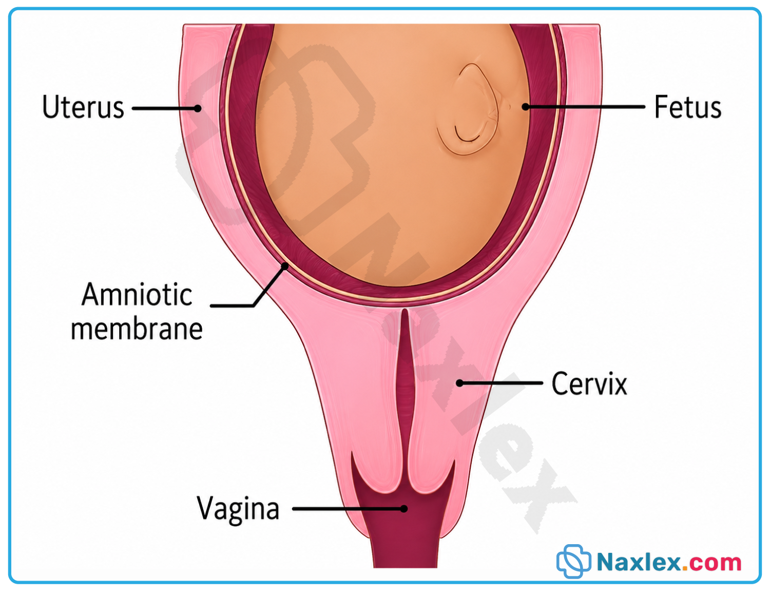
Nursing Insights
- The nurse must recognize that cervical change can occur without client awareness, emphasizing the importance of routine cervical assessment in high-risk clients.
- The nurse should teach clients with cervical insufficiency to report pelvic pressure immediately, as this may indicate advancing cervical change.
Clinical Manifestations And Diagnosis
8.1 Signs and Symptoms
- Clients with preterm labor often present with subtle, easily dismissed symptoms rather than overt labor pain.
- Common presenting symptoms include:
- Uterine contractions occurring at regular intervals, palpable or reported as tightening.
- Low, dull backache that is constant or intermittent, unrelieved by position change.
- Pelvic pressure described as a sensation of the fetus "pushing down."
- Menstrual-like cramping localized to the lower abdomen.
- Increased or changed vaginal discharge, becoming watery, mucoid, or blood-tinged.
- Abdominal cramping with or without diarrhea.
- A sensation of pelvic heaviness or fullness.
- Symptoms are frequently mistaken for normal pregnancy discomfort, delaying client presentation for evaluation.
- Vaginal bleeding, though less common, warrants immediate evaluation for placental abruption or previa.
- Rupture of membranes may present as a sudden gush or a continuous slow leakage of fluid.
Nursing Insights
- The nurse must educate clients on warning signs of preterm labor at every prenatal visit beginning at 20 weeks of gestation.
- The nurse should teach clients that any of these symptoms occurring more than four to six times per hour warrants immediate evaluation.
- The nurse must instruct clients to differentiate true labor backache, which is constant, from musculoskeletal back pain, which varies with position.
8.2 Diagnostic Criteria
- Diagnosis of preterm labor requires both regular uterine contractions and objective evidence of cervical change.
- Diagnostic criteria include:
- Gestational age between 20-0/7 and 36-6/7 weeks.
- Regular contractions at a frequency of ≥4 in 20 minutes or ≥8 in 60 minutes.
- Cervical dilation ≥2 cm or documented progressive change on serial examination.
- Cervical effacement ≥80% in the presence of regular contractions.
- A single cervical examination showing dilation without documented change may represent baseline cervical status rather than active labor.
- Clients with contractions but stable cervical findings are diagnosed with threatened preterm labor, requiring observation rather than aggressive intervention.
Nursing Insights
- The nurse must document baseline cervical findings early in pregnancy for clients with risk factors to allow comparison during suspected preterm labor.
- The nurse should recognize that digital cervical examination is contraindicated in the presence of active vaginal bleeding until placenta previa is excluded.
8.3 Diagnostic Tests and Procedures
- Diagnostic evaluation of suspected preterm labor includes the following tests and procedures:
- Transvaginal ultrasound – measures cervical length; a length <2.5 cm before 24 weeks indicates increased risk.
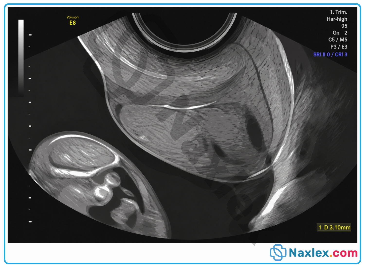
-
- Fetal fibronectin testing – a negative result between 22 and 34 weeks has a high negative predictive value for delivery within 14 days.
- Electronic fetal monitoring – assesses contraction frequency, duration, and fetal heart rate response.
- Sterile speculum examination – visualizes the cervix and assesses for rupture of membranes or bleeding source.
- Nitrazine paper test – detects alkaline amniotic fluid, turning blue when membranes have ruptured.
- Fern test – microscopic evaluation of fluid for characteristic ferning pattern, confirming amniotic fluid.
- Urinalysis and urine culture – rules out urinary tract infection as a contributing cause.
- Vaginal and rectal group B streptococcus culture – guides intrapartum antibiotic prophylaxis if delivery occurs.
- Complete blood count and C-reactive protein – evaluates for underlying infection or inflammation.
- Amniocentesis – may be performed to assess for intra-amniotic infection or fetal lung maturity in select cases.
- Fetal fibronectin testing should not be performed within 24 hours of intercourse, digital examination, or vaginal ultrasound due to false-positive risk.
Nursing Insights
- The nurse must ensure the fetal fibronectin swab is collected before digital cervical examination to prevent contamination and false-positive results.
- The nurse should recognize that a negative fetal fibronectin result allows for outpatient management in select stable clients, reducing unnecessary hospitalization.
- The nurse must obtain a sterile speculum examination, not a digital examination, when rupture of membranes is suspected, to reduce infection risk.
Management Of Preterm Labor
10.1 Tocolytic Therapy
- Tocolytics are pharmacologic agents used to suppress uterine contractions and delay delivery for 48 hours.
- The 48-hour window allows administration of corticosteroids to enhance fetal lung maturity before delivery.
- Tocolytic therapy is contraindicated in the presence of chorioamnionitis, severe preeclampsia, or non-reassuring fetal status.
- Commonly used tocolytic agents include the following:
|
Tocolytic Agent |
Mechanism of Action |
Key Nursing Considerations |
|
Nifedipine (calcium channel blocker) |
Blocks calcium influx into myometrial cells, reducing contractility |
Monitor blood pressure for hypotension; avoid concurrent use with magnesium sulfate |
|
Indomethacin (prostaglandin synthetase inhibitor) |
Inhibits prostaglandin synthesis, reducing uterine activity |
Limit use to <32 weeks; monitor for oligohydramnios and premature ductus arteriosus closure |
|
Terbutaline (beta-adrenergic agonist) |
Stimulates beta-2 receptors, causing myometrial relaxation |
Monitor for maternal tachycardia, palpitations, and pulmonary edema |
|
Magnesium sulfate (tocolytic use) |
Competes with calcium at the cellular level, reducing contractility |
Monitor deep tendon reflexes, respiratory rate, and urine output |
- Tocolytics do not prevent preterm birth long-term; they only delay delivery to allow corticosteroid administration or maternal transport.
- Combination tocolytic therapy is generally avoided due to increased risk of adverse maternal effects.
Nursing Insights
- The nurse must withhold indomethacin beyond 32 weeks of gestation due to the risk of premature closure of the fetal ductus arteriosus.
- The nurse should monitor a client receiving terbutaline for signs of pulmonary edema, including dyspnea, crackles, and oxygen desaturation.
- The nurse must recognize nifedipine and magnesium sulfate should not be administered concurrently due to risk of profound hypotension and neuromuscular blockade.
10.2 Corticosteroids for Fetal Lung Maturity
- Antenatal corticosteroids accelerate fetal surfactant production, reducing the incidence and severity of respiratory distress syndrome.
- Corticosteroids are recommended for clients between 24-0/7 and 33-6/7 weeks of gestation at risk for delivery within 7 days.
- A single repeat course may be considered for clients less than 34 weeks who received an initial course more than 14 days prior.
- Two corticosteroid regimens are commonly used:
- Betamethasone – 12 mg intramuscularly every 24 hours for 2 doses.
- Dexamethasone – 6 mg intramuscularly every 12 hours for 4 doses.
- Maximal fetal benefit occurs approximately 24 hours after the first dose and lasts up to 7 days.
- Corticosteroids also reduce the incidence of intraventricular hemorrhage and necrotizing enterocolitis in preterm neonates.
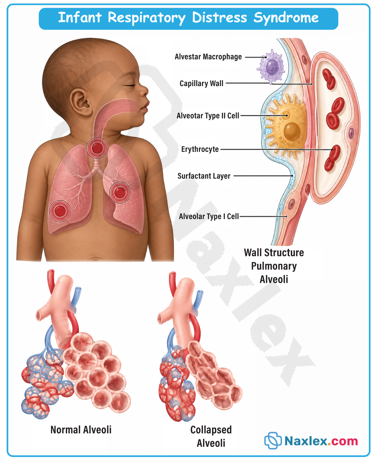
Nursing Insights
- The nurse must administer the corticosteroid dose as scheduled even if delivery appears imminent, since partial dosing still confers some fetal benefit.
- The nurse should document the exact time of administration to allow the neonatal team to anticipate the degree of lung maturity benefit.
- The nurse must monitor maternal blood glucose closely in clients with diabetes mellitus, as corticosteroids transiently elevate blood glucose levels.
10.3 Magnesium Sulfate for Fetal Neuroprotection
- Magnesium sulfate is administered for fetal neuroprotection to reduce the risk and severity of cerebral palsy in preterm infants.
- Neuroprotective magnesium sulfate is recommended for clients at risk of delivery before 32 weeks of gestation.
- A typical regimen includes a loading dose of 4 to 6 g intravenously over 20 to 30 minutes, followed by a maintenance infusion of 1 to 2 g/hr.
- Magnesium sulfate toxicity assessment requires monitoring of the following parameters:
- Deep tendon reflexes – absence indicates possible toxicity.
- Respiratory rate – should remain ≥12 breaths per minute.
- Urine output – should remain ≥30 mL/hr to ensure adequate renal clearance.
- Serum magnesium level – therapeutic range is 4 to 7 mEq/L; toxicity occurs above 8 mEq/L.
- Calcium gluconate 1 g intravenously is the antidote for magnesium sulfate toxicity and must be readily available at the bedside.
Nursing Insights
- The nurse must assess deep tendon reflexes, respiratory rate, and urine output hourly during magnesium sulfate infusion to detect early toxicity.
- The nurse should recognize that absent deep tendon reflexes combined with respiratory depression signals magnesium toxicity requiring immediate intervention.
- The nurse must have calcium gluconate readily accessible at the bedside whenever a client is receiving magnesium sulfate therapy.
10.4 Antibiotic Therapy
- Antibiotic therapy in preterm labor is indicated primarily for group B streptococcus prophylaxis and management of preterm premature rupture of membranes.
- Group B streptococcus prophylaxis is administered intrapartum to clients with unknown or positive culture status to prevent neonatal early-onset sepsis.
- Penicillin G is the first-line agent for group B streptococcus prophylaxis; clindamycin or vancomycin is used for penicillin-allergic clients based on sensitivity.
- Broad-spectrum antibiotics, such as ampicillin and azithromycin, are administered in confirmed preterm premature rupture of membranes to prolong latency.
- Antibiotics are not routinely indicated for preterm labor with intact membranes in the absence of confirmed infection.
Nursing Insights
- The nurse must verify group B streptococcus culture status upon admission and initiate prophylaxis promptly if the client is in active labor.
- The nurse should administer the first dose of antibiotic prophylaxis at least 4 hours before delivery to achieve adequate neonatal protection.
10.5 Activity Restriction and Bed Rest
- Modified activity restriction may be recommended for select clients with preterm labor, though evidence supporting strict bed rest is limited.
- Prolonged bed rest carries risks, including venous thromboembolism, muscle deconditioning, and psychological stress.
- Clients on activity restriction should be taught the following precautions:
- Perform range-of-motion exercises to reduce thromboembolism risk.
- Wear sequential compression devices or compression stockings as prescribed.
- Report calf pain, swelling, or shortness of breath immediately.
- Pelvic rest, including avoidance of sexual intercourse, is often recommended to reduce prostaglandin exposure and mechanical cervical stimulation.
Nursing Insights
- The nurse must educate the client on the risks of prolonged bed rest, including deep vein thrombosis, and encourage prescribed range-of-motion exercises.
- The nurse should recognize that current evidence does not support strict bed rest as an effective intervention to prevent preterm birth.
Nursing Management And Care
12.1 Nursing Assessment
- Comprehensive nursing assessment of the client with suspected preterm labor includes the following components:
- Obtain a detailed obstetric and gynecologic history, including prior preterm birth and cervical procedures.
- Assess vital signs, noting maternal temperature, which may indicate underlying infection.
- Palpate uterine contractions for frequency, duration, and intensity.
- Apply continuous electronic fetal monitoring to assess fetal heart rate and contraction pattern.
- Perform sterile speculum examination if rupture of membranes is suspected.
- Assess for vaginal bleeding, discharge characteristics, and pelvic pressure.
- Review laboratory and diagnostic results, including fetal fibronectin, cervical length, and group B streptococcus status.
- Ongoing assessment includes monitoring maternal and fetal response to tocolytic, corticosteroid, and magnesium sulfate therapy.
Nursing Insights
- The nurse must correlate maternal temperature elevation with fetal tachycardia, as both together suggest chorioamnionitis requiring prompt intervention.
- The nurse should reassess cervical status only as clinically indicated, minimizing digital examinations to reduce infection risk in clients with ruptured membranes.
12.2 Nursing Interventions
- Priority nursing interventions for the client with preterm labor include the following:
- Position the client in the left lateral position to optimize uteroplacental perfusion.
- Initiate intravenous access and administer intravenous fluids as prescribed to promote uterine relaxation.
- Administer tocolytic, corticosteroid, antibiotic, and magnesium sulfate therapy as prescribed, monitoring for adverse effects.
- Monitor intake and output strictly, particularly during magnesium sulfate infusion.
- Provide continuous fetal monitoring and report non-reassuring fetal heart rate patterns immediately.
- Prepare for possible neonatal intensive care unit admission by notifying the neonatal team in advance.
- Provide emotional support, acknowledging maternal anxiety and fear related to preterm birth.
- Nurses must coordinate interdisciplinary care involving obstetric, neonatal, and pharmacy teams for optimal outcomes.
Nursing Insights
- The nurse must position the client in the left lateral position to reduce compression of the inferior vena cava and improve placental blood flow.
- The nurse should anticipate the need for neonatal resuscitation equipment at delivery whenever preterm birth is imminent.
12.3 Client and Family Education
- Client education is essential to promote early recognition of preterm labor symptoms and adherence to the treatment plan.
- Key educational points include the following:
- Teach the client to recognize and report warning signs, including contractions, pelvic pressure, and fluid leakage.
- Explain the purpose, administration schedule, and potential side effects of prescribed medications.
- Instruct the client on activity modifications, including pelvic rest and limited exertion as prescribed.
- Prepare the client and family for the possibility of preterm delivery and neonatal intensive care unit admission.
- Encourage the client to verbalize fears and concerns, providing psychosocial support throughout the hospitalization.
- Provide information regarding signs of magnesium toxicity if the client will be monitored at home or transferred.
- Discharge teaching, if the client is stabilized, includes instructions on when to seek immediate care.
Nursing Insights
- The nurse must ensure the client and family understand the purpose of a neonatal intensive care unit tour, if available, to reduce anxiety before preterm delivery.
- The nurse should use teach-back methods to confirm the client's understanding of warning signs before discharge.
Complications Of Preterm Labor And Preterm Birth
14.1 Maternal Complications
- Maternal complications associated with preterm labor and its management include the following:
- Postpartum hemorrhage – uterine atony risk increases following prolonged tocolytic therapy and rapid labor progression.
- Infection – chorioamnionitis and endometritis risk increase with prolonged rupture of membranes and multiple cervical examinations.
- Pulmonary edema – associated with prolonged beta-adrenergic tocolytic use combined with intravenous fluid administration.
- Psychological distress – anxiety, depression, and post-traumatic stress symptoms are common following preterm birth experiences.
- Adverse effects of tocolytic therapy – including maternal tachycardia, hypotension, and, rarely, pulmonary edema or cardiac dysrhythmia.
- Failed tocolysis – progression to preterm birth despite pharmacologic intervention, often necessitating emergent delivery.
- Clients with cervical insufficiency requiring emergent cerclage face additional surgical risks, including membrane rupture and infection.
Nursing Insights
- The nurse must monitor fluid balance closely in clients receiving both intravenous tocolytics and corticosteroids due to combined fluid retention risk.
- The nurse should assess for signs of postpartum depression and anxiety at follow-up visits, as preterm birth significantly increases this risk.
- The nurse must recognize that failed tocolysis requires rapid transition of care priorities toward preparation for preterm delivery and neonatal support.
14.2 Neonatal Complications
- Preterm neonates are at risk for multiple organ system complications related to physiologic immaturity, including the following:
- Respiratory distress syndrome – results from surfactant deficiency, causing alveolar collapse and impaired gas exchange.
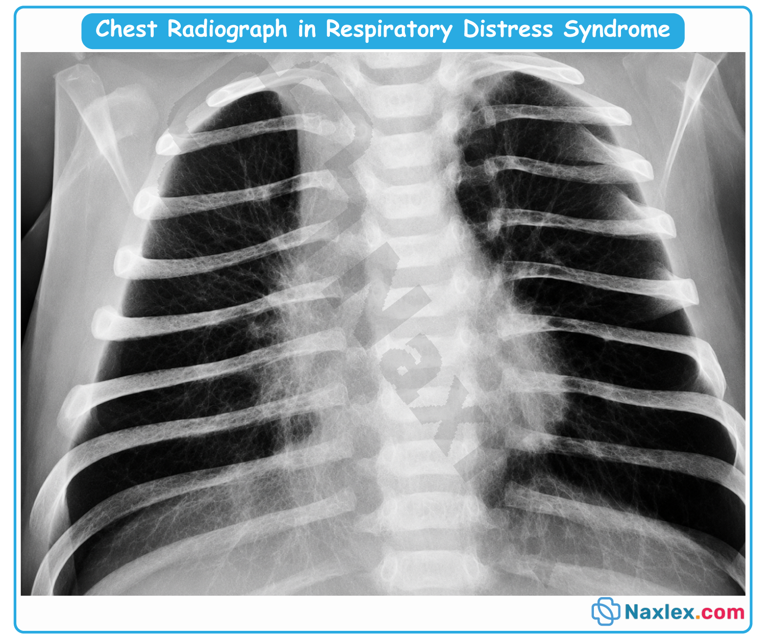
-
- Intraventricular hemorrhage – bleeding into the periventricular and intraventricular regions due to fragile cerebral vasculature.
- Necrotizing enterocolitis – ischemic and inflammatory bowel injury related to immature gastrointestinal function.
- Retinopathy of prematurity – abnormal retinal vascular development, often related to oxygen therapy exposure.
- Bronchopulmonary dysplasia – chronic lung disease resulting from prolonged mechanical ventilation and oxygen exposure.
- Patent ductus arteriosus – failure of ductal closure, common in extremely preterm infants.
- Thermoregulation impairment – due to decreased subcutaneous fat and immature hypothalamic function, predisposing to cold stress.
- Hypoglycemia – related to limited glycogen stores and immature glucose regulation.
- Hyperbilirubinemia – resulting from hepatic immaturity and reduced conjugation capacity.
- Sepsis – increased susceptibility due to immature immune function and invasive procedures in the neonatal intensive care unit.
- Long-term complications may include cerebral palsy, developmental delay, chronic lung disease, and vision or hearing impairment.
Nursing Insights
- The nurse must recognize that the earlier the gestational age at birth, the greater the risk and severity of neonatal complications.
- The nurse should anticipate immediate thermoregulation support, such as placement in a radiant warmer, for every preterm delivery.
- The nurse must monitor preterm neonates closely for feeding intolerance, abdominal distension, and bloody stools as early signs of necrotizing enterocolitis.
Prevention Strategies
15.1 Primary Prevention
- Primary prevention focuses on reducing modifiable risk factors before preterm labor develops in the general obstetric population.
- Key primary prevention strategies include the following:
- Promote early and consistent prenatal care beginning in the first trimester.
- Encourage smoking cessation, substance abuse treatment, and avoidance of alcohol during pregnancy.
- Screen and treat asymptomatic bacteriuria, periodontal disease, and sexually transmitted infections.
- Optimize maternal nutrition, addressing underweight status and micronutrient deficiencies.
- Educate clients on interpregnancy spacing, recommending at least 18 months between pregnancies.
- Provide psychosocial support and screen for intimate partner violence and depression.
Nursing Insights
- The nurse must initiate risk factor screening at the very first prenatal encounter to allow timely referral and intervention.
- The nurse should counsel every reproductive-age client on optimal interpregnancy interval as part of routine preconception counseling.
15.2 Secondary Prevention
- Secondary prevention targets clients already identified as high-risk for preterm labor based on history or clinical findings.
- Key secondary prevention strategies include the following:
- Progesterone supplementation – vaginal or intramuscular progesterone reduces recurrence risk in clients with prior spontaneous preterm birth.
- Cervical cerclage – surgical reinforcement of the cervix, indicated for clients with cervical insufficiency or shortened cervix on ultrasound.
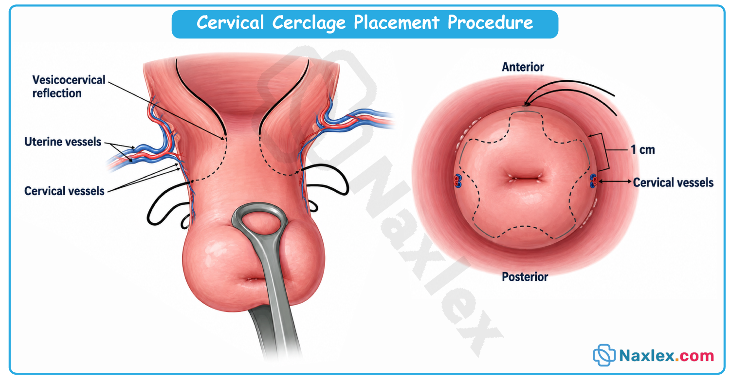
-
- Cervical pessary – a mechanical device redistributing uterine weight away from the cervix, used as an alternative to cerclage in select clients.
- Serial cervical length surveillance – transvaginal ultrasound monitoring in clients with prior preterm birth to detect early shortening.
- Patient education on symptom recognition – reinforced at each visit for clients identified as high-risk.
- Progesterone therapy is most effective when initiated between 16 and 20 weeks and continued through 36 weeks of gestation.
Nursing Insights
- The nurse must reinforce that cervical cerclage is typically placed between 12 and 14 weeks of gestation for prophylactic indications.
- The nurse should teach the client receiving vaginal progesterone proper insertion technique and the importance of consistent nightly administration.
- The nurse must recognize that cerclage is contraindicated in the presence of active labor, infection, or ruptured membranes.
Summary
- Preterm labor is defined as regular contractions with cervical change occurring between 20-0/7 and 36-6/7 weeks of gestation.
- Preterm birth remains a leading cause of neonatal morbidity and mortality worldwide, disproportionately affecting underserved populations.
- Etiology is multifactorial, encompassing maternal, fetal, placental, and behavioral risk factors that often overlap in a single client.
- Pathophysiology involves four overlapping pathways: infection/inflammation, decidual hemorrhage, uterine overdistension, and maternal-fetal stress.
- Diagnosis requires both regular contractions and objective cervical change, supported by transvaginal ultrasound and fetal fibronectin testing.
- Management centers on tocolysis to delay delivery, corticosteroids for fetal lung maturity, and magnesium sulfate for fetal neuroprotection.
- Nursing care emphasizes vigilant assessment, timely intervention, medication safety monitoring, and psychosocial support for the client and family.
- Complications range from maternal infection and hemorrhage to severe neonatal morbidities, including respiratory distress syndrome and intraventricular hemorrhage.
- Prevention strategies include primary risk reduction for all clients and secondary interventions, such as progesterone and cerclage, for high-risk clients.
- Comprehensive, evidence-based nursing management significantly improves maternal and neonatal outcomes in the setting of preterm labor.
Naxlex
Videos
Login to View Video
Click here to loginTake Notes on Preterm Labor
This filled cannot be empty
Join Naxlex Nursing for nursing questions & guides! Sign Up Now