
Chest Tube Insertion and Monitoring
Lessons
- Objectives
- Introduction
- Anatomy And Physiology Of The Pleural Space
- Indications For Chest Tube Placement
- Nursing Roles In Chest Tube Insertion
- Practice Exercise 1
- The Chest Drainage System
- Essential Nursing Assessment And Monitoring
- Critical Nursing Interventions And Troubleshooting
- Chest Tube Removal
- Summary
- Practice Exercise 2
Notes Highlighting is available once you sign in. Login Here.
Objectives
- Explain the normal anatomy and negative pressure dynamics of the pleural space.
- Identify the key clinical indications for chest tube.
- Detail the essential nursing steps for patient preparation and management before, during, and after insertion.
- Describe the function of the three chambers in the Chest Drainage System (CDS) and their associated nursing checks.
- Interpret the significance of bubbling and tidaling in the water-seal chamber.
- Execute critical, rapid nursing interventions for emergencies, such as chest tube dislodgement or disconnection.
- Outline the criteria and nursing role for safe chest tube removal.
Introduction
- A chest tube is a flexible tube inserted into the pleural space to remove air, fluid, or pus.
- It helps re-expand collapsed lungs and maintain proper lung function.
- Chest tubes are commonly used for pneumothorax, hemothorax, pleural effusions, or post-thoracic surgery drainage.
- The tube connects to a drainage system that may include a water seal and suction source.
- Nurses must monitor respiratory status, vital signs, and oxygen saturation closely.
- The insertion site should be checked regularly for bleeding, infection, or subcutaneous emphysema.
- Tubing should remain free of kinks and connections must be secure to ensure proper drainage.
- Patient education includes explaining the procedure, encouraging deep breathing, and teaching signs of complications.
Anatomy And Physiology Of The Pleural Space
The chest tube system is designed to manage conditions affecting the pleural space—the potential space between the visceral pleura and the parietal pleura. In a healthy state, this space contains a small amount of lubricating fluid (about 20-25 mL) that allows the lungs to slide smoothly against the chest wall during respiration. The pressure within the pleural space is normally negative, which helps maintain lung expansion.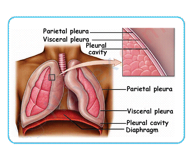
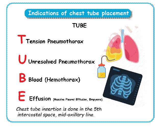
Indications For Chest Tube Placement
A chest tube, or thoracic catheter, is inserted into the pleural space to drain fluid, blood, or air, thereby restoring negative pressure and allowing the collapsed or partially collapsed lung to re-expand.
Common Conditions Requiring Chest Tubes:
- Pneumothorax: The presence of air in the pleural space, causing partial or complete lung collapse. This can be spontaneous, traumatic e.g., fractured rib, or iatrogenic e.g., biopsy complication.
- Tension Pneumothorax: A life-threatening emergency where air enters the pleural space but cannot exit, leading to rapidly increasing pressure that shifts the mediastinum (heart, trachea) and compresses the unaffected lung and great vessels.
- Hemothorax: The presence of blood in the pleural space, often due to trauma or surgery.
- Chylothorax: Accumulation of lymphatic fluid in the pleural space.
- Empyema: Collection of purulent fluid in the pleural space, usually secondary to pneumonia or infection.
- Post-Thoracic Surgery: Placement following procedures like thoracotomy or cardiac surgery (e.g., CABG) to drain residual fluid and air.
Nursing Roles In Chest Tube Insertion
The nurse plays a critical role in supporting the patient and assisting the provider throughout the entire insertion process to ensure patient comfort, safety, and system readiness.
Before Insertion
The primary goals are patient education, informed consent verification, and preparation of the sterile field and drainage system.
- Verify Orders and Consent: Confirm the physician’s order for chest tube insertion, including the side, size of the tube, and type of drainage system required (e.g., portable, standard CDS with suction). Verify that the patient or legally authorized representative has signed the informed consent document.
- Patient Education and Positioning: Explain the procedure in simple, clear terms, addressing patient anxiety. Position the patient appropriately: generally, the patient is placed in the Semi-Fowler's position or on the unaffected side with the arm raised above the head to expose the insertion site (usually the 4th or 5th intercostal space).
- Pain and Sedation: Administer ordered pre-procedure analgesics or sedatives as prescribed to ensure patient comfort during the procedure.
- Gather Equipment: Prepare the sterile chest tube tray, chest tube of the appropriate size, antiseptic solution, local anesthetic, sutures, sterile gloves and gowns for the provider, and an occlusive dressing kit.
- Prepare the Drainage System: Fill the water-seal chamber and the water-suction control chamber with the required amount of sterile water or saline. Keep the system capped and immediately available, below the level of the insertion site.
During Insertion
The nurse monitors the patient's vital signs and reaction while maintaining a sterile environment and assisting the provider.
- Maintain Sterility: Open the sterile supplies and assist the provider in donning sterile garb. Ensure the sterile field is not contaminated.
- Monitor Patient Status: Continuously monitor the patient’s vital signs, cardiac rhythm, respiratory rate, and oxygen saturation. Watch for signs of vasovagal response (bradycardia, hypotension) or pneumothorax exacerbation.
- Medication Administration: May need to administer additional local anesthetic or sedation upon the provider's request.
- Connection: As soon as the chest tube is inserted, and before the provider sutures the site, quickly and sterilely connect the chest tube to the prepared chest drainage system to re-establish negative pressure.
- Initial Assessment: Once connected, look for immediate drainage of fluid or air.
After Insertion
Immediate assessment and securing the system are paramount for effective therapy and complication prevention.
- Secure the System: Coil and secure the chest tube tubing to the patient's chest wall to prevent accidental dislodgement or tension on the insertion site. Ensure the tubing runs straight to the CDS without dependent loops.
- Apply Occlusive Dressing: Ensure the provider applies a clean, occlusive dressing (petrolatum gauze, followed by a sterile 4x4 or similar dressing) and tapes it securely to seal the insertion site.
- Initial Drainage Documentation: Note the immediate output (color, amount) in the collection chamber. Document the dressing integrity, the depth of the tube, and the presence of tidaling or bubbling in the water seal.
- Initiate Suction: Connect the drainage system to the wall suction and turn it on to the prescribed level (e.g., -20 cm H2O). Ensure gentle, continuous bubbling is present in the suction control chamber (if wet system).
- Verification: Obtain a stat chest X-ray immediately after insertion to verify the tube’s correct placement and to assess the degree of lung re-expansion.
- Reassess: Perform a full respiratory assessment, comparing pre- and post-insertion findings. Document the patient's pain level and administer analgesia as needed.
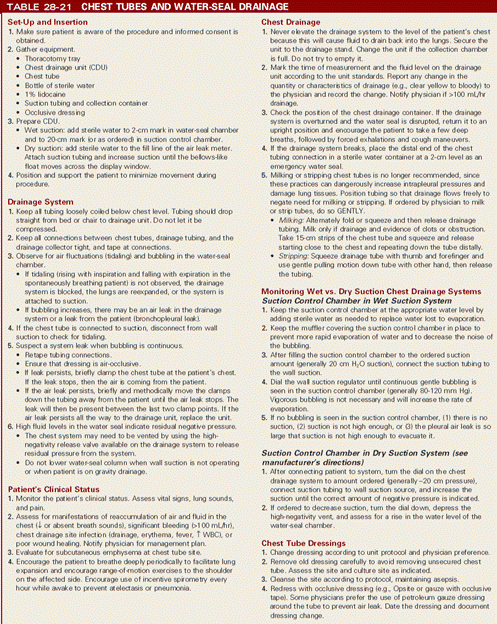
The Chest Drainage System
The Chest Drainage System (CDS), (often called a Pleur-Evac or Atrium device) is a sterile, closed, disposable unit that uses a three-chamber system to manage fluid, air, and pressure.
Chamber 1: Collection Chamber
- Function: Gathers fluid and air draining from the patient's chest.
- Nursing Focus:
- Monitor the color, consistency, and amount of drainage (output).
- Mark the fluid level (date, time, and initials) on the outside of the chamber at least every shift, and more frequently if drainage is heavy (e.g., hourly post-operatively).
- Report Immediate Concerns: Sudden increase (>100 mL/hr, especially if bright red) or a sudden decrease in drainage.
Chamber 2: Water-Seal Chamber
- Function: Acts as a one-way valve to prevent air and fluid from entering the pleural space while allowing them to escape. It contains 2 cm of sterile water.
- Nursing Focus:
- Air Leak Indicator: Bubbling in this chamber indicates an air leak.
- Intermittent/Tidaling Bubbling: Common in patients with a pneumothorax as air is actively escaping the lung.
- Continuous/Vigorous Bubbling: Indicates a large air leak, which could be from the patient (bronchopleural fistula) or a leak in the system (connections, insertion site, or damaged unit). This needs immediate investigation.
- Tidaling: Fluctuation of the water level with the patient's respirations.
- Inspiration: Water level rises - due to increased negative pressure.
- Expiration: Water level falls - due to decreased negative pressure.
- Significance: Tidaling is normal and indicates the system is working and the tubing is patent. If tidaling stops, the lung may be fully re-expanded, or the tube may be kinked or obstructed.
- Air Leak Indicator: Bubbling in this chamber indicates an air leak.
Chamber 3: Suction Control Chamber
- Function: Controls the amount of negative pressure applied to the chest cavity. Suction is typically ordered at -20 cm H2O.
- Types of Suction Control:
- Water Suction Control: Requires sterile water (usually 20 cm) to be added. Gentle, continuous bubbling in this chamber is the expected finding and confirms that the desired level of suction is being applied (often called a "good boil"). The amount of suction is determined by the height of the water, not the suction source setting.
- Dry Suction Control: Uses a rotary suction control dial to set the level (e.g., -20 cm H2O). It does not require water and has a visual bellows or float indicator to confirm suction is active. This type typically produces no bubbling sounds.
Essential Nursing Assessment And Monitoring
Respiratory Assessment
- Rate and Depth: Monitor for signs of respiratory distress (tachypnea, shallow breathing).
- Breath Sounds: Auscultate frequently. Note if breath sounds are improved, equal bilaterally, or diminished/absent over the affected lung.
- Oxygen Saturation: Maintain SpO2 per orders, often > 92%.
- Tracheal Alignment: Assess for tracheal deviation, a late but critical sign of tension pneumothorax.
Insertion Site Assessment
- Check the dressing for stability, security, and integrity.
- Assess the skin around the insertion site for subcutaneous emphysema.
- Ensure the chest tube is securely sutured and that the eyelet openings are not visible.
Tubing and Drainage System
- Tubing Patency: Check the tubing frequently for kinks, loops, or dependent pooling of fluid, which can obstruct drainage. Keep the tubing straight and secured to the patient's gown or bed linen.
- Drainage System Position: Always keep the drainage system unit below the level of the patient’s chest.
- Water Levels: Ensure the water seal and suction control chambers have the correct amount of sterile water.
- Bubbling/Tidaling: Continuously monitor the water-seal chamber for the presence or absence of tidaling and bubbling.
Critical Nursing Interventions And Troubleshooting
Troubleshooting Air Leaks
|
Finding |
Indication |
Nursing Intervention |
|
Air Leak in Water Seal |
Air is exiting the pleural space. |
Locate the source: Temporarily clamp the tubing close to the chest insertion site. If bubbling stops, the leak is inside the chest or at the insertion site. If bubbling continues, unclamp immediately and check the connection sites down the tubing and at the CDS unit. |
|
New, Continuous, Vigorous Bubbling |
Leak in the system (tubing disconnected/cracked unit) or a worsening leak in the patient. |
Check all connections and ensure the chest tube is secured to the chest wall. Notify the provider if the leak persists and the patient's respiratory status declines. |
|
No Bubbling in Suction Chamber (Wet System) |
Suction is not adequate or the suction source is off. |
Check the wall suction setting (it should be on and functioning). Ensure the water level in the suction control chamber is correct. |
Immediate Actions for Emergencies
|
Situation |
Nursing Action |
Rationale |
|
Tube Dislodgement (Pulled out of patient) |
1. Immediately apply pressure to the site. 2. Cover the site with a sterile gauze dressing secured on three sides (vent dressing). 3. Notify the Rapid Response Team/Provider. |
The three-sided dressing acts as a flutter valve: air can escape during exhalation but cannot enter during inhalation, preventing a tension pneumothorax. |
|
Tube Disconnection (From CDS) |
1. Instruct the patient to exhale fully and cough. 2. Submerge the end of the chest tube in 2 cm of sterile water or saline. 3. Clean the connection site with antiseptic and reconnect to the CDS. |
Re-establishes the water seal immediately to prevent air from rushing into the pleural space. |
|
Sudden, Massive Bright Red Drainage (>100 mL/hr) |
1. Check vital signs (BP, HR). 2. Notify the Provider and Rapid Response Team immediately. 3. Prepare for blood product administration. |
Suggests hemorrhage or bleeding from a major vessel—a surgical emergency. |
Clamping the Chest Tube
Chest tubes are generally never clamped without a direct order from the provider. Clamping a chest tube in the presence of an air leak can lead to a rapid tension pneumothorax because air cannot escape.
- Allowed Reasons for Brief Clamping (Under Provider Order):
- To quickly assess for the location of an air leak.
- To change the drainage unit.
- One hour before removal to assess the patient's tolerance.
Patient Positioning and Mobility
- Positioning: Encourage semi-Fowler's position to promote lung expansion and drainage.
- Ambulation: Ambulation and movement are encouraged to promote drainage and lung re-expansion. The CDS must be maintained below the chest level during transport or walking.
- Coughing/Deep Breathing: Instruct the patient to cough and deep breathe every 2 hours to help expand the lungs.
- Arm Exercises: Range-of-motion exercises for the shoulder on the affected side prevent shoulder stiffness (frozen shoulder).
Chest Tube Removal
The provider determines the time for removal when:
- The lung is fully re-expanded (confirmed by chest X-ray).
- Drainage has decreased to an acceptable amount (e.g., <50 mL in 24 hours).
- Bubbling/air leak has ceased.
- The patient has tolerated clamping of the tube for a prescribed period.
Nursing Role During Removal
- Pre-procedure: Administer ordered analgesic 30–60 minutes prior to the procedure. Gather necessary supplies (suture removal kit, petroleum gauze, 4x4 gauze, wide adhesive tape).
- Procedure:
- The patient takes a deep breath and performs a Valsalva maneuver or exhales fully and holds their breath while the provider rapidly removes the tube. This minimizes the risk of air entry into the pleural space.
- Post-procedure:
- Immediately apply an occlusive dressing (petroleum-coated gauze and a pressure dressing) to the site and secure with tape.
- Monitor for respiratory distress.
- Obtain a post-removal chest X-ray to confirm lung status.
- Document the procedure, patient tolerance, site appearance, and amount of drainage.
Summary
- Chest tubes are inserted into the pleural space to remove air, fluid, or pus.
- They help restore normal lung expansion and improve breathing.
- Indications include pneumothorax, hemothorax, pleural effusion, and post-surgical drainage.
- The tube is connected to a drainage system that may use a water seal or suction.
- Nurses monitor respiratory status, vital signs, and oxygen levels regularly.
- The insertion site must be assessed for bleeding, infection, or swelling.
- Proper tube placement, secure connections, and free-flowing tubing are essential for effectiveness.
- Patient education focuses on procedure explanation, deep breathing exercises, and recognizing complications.
