
Please set your exam date
Respiratory Diagnostic Procedures
Study Questions
Practice Exercise 1
A 92-year-old female patient is being admitted to the emergency department with severe shortness of breath. Being aware of the patient’s condition, what approach should the nurse use to assess the patient’s lungs? (Select all that apply.)
Explanation
The nurse should assess the lungs beginning at the bases and move upward toward the apices, including the lateral fields, and start on the posterior chest before moving to the anterior. This systematic approach ensures that areas most likely to develop abnormal breath sounds—such as the lower lobes—are assessed first, providing the most accurate evaluation of respiratory function in a patient experiencing severe shortness of breath.
Rationale for correct answers:
2. Base to apex. Beginning auscultation at the lung bases allows the nurse to identify early adventitious sounds, such as crackles, which often develop first in the lower lobes due to gravity-dependent fluid accumulation. This approach ensures that subtle changes associated with conditions like pulmonary edema or pneumonia are detected early in the assessment.
3. Lateral sequence. Including the lateral chest areas provides access to the right middle lobe and lingula of the left lung, which are not well heard from the anterior or posterior chest. Assessing these areas ensures a complete and thorough evaluation of all lung fields for possible localized abnormalities.
5. Posterior then anterior. The posterior chest contains a greater portion of lung tissue, making it the most effective area for identifying breath sounds and potential abnormalities. Starting posteriorly and then moving anteriorly provides a more comprehensive assessment of air movement and lung expansion.
Rationale for incorrect answers:
1. Apex to base. This method is commonly used for general assessments but may miss lower-lobe abnormalities in patients who are acutely short of breath. Because fluid and secretions often collect in the bases first, starting at the apex can delay the recognition of significant findings.
4. Anterior then posterior. Beginning the assessment on the anterior chest may overlook critical findings in the posterior lung fields where most adventitious sounds originate. In patients with respiratory distress, starting posteriorly allows for a clearer and more complete assessment of lung function.
Take-home points:
• Assessing from base to apex helps identify lower-lobe abnormalities early.
• Include the lateral lung fields to evaluate all lobes thoroughly.
• Begin posteriorly and move anteriorly for optimal assessment of breath sounds.
• A systematic approach ensures accurate detection of respiratory complications in patients with shortness of breath.
What keeps alveoli from collapsing?
Explanation
Surfactant is a phospholipid substance secreted by type II alveolar cells that reduces surface tension within the alveoli, preventing them from collapsing during exhalation. It helps maintain alveolar stability and promotes efficient gas exchange by keeping the alveoli open and compliant throughout the breathing cycle. Without adequate surfactant, alveolar collapse (atelectasis) can occur, leading to impaired oxygenation and respiratory distress.
Rationale for correct answer:
2. Surfactant. Surfactant lowers the surface tension of the alveolar fluid, which allows the alveoli to remain expanded even after exhalation. It is essential for normal lung compliance and effective gas exchange. A deficiency in surfactant, such as in premature infants with respiratory distress syndrome, leads to alveolar collapse and decreased oxygenation.
Rationale for incorrect answers:
1. Carina. The carina is the point where the trachea divides into the right and left main bronchi. It plays no role in maintaining alveolar stability; instead, it functions primarily as a landmark and a sensitive area that triggers coughing when irritated.
3. Empyema. Empyema refers to the accumulation of pus in the pleural space due to infection. Rather than preventing collapse, it can actually impair lung expansion and gas exchange by restricting lung movement.
4. Thoracic cage. The thoracic cage protects the lungs and supports the respiratory system structurally, but it does not directly prevent alveolar collapse. Its role is mechanical, aiding in ventilation rather than maintaining alveolar patency.
Take-home points:
• Surfactant prevents alveolar collapse by reducing surface tension within the lungs.
• Adequate surfactant is crucial for maintaining effective gas exchange and lung compliance.
• Deficiency of surfactant can lead to atelectasis and respiratory distress.
• Structural components like the thoracic cage support breathing but do not maintain alveolar stability.
What accurately describes the alveolar sacs?
Explanation
The alveolar sacs are the terminal structures of the respiratory tract where the process of gas exchange occurs. Each sac consists of clusters of alveoli surrounded by a dense network of pulmonary capillaries, allowing oxygen to diffuse into the blood and carbon dioxide to diffuse out. These sacs represent the endpoint of the bronchial tree and are critical for maintaining effective respiration.
Rationale for correct answer:
3. Terminal structures of the respiratory tract. The alveolar sacs mark the distal end of the respiratory system and are the primary sites of gas exchange between the air and the bloodstream. They contain numerous alveoli, which provide a large surface area and thin membrane interface essential for efficient oxygen and carbon dioxide diffusion. Damage or destruction of alveolar sacs, as seen in conditions such as emphysema, significantly impairs respiratory efficiency.
Rationale for incorrect answers:
1. Line the lung pleura. The alveolar sacs do not line the pleura; instead, the pleura are double-layered membranes that encase the lungs and line the thoracic cavity to reduce friction during breathing. The alveoli are located deep within the lung parenchyma, not along the pleural surface.
2. Warm and moisturize inhaled air. Warming and humidifying inspired air are functions of the upper respiratory tract, particularly the nasal passages and nasopharynx. The alveolar sacs are involved in gas exchange, not air conditioning.
4. Contain dead air that is not available for gas exchange. The alveolar sacs contain functional air that actively participates in gas exchange, not dead air. Dead space air is found in the conducting airways, such as the trachea and bronchi, where no exchange occurs.
Take-home points:
• The alveolar sacs are the final structures of the respiratory tract and the main sites of gas exchange.
• Their structure maximizes surface area for diffusion of oxygen and carbon dioxide.
• They do not warm or humidify air—that function occurs in the upper airway.
• Destruction of alveolar sacs reduces gas exchange efficiency, as seen in chronic lung diseases.
What covers the larynx during swallowing?
Explanation
The epiglottis is a leaf-shaped flap of cartilage located at the base of the tongue that covers the laryngeal opening during swallowing. Its primary function is to prevent food and liquids from entering the trachea by directing them instead into the esophagus. This protective mechanism ensures that the airway remains clear and prevents aspiration during the act of swallowing.
Rationale for correct answer:
2. Epiglottis. The epiglottis acts as a crucial safeguard for the lower respiratory tract by closing over the glottis when swallowing occurs. This action prevents aspiration of food, fluids, or secretions into the lungs, which could otherwise lead to choking or respiratory infections such as aspiration pneumonia. Its automatic, reflexive movement ensures airway protection without conscious effort.
Rationale for incorrect answers:
1. Trachea. The trachea, or windpipe, is the airway that carries air to and from the lungs. It does not play a role in covering the larynx; instead, it remains open to facilitate airflow except when protected by the epiglottis during swallowing.
3. Turbinates. Turbinates are structures within the nasal cavity that warm, filter, and humidify inspired air. They are part of the upper respiratory tract but have no role in protecting the airway during swallowing.
4. Parietal pleura. The parietal pleura is a membrane lining the inside of the thoracic cavity. It serves to reduce friction during respiration, not to protect the airway or cover the larynx.
Take-home points:
• The epiglottis prevents aspiration by closing over the larynx during swallowing.
• This reflex action directs food and fluids into the esophagus instead of the trachea.
• Structures like the trachea, turbinates, and pleura serve other respiratory functions but do not protect the airway during swallowing.
• Proper epiglottic function is essential for safe swallowing and airway protection.
When does the nurse record the presence of an increased anteroposterior (AP) diameter of the chest?
Explanation
An increased anteroposterior (AP) diameter of the chest is recorded when the width (transverse diameter) and the depth (AP diameter) of the chest appear approximately equal. This change gives the chest a rounded, barrel-like appearance and is most commonly associated with chronic obstructive pulmonary disease (COPD), particularly emphysema. The finding indicates long-term hyperinflation of the lungs and loss of normal chest elasticity.
Rationale for correct answer:
2. The width of the chest is equal to the depth of the chest. In a healthy adult, the normal ratio of the anteroposterior to transverse chest diameter is about 1:2. When this ratio approaches 1:1, the chest takes on a barrel shape, signifying chronic air trapping and overexpansion of the alveoli. This adaptation occurs gradually in diseases that cause long-standing respiratory effort, such as COPD, and reflects structural changes in the thoracic cavity due to hyperinflation.
Rationale for incorrect answers:
1. There is a prominent protrusion of the sternum. A protruding sternum is known as pectus carinatum, or pigeon chest, a congenital deformity unrelated to increased AP diameter. It involves abnormal chest wall structure rather than chronic pulmonary changes.
3. There is equal but diminished movement of the two sides of the chest. This finding suggests restricted chest wall movement, possibly due to neuromuscular weakness, pleural effusion, or severe respiratory fatigue, not an increased AP diameter.
4. The patient cannot fully expand the lungs because of kyphosis of the spine. Kyphosis causes a stooped posture and restricts chest expansion but does not result in an increased AP diameter. It leads to decreased lung expansion due to spinal curvature rather than barrel chest formation.
Take-home points:
• An increased AP diameter gives the chest a barrel-shaped appearance where width equals depth.
• This finding is most often seen in patients with COPD or emphysema due to chronic lung hyperinflation.
• Structural deformities like pectus carinatum or kyphosis alter chest shape differently and are not associated with increased AP diameter.
• Recognizing a barrel chest can help the nurse identify signs of chronic pulmonary disease during physical assessment.
Practice Exercise 2
A patient with a respiratory condition asks, “How does air get into my lungs?” The nurse bases the answer on knowledge that air moves into the lungs because of:
Explanation
Air moves into the lungs because of a decrease in intrathoracic pressure relative to pressure at the airway. During inspiration, the diaphragm contracts and moves downward while the intercostal muscles lift the rib cage, expanding the thoracic cavity. This expansion lowers the pressure within the lungs below atmospheric pressure, causing air to flow in until equilibrium is reached.
Rationale for correct answer:
4. Decrease in intrathoracic pressure relative to pressure at the airway. Inhalation occurs as a result of pressure gradients between the atmosphere and the lungs. When the thoracic cavity expands, the intrapulmonary pressure drops below atmospheric pressure, allowing air to move in passively. This principle, known as negative pressure ventilation, is fundamental to normal breathing and ensures that oxygen enters the alveoli for gas exchange.
Rationale for incorrect answers:
1. Contraction of the accessory abdominal muscles. These muscles are primarily involved in forced expiration, not inspiration. They assist in expelling air from the lungs during activities such as coughing or vigorous exercise, rather than drawing air in.
2. Increased carbon dioxide and decreased oxygen in the blood. While changes in blood gas levels stimulate the respiratory center in the brain to increase the rate and depth of breathing, they do not directly cause the mechanical movement of air into the lungs. They serve as a chemical trigger rather than a physical mechanism of ventilation.
3. Stimulation of the respiratory muscles by the chemoreceptors. Chemoreceptors detect changes in blood pH, CO₂, and O₂ levels and send signals to the respiratory center to adjust breathing. However, it is the muscular action that changes thoracic pressure, not the chemoreceptor activity itself, that causes air inflow.
Take-home points:
- Air enters the lungs when intrathoracic pressure falls below atmospheric pressure.
- This occurs through contraction of the diaphragm and intercostal muscles, creating negative pressure.
- Chemoreceptors regulate the rate and depth of breathing but do not directly move air.
- Understanding pressure gradients helps explain the mechanics of normal and assisted ventilation.
The nurse can best determine adequate arterial oxygenation of the blood by assessing:
Explanation
The nurse can best determine adequate arterial oxygenation of the blood by assessing the arterial oxygen tension (PaO₂). This value, obtained through arterial blood gas (ABG) analysis, measures the partial pressure of oxygen dissolved in arterial blood and directly reflects how well oxygen is able to move from the lungs into the bloodstream. A normal PaO₂ (typically 80–100 mm Hg in healthy adults) indicates effective oxygenation and gas exchange at the alveolar level.
Rationale for correct answer:
3. Arterial oxygen tension (PaO₂). PaO₂ is the most accurate indicator of arterial oxygenation because it quantifies the actual amount of oxygen available in the blood for tissue use. It reflects lung function efficiency in transferring oxygen from alveoli to the bloodstream. Monitoring PaO₂ is especially important in critically ill patients or those receiving oxygen therapy, as it provides a direct assessment of oxygenation status and helps guide respiratory interventions.
Rationale for incorrect answers:
1. Heart rate. Although tachycardia may occur in response to hypoxemia, heart rate alone is not a reliable indicator of oxygenation. Many factors, including anxiety, pain, or medications, can alter heart rate without reflecting true arterial oxygen levels.
2. Hemoglobin level. Hemoglobin is necessary for oxygen transport, but its quantity alone does not indicate oxygenation adequacy. A patient can have normal hemoglobin but still be hypoxemic if oxygen saturation or PaO₂ is low.
4. Arterial carbon dioxide tension (PaCO₂). PaCO₂ measures the partial pressure of carbon dioxide in the blood and reflects ventilatory status, not oxygenation. It helps evaluate how effectively the lungs remove CO₂ but does not indicate how much oxygen is available for tissue use.
Take-home points:
• PaO₂ is the most accurate measure of arterial oxygenation and gas exchange efficiency.
• Normal PaO₂ values range from 80–100 mm Hg, depending on age and health status.
• PaCO₂ reflects ventilation, while PaO₂ reflects oxygenation.
• Heart rate and hemoglobin are indirect indicators and should be interpreted alongside ABG results for a full respiratory assessment.
When teaching a patient about the most important respiratory defense mechanism distal to the respiratory bronchioles, which topic would the nurse discuss?
Explanation
The most important respiratory defense mechanism distal to the respiratory bronchioles is the action of alveolar macrophages. These specialized immune cells reside within the alveoli and serve as the primary defense against inhaled microorganisms and foreign particles that reach the lower respiratory tract. They engulf and digest pathogens and debris through the process of phagocytosis, helping maintain sterility in the distal airways where other defense mechanisms are no longer effective.
Rationale for correct answer:
1. Alveolar macrophages. Alveolar macrophages are large phagocytic cells that patrol the alveolar spaces, removing bacteria, dust particles, and other harmful substances that escape the upper airway defenses. Because the mucociliary clearance system ends at the level of the respiratory bronchioles, these macrophages provide a crucial line of defense in maintaining pulmonary health. They also release inflammatory mediators to recruit other immune cells when infection occurs, playing a vital role in immune surveillance within the lungs.
Rationale for incorrect answers:
2. Impaction of particles. Impaction occurs primarily in the larger airways where airflow changes direction rapidly, such as the nasal passages or bronchi. It helps trap particles in mucus before they reach the alveoli, but it is not a defense mechanism distal to the respiratory bronchioles.
3. Reflex bronchoconstriction. This reflex is triggered when irritants enter the airways, causing the bronchi to constrict and limit further entry of harmful substances. However, this response occurs in the upper and middle airways and does not operate within the alveolar region.
4. Mucociliary clearance mechanism. The mucociliary escalator moves mucus and trapped particles upward toward the throat for removal, but this system stops at the level of the terminal bronchioles. Beyond that point, alveolar macrophages take over as the primary defense mechanism.
Take-home points:
• Alveolar macrophages provide the main defense beyond the respiratory bronchioles.
• They remove pathogens and debris through phagocytosis to maintain alveolar sterility.
• Other defenses like the mucociliary clearance and reflex bronchoconstriction operate only in the upper and middle airways.
• Effective macrophage function is essential for preventing lower respiratory infections and maintaining pulmonary health.
A student nurse asks what can be measured by arterial blood gases (ABGs). The RN tells the student that the ABGs can measure: (Select all that apply.)
Explanation
Arterial blood gases (ABGs) provide critical information about a patient’s acid–base balance, oxygenation, and ventilation status. They measure the partial pressures of oxygen (PaO₂) and carbon dioxide (PaCO₂), blood pH (acidity), and the concentration of bicarbonate (HCO₃⁻), allowing clinicians to assess respiratory and metabolic function. These values help determine the presence of acidosis or alkalosis and whether the cause is respiratory or metabolic in origin.
Rationale for correct answers:
1. Acid–base balance. ABGs are the gold standard for assessing the body’s acid–base status by evaluating pH, PaCO₂, and HCO₃⁻ levels. These values help determine whether an imbalance is caused by a respiratory or metabolic disturbance, such as respiratory acidosis or metabolic alkalosis.
2. Oxygenation status. The PaO₂ value in ABG analysis measures the partial pressure of oxygen dissolved in arterial blood, directly reflecting how effectively the lungs are oxygenating the blood. This is critical in assessing hypoxemia and guiding oxygen therapy.
3. Acidity of the blood. The pH component of the ABG indicates the hydrogen ion concentration, showing whether the blood is acidic or alkaline. A pH below 7.35 signifies acidosis, while a value above 7.45 indicates alkalosis.
5. Bicarbonate (HCO₃⁻) in arterial blood. ABGs measure bicarbonate levels, which reflect the metabolic component of acid–base balance regulated by the kidneys. HCO₃⁻ acts as a buffer that neutralizes acids, maintaining stable blood pH.
Rationale for incorrect answer:
4. Glucose bound to hemoglobin. Glucose bound to hemoglobin refers to glycosylated hemoglobin (HbA1c), which is measured by a separate blood test used to assess long-term glucose control in diabetic patients. ABGs do not provide information about blood glucose or HbA1c levels.
Take-home points:
• ABGs assess oxygenation (PaO₂), ventilation (PaCO₂), and acid–base balance (pH and HCO₃⁻).
• They are essential for diagnosing and managing respiratory or metabolic acidosis and alkalosis.
• ABGs do not measure glucose or hemoglobin-bound glucose levels (HbA1c).
• Understanding ABG interpretation helps guide treatment decisions in patients with respiratory or metabolic disorders.
To detect early signs or symptoms of inadequate oxygenation, the nurse would examine the patient for:
Explanation
The earliest signs of inadequate oxygenation are apprehension and restlessness, which result from decreased oxygen delivery to the brain and other vital organs. These subtle behavioral and neurological changes often occur before physical manifestations like cyanosis appear. Recognizing these early signs allows for prompt intervention to prevent progression to severe hypoxemia and respiratory failure.
Rationale for correct answer:
2. Apprehension and restlessness. The brain is highly sensitive to low oxygen levels, and early hypoxemia often presents with changes in mental status such as anxiety, irritability, confusion, or restlessness. These symptoms precede measurable physiologic changes and indicate that tissues are not receiving sufficient oxygen. If unaddressed, they can progress to altered consciousness, tachycardia, and cyanosis.
Rationale for incorrect answers:
1. Dyspnea and hypotension. While dyspnea (shortness of breath) may occur as oxygen deprivation worsens, hypotension is typically a late sign of severe hypoxia or circulatory collapse. Early inadequate oxygenation usually presents with neurological symptoms before cardiovascular changes become evident.
3. Cyanosis and cool, clammy skin. Cyanosis appears only after significant oxygen desaturation has occurred, making it a late indicator of hypoxemia. Cool, clammy skin may result from peripheral vasoconstriction due to prolonged oxygen deprivation but is not an early warning sign.
4. Increased urine output and diaphoresis. Inadequate oxygenation generally leads to decreased, not increased, urine output as the kidneys receive less blood flow. Diaphoresis (sweating) can occur with stress or fever but is not a reliable indicator of early hypoxia.
Take-home points:
• Apprehension and restlessness are the earliest signs of inadequate oxygenation due to decreased cerebral oxygen levels.
• Early recognition of neurological symptoms allows for timely oxygen and respiratory support.
• Cyanosis and hypotension are late signs of hypoxemia and should prompt emergency intervention.
• Continuous monitoring of mental status and behavior is vital in detecting early respiratory compromise.
Practice Exercise 3
A nurse is caring for a client who is scheduled for a thoracentesis. Prior to the procedure, which of the following actions should the nurse take?
Explanation
Before a thoracentesis, the nurse should position the client in an upright position, leaning over the bedside table with arms and head supported. This position allows the fluid in the pleural space to pool at the lung bases and provides optimal access for the healthcare provider to insert the needle safely between the ribs to remove the fluid. Proper positioning also helps expand the intercostal spaces, reducing the risk of lung injury during the procedure.
Rationale for correct answer:
1. Position the client in an upright position, leaning over the bedside table. This position is the most effective in allowing pleural fluid to collect at the lower posterior thorax, where it can be easily accessed. It also helps stabilize the client and minimizes the chance of accidental movement during the procedure. If the client cannot sit up, a side-lying position with the affected side up may be used as an alternative.
Rationale for incorrect answers:
2. Explain the procedure to the client. While it is important for the procedure to be explained, this responsibility primarily belongs to the healthcare provider performing the thoracentesis. The nurse’s role is to reinforce the explanation, ensure understanding, and provide emotional support, not to obtain informed consent or perform the initial explanation.
3. Obtain arterial blood gases (ABGs) from the client. ABGs are not routinely required before a thoracentesis unless the provider specifically orders them to evaluate respiratory status. They are not part of standard pre-procedure preparation.
4. Administer benzocaine spray to the client. Benzocaine spray is a topical anesthetic used for throat procedures, not thoracentesis. Local anesthesia for a thoracentesis is administered by the healthcare provider using a sterile injectable anesthetic such as lidocaine.
Take-home points:
- The upright, leaning-forward position is the best for thoracentesis because it allows fluid to pool and provides safe access.
- The nurse’s role includes ensuring proper positioning, supporting the patient, and maintaining sterile technique.
- ABGs and benzocaine spray are not part of standard pre-procedure preparation for thoracentesis.
- Effective patient positioning reduces the risk of complications such as pneumothorax and ensures accurate fluid removal.
A nurse is assessing a client following a bronchoscopy. Which of the following findings should the nurse report to the provider?
Explanation
After a bronchoscopy, the nurse should report the presence of bronchospasms to the provider immediately. Bronchospasms indicate narrowing of the airways, which can cause severe respiratory distress and potentially lead to airway obstruction. This is a serious complication requiring prompt medical intervention to prevent hypoxia or respiratory failure.
Rationale for correct answer:
4. Bronchospasms. Bronchospasms are abnormal contractions of the bronchial muscles that can occur as a reaction to airway irritation from the bronchoscope or from an allergic response to medications used during the procedure. They compromise airflow and oxygen exchange, presenting with symptoms such as wheezing, shortness of breath, and difficulty breathing. This finding is abnormal and emergent, requiring immediate provider notification and treatment with bronchodilators or oxygen support.
Rationale for incorrect answers:
1. Blood-tinged sputum. A small amount of blood-tinged sputum is expected after bronchoscopy due to minor mucosal irritation from the scope. This is a normal finding unless the bleeding is excessive or persistent, which would then warrant reporting.
2. Dry, nonproductive cough. Mild coughing may occur as the airways recover from irritation, and this is generally expected after the procedure. The cough should subside as the airway heals.
3. Sore throat. A sore or scratchy throat is common following bronchoscopy because of mechanical irritation from the scope passing through the oropharynx. It typically resolves within a day or two and can be managed with mild comfort measures such as lozenges or warm fluids.
Take-home points:
- Bronchospasms are an emergency complication after bronchoscopy and require immediate intervention.
- Mild blood-tinged sputum, cough, and sore throat are normal post-procedure findings.
- Continuous monitoring of airway patency, breath sounds, and oxygen saturation is essential after bronchoscopy.
- Early recognition of airway compromise ensures patient safety and timely treatment.
After which test should the nurse observe the patient for symptoms of a pneumothorax?
Explanation
After a thoracentesis, the nurse should closely observe the patient for symptoms of a pneumothorax. This procedure involves inserting a needle into the pleural space to remove fluid or air, which carries the risk of accidentally puncturing the lung and causing air to leak into the pleural cavity. Early detection of pneumothorax is critical to prevent respiratory distress and further complications.
Rationale for correct answer:
1. Thoracentesis. During thoracentesis, a needle or catheter is introduced into the pleural space to withdraw fluid or air. If the lung is inadvertently punctured, air can enter the pleural cavity, leading to partial or complete lung collapse (pneumothorax). The nurse must monitor for signs such as sudden shortness of breath, decreased or absent breath sounds on one side, tachycardia, and tracheal deviation. Immediate provider notification and a chest x-ray are required if pneumothorax is suspected.
Rationale for incorrect answers:
2. Pulmonary function test. This is a noninvasive diagnostic test that measures lung volumes and airflow to assess respiratory function. It does not involve penetration of the chest wall, so the risk of pneumothorax is nonexistent.
3. Ventilation–perfusion scan. This imaging test evaluates airflow and blood flow in the lungs using radioactive tracers. It is noninvasive and does not place the patient at risk for pneumothorax.
4. Positron emission tomography (PET) scan. A PET scan uses a radioactive tracer to evaluate metabolic activity in lung tissue and detect malignancies. It is noninvasive and does not involve needle insertion into the pleural space, so pneumothorax is not a concern.
Take-home points:
- Pneumothorax is a potential complication following thoracentesis due to possible lung puncture.
- Key symptoms include sudden dyspnea, chest pain, and diminished breath sounds on the affected side.
- Post-thoracentesis monitoring and a follow-up chest x-ray help confirm lung re-expansion and patient safety.
- Noninvasive respiratory tests such as PFTs, V/Q scans, and PET scans do not pose a risk for pneumothorax.
A client hospitalized for a severe case of pneumonia asks a nurse why a sputum sample is needed. The nurse should reply that the primary reason is to:
Explanation
When a client is hospitalized for severe pneumonia, obtaining a sputum sample is essential to help select the appropriate antibiotic. The sputum is analyzed to identify the specific microorganism causing the infection and determine its sensitivity to various antibiotics. This ensures that treatment is targeted and effective, reducing the risk of complications or antibiotic resistance.
Rationale for correct answer:
4. Help select the appropriate antibiotic. A sputum culture and sensitivity test identifies the bacteria responsible for pneumonia and reveals which antibiotics are most effective against it. This allows the healthcare provider to tailor therapy rather than relying solely on broad-spectrum antibiotics. Early identification of the causative organism promotes faster recovery, prevents the spread of infection, and minimizes antibiotic resistance.
Rationale for incorrect answers:
1. Complete the first of three samples to be collected. Collecting three sputum samples is typically required for tuberculosis (TB) testing, not for bacterial pneumonia. Pneumonia usually requires only a single sputum sample for culture and sensitivity analysis.
2. Differentiate between pneumonia and atelectasis. A sputum sample cannot distinguish between pneumonia and atelectasis because atelectasis is a collapse of lung tissue, not an infection. Diagnosis of atelectasis is based on imaging studies such as a chest x-ray, not sputum analysis.
3. Encourage expectoration of secretions. While coughing up sputum can help clear the airways, this is not the purpose of obtaining a sputum sample. The collection is specifically for laboratory analysis, not therapeutic secretion clearance.
Take-home points:
• A sputum culture identifies the infectious organism and determines antibiotic sensitivity.
• This test guides targeted antibiotic therapy, improving treatment outcomes.
• Only one sputum sample is generally required for pneumonia diagnosis.
• Differentiating conditions like atelectasis requires imaging, not sputum testing.
A client has undergone thoracentesis and has been ordered to undergo a chest radiograph. Which of the following would the nurse identify for the client as the rationale for the radiograph?
Explanation
After a thoracentesis, a chest radiograph is ordered primarily to check for pneumothorax. This imaging test helps confirm that the lung has not been punctured and that there is no accumulation of air in the pleural space following fluid removal. Detecting a pneumothorax early prevents respiratory complications and ensures patient safety after the procedure.
Rationale for correct answer:
4. To check for pneumothorax. During a thoracentesis, a needle or catheter is inserted into the pleural cavity to remove fluid or air. There is a small but significant risk that the lung may be accidentally punctured, leading to air leaking into the pleural space and causing lung collapse (pneumothorax). A post-procedure chest x-ray verifies lung re-expansion and the absence of pneumothorax, allowing prompt treatment if complications are detected.
Rationale for incorrect answers:
1. To evaluate for pulmonary edema. Pulmonary edema involves fluid accumulation in the lung tissue and alveoli, which is not a complication of thoracentesis. This condition is typically associated with cardiac or renal causes, not pleural drainage.
2. To rule out emphysema. Emphysema is a chronic lung disease characterized by alveolar damage and hyperinflation, diagnosed through pulmonary function tests or CT scans, not post-thoracentesis imaging.
3. To assess for any cardiac distress. While cardiac monitoring may be appropriate for some patients, a chest x-ray does not evaluate for cardiac distress directly. The focus after thoracentesis is on respiratory complications, not cardiac assessment.
Take-home points:
- A chest x-ray after thoracentesis confirms that no pneumothorax has occurred.
- Signs of pneumothorax include sudden dyspnea, decreased breath sounds, and chest pain.
- Pulmonary edema, emphysema, and cardiac distress are unrelated to thoracentesis follow-up imaging.
- Early radiographic evaluation ensures safe recovery and timely management of potential complications.
Comprehensive Questions
A nurse is assessing a client who is in respiratory distress. The nurse should recognize that which of the following can cause a low pulse oximetry reading? (Select all that apply.)
Explanation
A low pulse oximetry reading may occur due to factors that interfere with accurate detection of oxygen saturation, such as nail polish, inadequate peripheral circulation, and edema. These conditions can prevent the oximeter’s light sensor from properly detecting arterial oxygen levels, leading to falsely low readings even when the patient’s oxygenation is normal.
Rationale for correct answers:
1. Nail polish. Dark-colored nail polish or artificial nails can block or distort the light signal used by the pulse oximeter, resulting in inaccurate or falsely low readings. The nurse should remove nail polish or use an alternate sensor site, such as the earlobe or toe, to obtain a reliable measurement.
2. Inadequate peripheral circulation. Poor blood flow, as seen in hypotension, hypothermia, or peripheral vascular disease, reduces the pulsatile flow required for accurate pulse oximetry readings. This can cause the device to register a lower saturation level than the actual arterial oxygen concentration.
5. Edema. Tissue swelling, especially in the fingers, interferes with the light transmission and absorption used by the oximeter. This results in unreliable or falsely low readings because the sensor cannot effectively detect the pulsating arterial blood.
Rationale for incorrect answers:
3. Hyperthermia. Elevated body temperature typically increases peripheral circulation and does not contribute to a low oxygen saturation reading. In some cases, it may even improve signal accuracy.
4. Increased hemoglobin (Hgb) level. A higher hemoglobin concentration enhances the blood’s oxygen-carrying capacity and does not lower pulse oximetry readings. In fact, pulse oximetry measures the percentage of hemoglobin saturated with oxygen, not the total hemoglobin level.
Take-home points:
- Nail polish, poor circulation, and edema can lead to falsely low pulse oximetry readings.
- Always verify low readings by reassessing with a clean, well-perfused site or using arterial blood gases (ABGs) for accuracy.
- Pulse oximetry readings should always be interpreted in conjunction with the patient’s overall clinical presentation.
The nurse is giving instructions to a client having pulmonary angiography. Which of the following statements is the best evidence that the client understands what will take place during the diagnostic procedure? (Select all that apply.)
Explanation
A client who understands the pulmonary angiography procedure recognizes that pressure and mild bleeding can occur at the catheter insertion site and that a warm, flushed feeling and urge to cough may be experienced when the contrast dye is injected. These are expected responses and indicate an accurate understanding of the procedure and its sensations.
Rationale for correct answers:
1. “I may feel some pressure at the site.” During pulmonary angiography, a catheter is inserted—usually into the femoral vein—and guided into the pulmonary artery. The client may feel mild pressure or discomfort at the insertion site, which is normal and indicates proper procedural awareness.
2. “I may have bleeding at the site following the procedure.” After the procedure, minor bleeding or oozing at the insertion site can occur due to vascular puncture. The nurse must apply pressure and monitor the site for hematoma formation. Recognizing this possibility demonstrates that the client understands a potential post-procedure concern.
4. “I will sense a warm, flushed feeling and an urge to cough when the dye is injected.” The contrast dye used during angiography often causes transient sensations such as warmth, flushing, or a brief urge to cough. These are expected physiological reactions to the contrast medium and are not harmful. The client’s acknowledgment of this indicates appropriate preparation and understanding.
Rationale for incorrect answer:
3. “I will be able to go to the bathroom when I return from the test.” After pulmonary angiography, the client is usually placed on bed rest for several hours to prevent bleeding from the catheter insertion site. Movement, including getting up to use the bathroom, is restricted until the insertion site is stable and vital signs are normal. This statement demonstrates a misunderstanding of post-procedure care.
Take-home points:
- Pressure, minor bleeding, and warmth from dye injection are normal experiences during pulmonary angiography.
- Clients should remain on bed rest after the procedure to prevent bleeding at the insertion site.
- Educating the client on expected sensations and post-procedure precautions promotes comfort, safety, and cooperation.
To promote the release of surfactant, the nurse encourages the patient to:
Explanation
To promote the release of surfactant, the nurse should encourage the patient to take deep breaths. Deep inspiration stretches the alveoli, stimulating type II alveolar cells to produce and secrete surfactant, which reduces surface tension and prevents alveolar collapse, thereby improving gas exchange and lung compliance.
Rationale for correct answer:
1. Take deep breaths. Deep breathing increases alveolar expansion, which triggers the production and release of pulmonary surfactant. Surfactant coats the alveolar surfaces, reducing surface tension and helping to keep alveoli open during exhalation. This mechanism is especially important for preventing atelectasis (alveolar collapse) and maintaining effective ventilation-perfusion balance.
Rationale for incorrect answers:
2. Cough five times per hour to prevent alveolar collapse. While coughing helps clear secretions and promote airway patency, it does not directly stimulate surfactant production. Deep breathing, not coughing, is the physiologic action that enhances surfactant release by expanding the alveoli.
3. Decrease fluid intake to reduce fluid accumulation in the alveoli. Limiting fluid intake does not influence surfactant production and could even lead to dehydration, thickened secretions, and impaired airway clearance. Surfactant release depends on alveolar expansion, not fluid restriction.
4. Sit with head of bed elevated to promote air movement through the pores of Kohn. Elevating the head of the bed aids ventilation but does not directly stimulate surfactant release. The pores of Kohn allow for collateral air movement between alveoli, but this process is not affected by patient positioning alone.
Take-home points:
- Deep breathing enhances surfactant release, maintaining alveolar stability and improving oxygenation.
- Coughing and positioning aid airway clearance but do not replace the benefits of alveolar expansion.
- Encouraging incentive spirometry or deep breathing exercises is vital for preventing atelectasis in postoperative and immobile patients.
When assessing activity–exercise patterns related to respiratory health, the nurse inquires about:
Explanation
When assessing activity–exercise patterns related to respiratory health, the nurse should inquire about dyspnea during rest or exercise. This information helps determine how well the patient’s respiratory system meets oxygen demands during physical activity and identifies early signs of respiratory compromise.
Rationale for correct answer:
1. Dyspnea during rest or exercise. Asking about shortness of breath (dyspnea) during physical activity or even at rest provides valuable data on the patient’s functional respiratory capacity. The severity and onset of dyspnea help the nurse assess disease progression, tolerance to activity, and the need for interventions such as pacing, oxygen therapy, or pulmonary rehabilitation. This assessment directly reflects the patient’s ability to meet metabolic demands through adequate ventilation.
Rationale for incorrect answers:
2. Recent weight loss or weight gain. Although changes in weight can provide important information about overall health and nutrition, they are not specific to activity–exercise patterns. Weight changes may indicate metabolic or cardiac issues rather than directly assessing respiratory function.
3. Ability to sleep through the entire night. This relates more to the sleep–rest pattern than to the activity–exercise pattern. While nighttime breathing difficulties (e.g., orthopnea) are important, they are assessed under a different functional health category.
4. Willingness to wear oxygen equipment in public. This question addresses the patient’s coping and self-concept rather than their activity–exercise pattern. Although it may influence treatment adherence, it does not evaluate respiratory tolerance to physical activity.
Take-home points:
- Dyspnea during activity or rest is the most relevant indicator when assessing activity–exercise patterns in respiratory health.
- Evaluating tolerance to exertion helps determine disease severity and functional limitations.
- Other aspects, such as sleep, nutrition, or coping, fall under separate health pattern assessments but remain important for holistic care.
When auscultating the chest of an older patient in respiratory distress, it is best to:
Explanation
When auscultating the chest of an older patient in respiratory distress, it is best to begin listening at the lung bases. Air movement in the lower lobes is often the first area affected by fluid accumulation, atelectasis, or other pathological changes, making this the most effective starting point for detecting abnormal breath sounds.
Rationale for correct answer:
2. Begin listening at the lung bases. The lung bases are the most dependent areas and are commonly involved in early respiratory problems such as crackles from fluid overload, pneumonia, or atelectasis. Starting here ensures that the nurse identifies abnormal sounds where they are most likely to appear first. From there, the nurse moves upward to assess the full lung field for comparison and progression of sounds. This systematic approach helps detect even subtle changes in ventilation.
Rationale for incorrect answers:
1. Begin listening at the apices. Although the apices should be assessed, starting here may cause the nurse to miss early signs of congestion or collapse at the bases. The apices are less commonly affected in early respiratory distress.
3. Begin listening on the anterior chest. While anterior assessment is necessary, lung sounds—especially in conditions like heart failure or pneumonia—are often more prominent and diagnostically significant on the posterior side, particularly at the bases.
4. Ask the patient to breathe through the nose with the mouth closed. During auscultation, the patient should breathe slowly and deeply through the mouth, not the nose, to ensure maximal air movement and clearer breath sounds. Breathing through the nose may muffle or obscure important respiratory findings.
Take-home points:
- Start auscultation at the lung bases, where abnormalities are most likely to be detected first.
- Assess both posterior and anterior chest fields systematically for comparison.
- Instruct patients to breathe deeply through the mouth to enhance sound clarity.
- This method improves early detection of respiratory compromise in older adults, who are more prone to lower-lobe complications.
Which assessment finding of the respiratory system does the nurse interpret as abnormal?
Explanation
The nurse should interpret bronchial breath sounds in the lower lung fields as an abnormal finding. Normally, bronchial sounds are heard only over the trachea and mainstem bronchi. When these harsh, high-pitched sounds are heard in the peripheral lung areas, it indicates that lung tissue has become consolidated or filled with fluid, as seen in conditions such as pneumonia.
Rationale for correct answer:
4. Bronchial breath sounds in the lower lung fields. These sounds are characterized by a loud, tubular quality with a longer expiratory phase. In healthy lungs, the alveoli dampen bronchial sounds, so they should not be heard in the lower fields. Their presence there suggests abnormal air-to-fluid or air-to-solid ratios, typically caused by lung consolidation, atelectasis, or fibrosis. This finding warrants prompt investigation to identify the underlying pathology.
Rationale for incorrect answers:
1. Inspiratory chest expansion of 1 in. Normal chest expansion during inspiration is about 1 inch (2.5 cm) and should be symmetric. This finding is expected and indicates adequate lung inflation.
2. Percussion resonance over the lung bases. Resonance is the normal percussion tone over healthy lung tissue, reflecting air-filled alveoli. It indicates normal lung aeration and is not abnormal.
3. Symmetric chest expansion and contraction. Equal movement of both sides of the chest during breathing reflects normal respiratory mechanics and adequate bilateral lung expansion.
Take-home points:
- Bronchial breath sounds heard in peripheral or lower lung fields are a key sign of abnormal lung consolidation or fluid accumulation.
- Normal findings include resonant percussion tones and symmetric chest expansion.
- Recognizing changes in breath sound location or quality is essential for early detection of respiratory pathology.
- Document abnormal findings clearly and notify the provider for further evaluation, such as imaging or sputum testing.
The nurse is preparing the patient for a diagnostic procedure to remove pleural fluid for analysis. The nurse would prepare the patient for which test?
Explanation
The nurse would prepare the patient for a thoracentesis, which is the diagnostic procedure used to remove pleural fluid for analysis. This test helps determine the cause of pleural effusion, such as infection, malignancy, or inflammatory conditions, and can also relieve pressure on the lungs caused by excess fluid accumulation.
Rationale for correct answer:
1. Thoracentesis. Thoracentesis involves inserting a needle into the pleural space between the chest wall and the lungs to withdraw pleural fluid for diagnostic or therapeutic purposes. The fluid is analyzed for cell count, protein, glucose, culture, and cytology to identify infection, cancer, or other causes of pleural effusion. The procedure also helps alleviate symptoms such as dyspnea by reducing lung compression.
Rationale for incorrect answers:
2. Bronchoscopy. A bronchoscopy involves inserting a flexible tube through the nose or mouth into the bronchi to directly visualize the airways, obtain tissue biopsies, or remove secretions. It does not involve removal of pleural fluid from the pleural space.
3. Pulmonary angiography. This test involves injecting contrast dye into the pulmonary arteries to evaluate blood flow and detect pulmonary embolism or vascular abnormalities. It is not used for fluid removal or pleural fluid analysis.
4. Sputum culture and sensitivity. A sputum test analyzes mucus expectorated from the lungs to identify microorganisms causing infection, such as pneumonia or tuberculosis. It evaluates airway secretions, not pleural fluid from the pleural cavity.
Take-home points:
- Thoracentesis is the correct procedure for removing and analyzing pleural fluid.
- The test helps diagnose infection, malignancy, or inflammatory causes of pleural effusion.
- The nurse should position the patient upright and leaning forward, monitor for respiratory distress, and obtain a post-procedure chest x-ray to rule out pneumothorax.
- Other respiratory tests like bronchoscopy, angiography, or sputum culture serve different diagnostic purposes and do not access the pleural space.
A patient’s ABGs include PaO2 88 mm Hg and PaCO2 38 mm Hg; mixed venous gases include PvO2 40 mm Hg and PvCO2 46 mm Hg. What do these findings indicate?
Explanation
The given arterial and mixed venous blood gas values indicate normal capillary oxygen–carbon dioxide exchange. The values show that oxygen is adequately delivered to the tissues and that normal gas exchange is occurring between arterial and venous blood.
Rationale for correct answer:
4. Normal capillary oxygen–carbon dioxide exchange. Normal arterial blood gas (ABG) values include a PaO₂ of 80–100 mm Hg and a PaCO₂ of 35–45 mm Hg. The mixed venous gases (PvO₂ around 40 mm Hg and PvCO₂ around 46 mm Hg) also fall within expected ranges. The difference between arterial and venous oxygen levels (A–V difference) demonstrates that oxygen is being delivered to tissues and carbon dioxide is being effectively removed — a sign of healthy pulmonary and circulatory function.
Rationale for incorrect answers:
1. Impaired cardiac output. Low cardiac output would reduce tissue perfusion, leading to decreased PvO₂ and increased PvCO₂ due to poor oxygen delivery and CO₂ accumulation. However, the normal venous values here indicate that cardiac output is sufficient.
2. Unstable hemodynamics. Unstable hemodynamics, such as hypotension or shock, would alter tissue oxygen extraction and cause abnormal mixed venous gas values. These results show stable gas exchange and therefore do not suggest hemodynamic instability.
3. Inadequate delivery of oxygen to the tissues. Inadequate oxygen delivery would result in low PvO₂ (<30 mm Hg) and high PvCO₂, reflecting tissue hypoxia. The normal values here (PvO₂ 40 mm Hg, PvCO₂ 46 mm Hg) indicate that oxygen delivery is appropriate and tissue oxygenation is adequate.
Take-home points:
- Normal ABG and mixed venous gas values reflect effective oxygen delivery and carbon dioxide removal.
- A PaO₂ of 88 mm Hg and PaCO₂ of 38 mm Hg are within normal arterial limits.
- PvO₂ of 40 mm Hg and PvCO₂ of 46 mm Hg are consistent with normal venous return after tissue gas exchange.
- These findings confirm normal cardiopulmonary function and efficient capillary gas exchange.
Pulse oximetry may not be a reliable indicator of oxygen saturation in which patient?
Explanation
Pulse oximetry may not be a reliable indicator of oxygen saturation in a patient in hypovolemic shock, because this condition leads to severely reduced blood volume and peripheral perfusion. When there is inadequate circulation to the extremities, the pulse oximeter cannot detect strong pulsatile blood flow, resulting in inaccurate or falsely low readings. Poor perfusion interferes with the sensor’s ability to measure oxygen saturation correctly, making the results unreliable for assessing true arterial oxygenation.
Rationale for correct answer:
3. Patient in hypovolemic shock. In hypovolemic shock, severe fluid loss decreases circulating blood volume and compromises peripheral circulation. Because pulse oximeters rely on detecting pulsatile arterial blood flow, poor perfusion produces erratic or inaccurate oxygen saturation readings. Therefore, SpO₂ results should be interpreted cautiously, and arterial blood gases (ABGs) may be required for accurate assessment.
Rationale for incorrect answers:
1. Patient with a fever. Fever increases oxygen demand but does not affect pulse oximeter accuracy as long as perfusion is adequate.
2. Patient who is anesthetized. Pulse oximetry remains accurate during anesthesia with proper probe placement and perfusion.
4. Patient receiving oxygen therapy. Oxygen therapy raises SpO₂ but does not make readings unreliable; values still reflect actual oxygen saturation.
Take-home points:
- Pulse oximetry requires adequate perfusion and pulsatile flow for accuracy.
- Conditions like shock, hypothermia, or vasoconstriction may cause false or low readings.
- In hypovolemia, ABG analysis offers a more precise measure of oxygenation.
- Always interpret SpO₂ in context with the patient’s clinical condition and perfusion status.
Why does a patient’s respiratory rate increase when there is an excess of carbon dioxide in the blood?
Explanation
A patient’s respiratory rate increases when there is an excess of carbon dioxide in the blood because CO₂ combines with water to form carbonic acid, which lowers the pH of cerebrospinal fluid (CSF). This acidic change stimulates the central chemoreceptors in the medulla oblongata, which respond by increasing the rate and depth of respiration to expel excess CO₂ and restore normal pH balance.
Rationale for correct answer:
3. CO₂ combines with water to form carbonic acid, which lowers the pH of cerebrospinal fluid. When CO₂ levels rise in the blood (hypercapnia), it diffuses across the blood–brain barrier and reacts with water to form carbonic acid (H₂CO₃). The acid dissociates into hydrogen ions (H⁺), decreasing CSF pH. This stimulates central chemoreceptors in the medulla, triggering an increase in respiratory rate and tidal volume to eliminate excess CO₂ and maintain acid–base balance.
Rationale for incorrect answers:
1. CO₂ displaces oxygen on hemoglobin, leading to decreased PaO₂. While CO₂ can bind to hemoglobin, it does not directly displace oxygen to the extent that it causes increased respiratory drive; the primary mechanism involves pH changes in CSF.
2. CO₂ causes an increase in the amount of hydrogen ions available in the body. Although this is true, it is incomplete — the lowered CSF pH, not simply the presence of hydrogen ions, is what stimulates the respiratory center.
4. CO₂ directly stimulates chemoreceptors in the medulla to increase respiratory rate and volume. CO₂ itself is not the direct stimulant; rather, the acidic pH resulting from CO₂ conversion to carbonic acid activates the chemoreceptors.
Take-home points:
- Excess CO₂ leads to the formation of carbonic acid, lowering CSF pH.
- Central chemoreceptors in the medulla detect this pH drop and increase respiration.
- The body compensates by expelling CO₂, restoring pH homeostasis.
- The respiratory drive is primarily controlled by CO₂ levels and CSF pH, not oxygen concentration.
Which respiratory defense mechanism is most impaired by smoking?
Explanation
The respiratory defense mechanism most impaired by smoking is mucociliary clearance. Smoking damages the cilia lining the respiratory tract and increases mucus production, which hinders the movement of mucus and trapped particles out of the airways. This impairment allows pathogens and debris to accumulate in the lungs, increasing the risk for infections and chronic respiratory conditions such as bronchitis and COPD.
Rationale for correct answer:
3. Mucociliary clearance. The mucociliary escalator is a key respiratory defense that traps and removes inhaled particles through coordinated ciliary movement and mucus transport. Cigarette smoke paralyzes and destroys cilia, thickens mucus, and decreases its clearance efficiency. As a result, irritants and pathogens remain in the airways, leading to chronic inflammation, infection, and airway obstruction over time.
Rationale for incorrect answers:
1. Cough reflex. Although chronic smoking may eventually dull the cough reflex, its initial and most significant effect is on the cilia and mucus transport system, not the cough reflex itself.
2. Filtration of air. Filtration primarily occurs in the nasal passages through hairs and turbinates, which are less affected by smoking compared to the ciliary mechanism in the lower airways.
4. Reflex bronchoconstriction. This reflex protects the airways from irritants by narrowing the bronchi; while smoke can trigger bronchoconstriction, it does not impair this reflex as consistently as it damages cilia.
Take-home points:
- Smoking destroys cilia and thickens mucus, impairing mucociliary clearance.
- This leads to mucus retention, infection risk, and chronic airway inflammation.
- Effective defense depends on intact ciliary function to remove debris and pathogens.
- Smoking cessation is essential to allow partial recovery of ciliary function and airway defense.
Which age-related changes in the respiratory system cause decreased secretion clearance? (Select all that apply.)
Explanation
Age-related changes that lead to decreased secretion clearance include decreased functional cilia, decreased force of cough, and decreased functional immunoglobulin A (IgA). These physiological changes impair the respiratory system’s ability to remove mucus and trapped pathogens, increasing the risk of airway obstruction and infection in older adults.
Rationale for correct answers:
1. Decreased functional cilia. With aging, the number and activity of cilia decline, reducing mucociliary clearance. This makes it harder for mucus and debris to move upward and out of the airways, predisposing older adults to respiratory infections.
2. Decreased force of cough. Age-related weakening of respiratory muscles and reduced chest wall elasticity decrease cough strength, limiting the ability to clear secretions effectively. This contributes to mucus retention and airway compromise.
5. Decreased functional immunoglobulin A (IgA). IgA helps defend against pathogens on mucosal surfaces. A decline in IgA levels and effectiveness weakens immune protection, allowing microorganisms to proliferate in retained secretions.
Rationale for incorrect answers:
3. Decreased chest wall compliance. Although reduced compliance affects breathing mechanics and expansion, it does not directly influence the body’s ability to clear secretions.
4. Small airway closure earlier in expiration. This contributes to air trapping and reduced ventilation efficiency, but it does not directly cause decreased secretion clearance.
Take-home points:
- Aging decreases cilia activity, cough strength, and IgA function, impairing secretion clearance.
- Impaired clearance increases the risk of infection, mucus retention, and atelectasis.
- Encourage hydration, pulmonary hygiene, deep breathing, and coughing exercises to promote secretion removal.
- Preventive care such as vaccinations and respiratory monitoring helps reduce complications in older adults.
Palpation is the assessment technique used to find which abnormal findings? (Select all that apply.)
Explanation
Palpation is used to assess finger clubbing, tracheal deviation, limited chest expansion, and increased tactile fremitus. These findings provide important information about underlying respiratory or cardiac abnormalities and help identify structural or functional changes in the lungs and thorax.
Rationale for correct answers:
2. Finger clubbing. Palpation of the nail beds can confirm the presence of clubbing, which indicates chronic hypoxemia associated with conditions such as lung cancer, bronchiectasis, or chronic obstructive pulmonary disease (COPD).
3. Tracheal deviation. By gently palpating the trachea at the suprasternal notch, the nurse can detect deviation from the midline, which may occur with pneumothorax, pleural effusion, or atelectasis.
4. Limited chest expansion. Placing hands on the posterior chest wall allows the nurse to assess for symmetry and depth of chest movement during respiration. Decreased or asymmetric expansion may indicate lung collapse, pleural effusion, or pneumonia.
5. Increased tactile fremitus. Palpation while the patient repeats a phrase (e.g., “ninety-nine”) can detect vibration transmission through the chest wall. Increased fremitus suggests lung consolidation, as in pneumonia, where sound waves travel more efficiently through dense tissue.
Rationale for incorrect answers:
1. Stridor. Stridor is a high-pitched inspiratory sound heard primarily with auscultation, not palpation.
6. Use of accessory muscles. The use of neck and shoulder muscles during breathing is observed visually, not palpated.
Take-home points:
- Palpation assesses structural alignment, symmetry, movement, and tactile vibrations of the chest.
- Key abnormal findings include tracheal shift, limited expansion, fremitus changes, and clubbing.
- Auscultation and inspection are used for detecting breath sounds and muscle use, not palpation.
- Combined use of inspection, palpation, percussion, and auscultation ensures a complete respiratory assessment.
A nurse has been exposed to tuberculosis (TB) during care of a patient with TB and has a TB skin test performed. When is the nurse considered infected?
Explanation
A nurse is considered infected with tuberculosis (TB) when the tuberculin skin test (TST) reveals an induration of 10 mm or greater at the injection site, indicating prior exposure to Mycobacterium tuberculosis. The reaction reflects an immune response to TB antigens injected under the skin, signifying latent or active infection. Because health care workers are at increased occupational risk, this threshold is used to determine infection and guide further evaluation, including a chest x-ray and confirmatory testing.
Rationale for correct answer:
4. Testing causes a 10-mm red, indurated area at the injection site.
An induration (raised, firm area—not redness) of 10 mm or more is considered positive in individuals such as health care workers, recent immigrants, or those with frequent exposure to TB. This indicates infection with Mycobacterium tuberculosis and the need for further evaluation, including a chest x-ray and possible interferon-gamma release assay (IGRA).
Rationale for incorrect answers:
1. There is no redness or induration at the injection site.
A lack of induration means the test is negative, indicating no infection or no immune response.
2. There is an induration of only 5 mm at the injection site.
An induration of 5 mm is considered positive only in high-risk groups (e.g., HIV-positive individuals, recent TB contacts, or immunosuppressed patients). For healthy nurses, 10 mm is the diagnostic threshold.
3. A negative skin test is followed by a negative chest x-ray.
A negative result on both tests suggests no infection, but does not meet the criteria for being “infected.”
Take-home points:
- Induration, not redness, determines TST results.
- ≥10 mm is considered positive for health care workers.
- A positive TST requires follow-up with a chest x-ray and possible TB blood test.
- Annual TB screening helps protect healthcare personnel and patients.
What is a primary nursing responsibility after obtaining a blood specimen for ABGs?
Explanation
The primary nursing responsibility after obtaining a blood specimen for arterial blood gases (ABGs) is to take the specimen immediately to the laboratory in an iced container. This prevents ongoing metabolism by red blood cells, which can alter gas values and lead to inaccurate results for pH, PaCO₂, and PaO₂ levels.
Rationale for correct answer:
3. Taking the specimen immediately to the laboratory in an iced container.
ABG samples must be transported on ice to slow down cellular metabolism and preserve the accuracy of gas measurements. Delays or warm temperatures can falsely lower PaO₂ and raise PaCO₂ due to ongoing cellular activity. Prompt delivery ensures valid results for accurate assessment of the patient’s respiratory and metabolic status.
Rationale for incorrect answers:
1. Adding heparin to the blood specimen.
Heparin is already present in the syringe before sampling to prevent clotting; adding more after collection is unnecessary and could dilute the specimen.
2. Applying pressure to the puncture site for 2 full minutes.
Pressure should be applied for at least 5 minutes (or longer if the patient is on anticoagulants) to prevent bleeding or hematoma formation.
4. Avoiding any changes in oxygen intervention for 20 minutes following the procedure.
Oxygen interventions should not be altered before the ABG draw, but this restriction does not apply after the sample has been collected.
Take-home points:
- ABG samples must be iced and promptly delivered to maintain accuracy.
- Heparinized syringes prevent clotting during collection.
- Firm pressure for 5 minutes reduces bleeding risk at the puncture site.
- Accurate ABG results are critical for evaluating oxygenation, ventilation, and acid–base balance.
Exams on Respiratory Diagnostic Procedures
Custom Exams
Login to Create a Quiz
Click here to loginLessons
Notes Highlighting is available once you sign in. Login Here.
Objectives
- Differentiate among the structures and functions of the upper respiratory tract, the lower respiratory tract, and the chest wall.
- Describe the process that initiates and controls inspiration and expiration.
- Describe the process of gas diffusion within the lungs.
- Identify the respiratory defense mechanisms.
- Describe the significance of arterial blood gas values in relation to respiratory function.
- Relate the signs and symptoms of inadequate oxygenation to the implications of these findings.
- Describe the purpose, significance of results, and nursing responsibilities related to diagnostic studies of the respiratory system
Introduction
The respiratory system is one of the body’s most vital systems, playing a central role in sustaining life by delivering oxygen to tissues and removing carbon dioxide. Structurally, it is divided into the upper and lower respiratory tracts, which include the nose, pharynx, larynx, trachea, bronchi, and lungs. The primary function of this system is gas exchange, which occurs within the alveoli, where oxygen diffuses into the bloodstream and carbon dioxide is eliminated. This process is essential for cellular metabolism, energy production, and overall homeostasis.
Breathing is controlled through both voluntary and involuntary mechanisms, coordinated by the brainstem and influenced by chemoreceptors that monitor blood gas levels. The respiratory system works in close partnership with the cardiovascular system, forming the cardiopulmonary unit, to ensure oxygen-rich blood is circulated throughout the body. In addition to facilitating gas exchange, the respiratory system provides a line of defense against harmful pathogens, dust, and pollutants through protective mechanisms such as mucus, cilia, and immune cells.
Respiratory disorders remain significant global health challenges. Healthcare professionals rely on diagnostic tools such as arterial blood gases, chest radiography, pulmonary function tests, and oximetry to evaluate respiratory health and guide clinical decision-making. Equally important is the ability to recognize early signs of respiratory distress, such as dyspnea, cyanosis, and abnormal breath sounds.
Nursing care focuses on maintaining airway clearance, ensuring adequate oxygenation, and preventing complications such as infection and hypoxia. Lifestyle factors including smoking, environmental pollution, and occupational hazards play a critical role in respiratory health and disease prevention. Preventive strategies such as smoking cessation programs, vaccinations, and pulmonary rehabilitation have proven essential in promoting respiratory wellness.
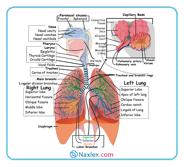
Anatomy Of The Respiratory System
Upper Respiratory Tract
The upper tract includes the nose, mouth, pharynx, epiglottis, larynx, and upper trachea. Its main role is to filter, warm, and moisten the air.
- Nose: Contains turbinates (conchae) that increase the surface area to condition the air.
- Epiglottis: A small, leaf-shaped cartilage flap that covers the opening of the larynx (glottis) during swallowing, protecting the lower airway from aspiration.
- Larynx: The "voice box" containing the vocal cords and acting as a critical point of airway protection.
Lower Respiratory Tract
The lower tract is considered the sterile environment for gas exchange, consisting of the trachea, bronchi, bronchioles, and alveoli.
- Trachea: Extends to the chest, where it divides into the right and left mainstem bronchi at the carina.
- Clinical Note: The Right Mainstem Bronchus is shorter, wider, and straighter than the left, making it the more common site for aspiration or accidental intubation.
- Bronchi and Bronchioles: The mainstem bronchi branch into progressively smaller airways, ending in the tiny, non-cartilaginous bronchioles.
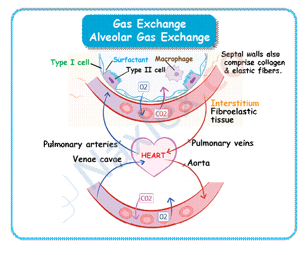
- Alveoli: These small, thin-walled sacs (about 300 million) are the primary sites of gas exchange, surrounded by an alveolar-capillary membrane.
- Surfactant: A lipoprotein that lines the alveoli, reducing surface tension and preventing alveolar collapse (atelectasis).
Anatomy of the Respiratory System
Physiology Of Respiration
Ventilation (Air Movement)
Ventilation is the mechanical process of moving air. It relies on pressure changes governed by muscle action.
- Inspiration (Inhalation): This is an active process. The diaphragm and intercostal muscles contract, expanding the chest. This makes intrathoracic pressure negative (subatmospheric), causing air to rush in.
- Expiration (Exhalation): This is normally a passive process. The muscles relax, and the elastic recoil of the lungs and chest wall pushes air out.
Diffusion and Gas Transport
Gas exchange across the alveolar-capillary membrane occurs via diffusion, moving gases from high partial pressure to low partial pressure.
- O2 Transport: O2 diffuses from the alveoli (PaO2≈100 mm Hg) into the blood and is primarily carried by hemoglobin.
- CO2 Transport: CO2 diffuses from the blood (PvCO2≈45 mm Hg) into the alveoli to be exhaled.
Control of Breathing
Respiration is an involuntary process regulated by the nervous system in response to chemical and mechanical signals.
- Central Chemoreceptors (Medulla): These are the main regulators. They respond to the concentration of H+ (acid-base balance), which is driven by PaCO2. An increase in PaCO2 is the primary stimulus for normal breathing.
- Peripheral Chemoreceptors (Carotid Arteries & Aortic Arch): These respond mainly to a significant decrease in PaO2 (arterial O2 concentration).
- Clinical Warning: In patients with chronic CO2 retention (e.g., severe COPD), their central receptors are desensitized. Their breathing is driven by the low PaO2 stimulus. Giving too much supplemental O2 can remove this drive and cause dangerous hypoventilation.
Respiratory Defense Mechanisms
The body has multiple defenses to protect the lower airways from particles and pathogens.
- Mucociliary Clearance System: This is a vital defense. Cilia (hair-like projections) beat rhythmically to propel the mucus layer (secreted by goblet cells) upward—the mucociliary escalator—trapping and removing debris.
- Impairment: Smoking, dehydration, and certain drugs can damage or slow ciliary action.
- Cough Reflex: A protective reflex that clears the airways using a high-velocity expulsion of air, acting as a backup to the mucociliary system.
- Alveolar Macrophages: At the alveolar level, these immune cells engulf inhaled bacteria and foreign particles (e.g., coal dust).
- Reflex Bronchoconstriction: The airways constrict in response to irritants (dust, cold air) to prevent their entry into the lungs.
Manifestations of Inadequate Oxygenation
Nurses must recognize the clinical signs of hypoxia (inadequate tissue oxygenation) by noting early versus late signs across body systems.
|
Body System |
Early Manifestations (Subtle/Compensated) |
Late Manifestations (Decompensated/Severe) |
|
Central Nervous System |
Restlessness, agitation, unexplained apprehension, confusion, irritability. |
Unresponsiveness, lethargy, coma. |
|
Cardiovascular |
Tachycardia, mild hypertension, dysrhythmias. |
Hypotension, severe dysrhythmias, cool/clammy skin. |
|
Respiratory |
Tachypnea, dyspnea on exertion, use of accessory muscles. |
Dyspnea at rest, retractions, cyanosis (late and unreliable sign). |
|
Critical Values |
SpO2>92% (PaO2>70 mm Hg) |
SpO2<90% (PaO2<60 mm Hg) - DANGER ZONE |
Arterial Blood Gas (ABG) Normal Values (Sea Level)
|
Laboratory Value |
Normal Sea Level (760 mm Hg) |
|
pH |
7.35–7.45 |
|
PaO2 |
80–100 mm Hg |
|
PaCO2 |
35–45 mm Hg |
|
HCO3− |
22–26 mEq/L |
Diagnostic Studies
|
Study |
Description and Purpose |
Nursing Responsibility |
|
Blood studies |
||
|
Hemoglobin |
Test reflects amount of hemoglobin available for combination with O2. Venous blood is used. Male: 13.2–17.3 g/dL (132–173 g/L). Female: 11.7–16.0 g/dL (117–160 g/L). |
Explain procedure and its purpose. |
|
Hematocrit |
Test reflects ratio of red blood cells to plasma. Increased hematocrit (polycythemia) found in chronic hypoxemia. Venous blood is used. Male: 39%–50% (0.39–0.50). Female: 35%–47% (0.35–0.47). |
Explain procedure and its purpose. |
|
Arterial blood gases (ABGs) |
Arterial blood is obtained through puncture of radial or femoral artery or through arterial catheter. Performed to assess acid-base balance, ventilation status, need for O2 therapy, change in O2 therapy, or change in ventilator settings. † Continuous ABG monitoring is also possible via a sensor or electrode inserted into arterial catheter. |
Indicate whether patient is using O2 (percentage, L/min). Avoid change in O2 therapy or interventions (e.g., suctioning, position change) for 20 min before obtaining sample. Assist with positioning (e.g., palm up, wrist slightly hyperextended if radial artery is used). Collect blood in heparinized syringe. To ensure accurate results, expel all air bubbles and place sample in ice, unless it will be analyzed in <1 min. Apply pressure to artery for at least 5 min after specimen is obtained to prevent hematoma at the arterial puncture site. |
|
Oxygen monitoring |
||
|
O2 Oximetry |
Monitors arterial or venous O2 saturation. Probe attaches to finger, toe, earlobe, bridge of the nose for SpO2 monitoring (see Fig. 26−2) or is contained in a pulmonary artery catheter for SvO2 monitoring. Oximetry is used for intermittent or continuous monitoring and exercise testing. |
Apply probe. When interpreting SpO2 and SvO2 values, first assess patient status and presence of factors that can alter accuracy of pulse oximeter reading. For SpO2, these include motion, low perfusion, cold extremities, bright lights, acrylic nails, dark skin color, carbon monoxide, and anemia. For SvO2, these include change in O2 delivery or O2 consumption. |
|
CO2 monitoring |
||
|
Monitoring End-tidal CO2 (PETCO2) (capnography) |
Assesses the level of CO2 in exhaled air. Graphically displays partial pressure of CO2. Expired gases are sampled from the patient's airway and are analyzed by a CO2 sensor that uses infrared light to measure exhaled CO2. The sensor may be attached to an adapter on the endotracheal or tracheostomy tube. A nasal cannula with a sidestream capnometer can be used in patients without an artificial airway. Can be used as a diagnostic measure to detect lung disease and for monitoring patients. Normal difference between PaCO2 and PETCO2 is 2−5 mm Hg (PaCO2: 35–45 mm Hg; PETCO2: 37–50 mm Hg). |
Teach patient and caregiver about the purpose of capnography monitoring, emphasizing the benefit of continuous monitoring. Make sure that sensor is properly attached. Record and document data per institution policy. |
|
Sputum Studies |
||
|
Culture and sensitivity |
Purpose is to diagnose bacterial infection, select antibiotic, and evaluate treatment. Sputum specimen is collected in a sterile container. Takes 48–72 hr for results. |
Instruct patient on how to produce a good specimen (see Gram stain). If patient cannot produce specimen, bronchoscopy may be used. |
|
Gram stain |
Staining of sputum permits classification of bacteria into gram-negative and gram-positive types. Results guide therapy until culture and sensitivity results are obtained. |
Instruct patient to expectorate sputum into container after coughing deeply. Obtain sputum innoculation, not saliva. Obtain specimen in early morning after mouth care because secretions collect during night. If unsuccessful, try increasing oral fluid intake unless fluids are restricted. Collect sputum in sterile container (sputum trap) during suctioning or by aspirating secretions from the trachea. Send specimen to laboratory promptly. |
|
Acid-fast smear and culture |
Assesses sputum for acid-fast bacilli (e.g., Mycobacterium tuberculosis). A series of three early-morning specimens is used. |
Instruct patient how to produce a good specimen (see Gram stain). Cover specimen and send to laboratory for analysis. |
|
Cytology |
Determines presence of abnormal cells that may indicate a malignant condition. Single sputum specimen is collected in special container with fixative solution. |
Instruct patient on how to produce a good specimen (see Gram stain). If patient cannot produce specimen, bronchoscopy may be used. Send specimen to laboratory promptly. |
|
Radiology |
|
|
|
Chest x-ray |
Used to screen, diagnose, and evaluate changes in respiratory system. Most common views are anteroposterior (AP) and lateral. |
Instruct patient to undress to waist, put on gown, and remove any metal between neck and waist. |
|
Computed tomography (CT) |
Performed for diagnosis of lesions difficult to assess (e.g., mediastinum, hilum, pleura) by conventional x-ray. Various common types are helical or spiral CT (contrast medium is usually used) and high-resolution CT scan (contrast medium is not used). Spiral CT used to diagnose a pulmonary embolism. |
Same as for chest x-ray. Contrast medium may be given IV. Evaluation of BUN and serum creatinine is done before contrast to assess renal function. Assess if patient is allergic to shellfish (iodine), since the contrast is iodine based. Be sure the patient is well hydrated before and after procedure (to excrete contrast). Warn patient that contrast injection may cause a feeling of being warm and flushed. Instruct the patient that he or she will need to lie still on a hard table and the scanner will revolve around the body with clicking noises. |
|
Magnetic resonance imaging (MRI) |
Used for diagnosis of lesions difficult to assess by CT scan (e.g., lung apex) and for distinguishing vascular from nonvascular structures. |
Same as for chest x-ray and CT scan, except contrast medium is not iodine based. A closed MRI is used and patient has claustrophobia, provide with relaxation or other modes to cope. Patient must remove all metal (e.g., jewelry, watch) before test. Patients with pacemakers and unidentifiable cardioverter-defibrillators may not be able to have MRI. |
|
Ventilation-perfusion (V/Q) scan |
Used to assess ventilation and perfusion of lungs. IV radioisotope given to assess perfusion. For the ventilation portion, the patient inhales a radioactive gas (xenon or krypton), which outlines the alveoli. Normal scan shows homogeneous radioactivity. Mismatched of absent radioactivity suggests lack of perfusion or airflow. Ventilation without perfusion suggests a pulmonary embolism. |
Same as for chest x-ray. Precautions needed afterward when the isotope and isotope-traced radioactivity for only a brief interval. |
|
Pulmonary angiogram |
Used to visualize pulmonary vasculature and locate obstruction or pathologic conditions (e.g., pulmonary embolism, contrast medium is injected through a catheter threaded into pulmonary artery to right side of the heart. Series of x-rays are taken after contrast medium is injected into pulmonary artery. Chest CT is replacing angiography, since it is less invasive. |
Same as for chest x-ray. (See CT scan for contrast media precautions.) Check pressure dressing site after procedure. Monitor blood pressure, pulse, and circulation distal to injection site. Report and record significant changes. |
|
Positron emission tomography (PET) scan |
Used to distinguish benign and malignant pulmonary nodules, because malignant lung cells use an increased uptake of glucose, the PET scan, which uses an IV radioactive glucose preparation, can demonstrate increased uptake of glucose in malignant lung cells. |
Pre-procedure: Check the blood glucose levels as high levels may interfere with test. Food and fluids other than water may be restricted for 4–6 hr. Post-procedure: Encourage fluids to excrete radioactive substance. |
|
Endoscopy |
||
|
Bronchoscopy
|
Flexible fiberoptic scope is used for diagnosis, biopsy, specimen collection, or assessment of changes. It may also be done to suction mucous plugs, lavage the lungs, or remove foreign objects. |
Instruct patient to be on NPO status for 6–12 hr. before the test. Obtain signed permit. Give sedative if ordered. After procedure, keep patient NPO until gag reflex returns. Monitor for recovery from sedation. Monitor blood-tinged sputum is normal. If biopsy was done, monitor for hemorrhage and pneumothorax. |
|
Mediastinoscopy |
Scope is inserted through a small incision in the suprasternal notch and advanced into the mediastinum to inspect and biopsy lymph nodes. Used to diagnose lung cancer, non-Hodgkin’s lymphoma, granulomatous infections, and sarcoidosis. |
Prepare patient for surgical intervention. Obtain signed informed consent. Performed in the operating room (OR) using a general anesthetic. Afterward, monitor as for bronchoscopy. |
|
Other Studies |
||
|
Lung Biopsy |
Specimens may be obtained by transbronchial or percutaneous biopsy or via transthoracic needle aspiration (TTNA), video-assisted thoracoscopic surgery (VATS), or open lung biopsy. Transbronchial biopsy and VATS can be performed in the bronchoscopy suite. TTNA is done under CT guidance in radiology department. Open lung is performed in the OR. VATS can also be done in the OR. These tests are used to obtain specimens for laboratory analysis. |
Same as bronchoscopy if procedure done with bronchoscope, and same as thoracotomy if open lung biopsy done. With TTNA, check breath sounds q4hr for 24 hr. and report any respiratory distress. Check incision site for bleeding. A chest x-ray should be done after TTNA or transbronchial biopsy to check for pneumothorax. With VATS a chest tube may be in post-procedure until lung has expanded. Monitor breath sounds to follow chest re-expansion. Encourage deep breathing for lung reinflation. Obtain signed informed consent for all procedures. |
|
Thoracentesis
|
Used to obtain specimen of pleural fluid for diagnosis, to remove pleural fluid, or to instill medication. Chest x-ray is always obtained afterward for procedure to check for pneumothorax. |
Explain procedure to patient and obtain signed informed consent before procedure, which is usually performed in the patient’s room. Position patient upright with elbows on an overbed table and feet supported. Instruct the patient not to talk or cough, and assist during procedure. Observe for signs of hypoxia and pneumothorax, and verify breath sounds in all fields after procedure. Encourage deep breaths to expand lungs. Send labeled specimens to laboratory. |
|
Pulmonary Function Tests |
Used to evaluate lung function. Involves use of spirometer to assess air movement as patient performs prescribed respiratory maneuvers. † |
Avoid scheduling immediately after mealtime. Avoid administration of inhaled bronchodilator 6 hr. before procedure. Explain procedure to patient. Assess for respiratory distress before procedure and report. Provide rest after the procedure. |
|
Exercise Testing |
Used in diagnosis and in determining exercise capacity. A complete test involves walking on a treadmill while expired O2 and CO2, respiratory rate, heart rate, and heart rhythm are monitored. In a modified test (desaturation test) only SpO2 is monitored. |
Instruct patient to wear comfortable shoes. Encourage patient to walk as quickly as possible. |
|
6-Min walk test |
Used to measure functional capacity and response to treatment in patients with heart or lung disease. Pulse oximetry is usually monitored during the walk. The distance walked is measured and used to monitor progression of disease or improvement after rehabilitation. |
The patient is instructed by a trained practitioner to walk as far as possible during 6 min, stopping when short of breath and continuing when able. |
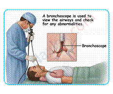
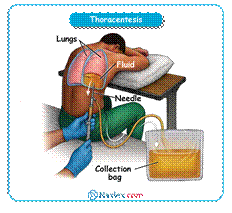
Summary
- The respiratory system is divided into upper and lower tracts responsible for air conduction and gas exchange.
- The chest wall, diaphragm, and intercostal muscles enable breathing through pressure changes.
- Inspiration is an active process of air entry, while expiration is normally passive due to lung recoil.
- Gas exchange occurs in the alveoli where oxygen diffuses into the blood and carbon dioxide diffuses out.
- Breathing is controlled by the medulla and chemoreceptors responding to CO₂, pH, and O₂ levels.
- Respiratory defenses include the mucociliary escalator, cough reflex, macrophages, and bronchoconstriction.
- Early signs of hypoxia include restlessness and tachycardia, while late signs include cyanosis and confusion.
- Normal ABG values help assess ventilation, oxygenation, and acid–base balance.
- Common respiratory diagnostics include blood tests, imaging, sputum studies, and pulmonary function tests.
- Nurses play key roles in patient education, test preparation, monitoring, and promoting effective oxygenation.
Naxlex
Videos
Login to View Video
Click here to loginTake Notes on Respiratory Diagnostic Procedures
This filled cannot be empty
Join Naxlex Nursing for nursing questions & guides! Sign Up Now
