





 Video Library


Please set your exam date
Cardiovascular Diagnostic and Therapeutic Procedures
Study Questions
Practice Exercise 1
A client arrives in the emergency department reporting crushing substernal chest pain that began approximately 90 minutes ago. The provider prescribes serial cardiac biomarker testing. Which nursing action is the priority?
Explanation
Acute myocardial infarction involves myocardial necrosis secondary to prolonged ischemia resulting from coronary artery occlusion. Diagnostic protocols prioritize rapid biomarker collection and reperfusion therapy to minimize irreversible damage. Prompt intervention is critical because cardiomyocyte death initiates within minutes of complete arterial blockage, necessitating immediate evaluation of troponin levels to guide management.
Rationale for correct answer
2. Rapid biomarker collection is the highest priority when evaluating acute coronary syndrome. Serial testing identifies cardiac enzyme elevation indicating myocardial damage. Timely blood sampling facilitates urgent diagnostic confirmation and subsequent therapeutic decision making. This immediate action optimizes patient outcomes.
Rationale for incorrect answers
1. Statins are typically initiated after confirming Acute myocardial infarction or as part of long-term management. Administering medication before establishing a clear diagnosis diverts attention from lifesaving triage. This intervention is secondary to urgent cardiac diagnostics. Immediate stabilization is the priority.
3. A fasting lipid profile provides information regarding long term cardiovascular risk factors and hyperlipidemia. It is not an urgent diagnostic tool for acute ischemia. Obtaining this specimen does not influence the immediate management of acute chest pain. It remains clinically irrelevant.
4. Educating on dietary modifications addresses long term lifestyle management for chronic disease states. This nursing intervention is inappropriate during the acute phase of a suspected cardiac emergency. The client requires immediate physiologic stabilization rather than nutritional counseling. Diet education is irrelevant here.
Test-taking strategy
- Analyze the Scenario/Question: The client presents with crushing substernal chest pain, suggesting Acute myocardial infarction. The nursing priority is identifying life threatening cardiac injury through diagnostic testing.
- Apply the Airway, Breathing, and Circulation (ABC) and Prioritization Principles:
- Prioritize Diagnostics: In emergency cardiac care, establishing a definitive diagnosis to initiate reperfusion is the most urgent action.
- Rule out 1: Administering Statins for maintenance is secondary to diagnostic confirmation.
- Rule out 3: A fasting lipid profile is not diagnostic for active cardiac muscle necrosis.
- Rule out 4: Education regarding dietary modifications is non urgent during an acute physiological crisis.
- Time-Sensitive Care: The 90-minute window requires immediate blood sampling to assess biomarkers like troponin.
- Select 2: Obtaining the specimen for biomarker collection is the essential first step for clinical decision making.
Take home points
- Troponin levels are the gold standard cardiac biomarkers for diagnosing myocardial infarction.
- Immediate diagnostic testing is required for all clients presenting with acute substernal chest pain.
- Priority nursing care in the emergency department focuses on stabilization and rapid diagnostic identification.
- Long term management strategies such as dietary changes and lipid testing are secondary to acute lifesaving interventions.
The nurse reviews the laboratory results of a client admitted with suspected acute coronary syndrome. Which finding is most consistent with ongoing myocardial injury?
Explanation
Acute myocardial infarction involves cellular death within the myocardium triggered by persistent ischemia. When myocytes undergo necrosis, intracellular proteins are released into the systemic circulation. Monitoring specific biomarker collection patterns, particularly cardiac-specific proteins, is essential for confirming tissue damage. These levels rise rapidly following an ischemic event, serving as the primary diagnostic indicator for acute cardiac injury.
Rationale for correct answer
2. Elevated troponin I level is the definitive biomarker for myocardial injury. This protein is highly specific to cardiac tissue, and its presence in the blood indicates active cellular damage. Prompt identification is critical for initiating reperfusion. Elevated levels confirm the diagnosis.
Rationale for incorrect answers
1. Elevated HDL cholesterol is generally considered a cardioprotective finding, not a marker of acute injury. It reflects long term lipid metabolism and does not indicate Acute myocardial infarction. This laboratory value does not necessitate emergent intervention or acute management.
3. An elevated triglyceride level is a risk factor for coronary artery disease but does not confirm Acute myocardial infarction. This finding relates to chronic metabolic status and lipid transport. It provides no information regarding current myocardial tissue necrosis or damage.
4. An elevated total cholesterol level indicates a potential predisposition for atherosclerosis. However, it is not a diagnostic marker for ongoing biomarker collection or acute cardiac muscle injury. It is irrelevant to the immediate management of a suspected cardiac event.
Test-taking strategy
- Analyze the Scenario/Question: The client is suspected of having Acute myocardial infarction. The question asks to identify a laboratory finding that specifically confirms ongoing myocardial cell death and injury.
- Apply Diagnostic Specificity and Clinical Relevance:
- Identify Cardiac-Specific Markers: Only cardiac-specific proteins provide evidence of current muscle damage.
- Rule out 1: Elevated HDL cholesterol is a lipid profile component, not a sign of acute damage.
- Rule out 3: Elevated triglyceride level indicates long term risk, not acute necrosis.
- Rule out 4: Elevated total cholesterol level does not measure cellular injury from biomarker collection.
- Prioritize Pathological Indicators: Focus on biomarkers that change due to cell membrane leakage during ischemia.
- Select 2: The troponin I level is the gold standard diagnostic test for myocardial injury. It is not influenced by dietary modifications or recent intake, making it the most consistent finding.
Take home points
- Cardiac troponin I is the most sensitive and specific biomarker for diagnosing myocardial injury.
- Lipid panels provide information about chronic risk factors but cannot diagnose acute myocardial infarction.
- Ongoing myocardial necrosis is characterized by the sustained release of intracellular proteins into the blood.
- Accurate diagnosis of acute coronary syndrome requires serial biomarker assessment to monitor the progression of injury.
A client is scheduled for a transesophageal echocardiogram (TEE). Which nursing interventions are appropriate before the procedure? Select all that apply
Explanation
A transesophageal echocardiogram is an invasive diagnostic procedure requiring the insertion of an endoscopic probe into the esophagus to visualize posterior cardiac structures. Because the procedure involves administration of conscious sedation and manipulation of the pharynx, strict adherence to safety protocols is mandatory. Nursing care focuses on protecting the airway, ensuring procedural legality through informed consent, and preparing the client physically to prevent severe complications during instrumentation.
Rationale for correct answer
1. Verify informed consent has been obtained. This legal requirement confirms the client understands the specific risks, benefits, and procedural alternatives. The nurse must validate that the provider has obtained this before any preoperative sedation is administered.
2. Instruct the client to remain NPO for the prescribed period. This critical safety measure minimizes the risk of pulmonary aspiration during the procedure. Maintaining an empty stomach is essential when the protective pharyngeal and gag reflexes are suppressed by medications.
5. Explain that conscious sedation may be administered. Providing education reduces procedural anxiety and increases client cooperation. The client must understand they will be monitored continuously for respiratory status while remaining responsive to verbal or physical stimuli.
Rationale for incorrect answers
3. Retain dentures and removable oral prostheses. These items are strictly contraindicated because they can become dislodged during probe insertion, causing severe airway obstruction or oral trauma. They must be removed prior to the procedure to ensure a clear pathway.
4. Encourage the client to eat a light meal before the procedure. This is unsafe and increases the risk of aspiration during endoscopic probe insertion. The client must remain fasting to keep the gastric contents minimal and prevent potential vomiting or regurgitation complications.
Test-taking strategy
- Analyze the Scenario/Question: The client is undergoing a transesophageal echocardiogram, an invasive procedure involving sedative medications and instrumentation of the oropharynx. The question requires identifying safety and preparation steps to prevent adverse outcomes.
- Apply Safety and Risk Reduction Principles:
- Prioritize Airway and Safety:
- Rule in 2: Maintaining NPO status is the most critical intervention to prevent aspiration.
- Rule in 5: Providing education regarding conscious sedation is essential for patient cooperation and safety.
- Apply Legal and Procedural Requirements:
- Rule in 1: Informed consent is a mandatory prerequisite for invasive diagnostic procedures to ensure ethical compliance.
- Rule out 3 and 4: Both actions directly endanger the client’s airway. Prostheses must be removed, and dietary intake is strictly prohibited before procedures requiring sedation.
Take home points
- Transesophageal echocardiogram requires strictly maintained NPO status to prevent dangerous aspiration pneumonia.
- Informed consent must be documented before the administration of any sedative or anesthetic medications.
- Airway management remains the primary nursing priority during procedures involving the use of conscious sedation.
- All oral appliances must be removed to prevent mechanical airway obstruction during the probe advancement phase.
A client returns to the unit immediately after a transesophageal echocardiogram. Which assessment should the nurse perform first?
Explanation
A transesophageal echocardiogram involves endoscopic instrumentation of the esophagus, which necessitates topical pharyngeal anesthesia. This pharmacological blockade temporarily suppresses the protective gag reflex, creating a significant risk for post-procedural aspiration. The nurse must prioritize monitoring the airway and pharyngeal status before initiating any activities that involve the oral cavity or require patient mobility.
Rationale for correct answer
1. Determine whether the gag reflex has returned. This is the highest priority because the pharynx was anesthetized during the procedure. Assessing the ability to swallow safely prevents aspiration pneumonia, which can occur if the client attempts to ingest substances before the reflex recovers.
Rationale for incorrect answers
2. Assess bowel sounds. Auscultating bowel sounds is not required post-procedure because the gastrointestinal tract is not the primary site of intervention. The procedure is limited to the esophagus and does not affect intestinal motility or indicate the need for this assessment.
3. Assist the client with ambulation. Attempting to move the client before ensuring they are hemodynamically stable from the conscious sedation is inappropriate. The nurse must first confirm the client is fully alert and maintains a patent airway before initiating any activity.
4. Encourage oral fluid intake. This is contraindicated until the nurse confirms the return of the gag reflex. Introducing liquids before the pharyngeal response is restored places the client at high risk for bronchial aspiration, which is a life-threatening complication.
Test-taking strategy
- Analyze the Scenario/Question: The client has just returned from a transesophageal echocardiogram. The nurse must determine the most urgent assessment to ensure patient safety immediately after sedation and pharyngeal anesthesia.
- Apply the Airway, Breathing, and Circulation (ABC) and Risk Reduction Principles:
- Prioritize Airway:
- Rule in 1: Evaluating the gag reflex is essential to maintain a patent airway and prevent aspiration.
- Rule out 2: Bowel sounds are not relevant to the procedure and do not address immediate patient safety.
- Rule out 3: Ambulation is secondary to stabilization and ensuring the airway is protected from potential choking or vomiting.
- Rule out 4: Encouraging fluids is dangerous without confirming the return of the protective reflex, as it would cause immediate aspiration risk.
Take home points
- The return of the gag reflex is the most critical assessment following a transesophageal echocardiogram.
- Oral intake is strictly prohibited until the nurse verifies that the pharyngeal and gag reflexes have fully returned.
- Aspiration pneumonia is a serious, preventable complication resulting from premature oral ingestion after pharyngeal anesthesia.
- Nursing priorities post-procedure focus on airway maintenance, hemodynamic stability, and the safe recovery from conscious sedation.
The nurse is reviewing lipid profile results for four clients. Which client has the greatest risk for developing coronary artery disease?
Explanation
Coronary artery disease development is strongly correlated with an imbalanced lipid profile, characterized by elevated low-density lipoproteins and low high-density lipoproteins. The interplay between these lipid fractions dictates the rate of atherosclerosis and arterial plaque formation. A high concentration of circulating LDL promotes endothelial lipid deposition, while inadequate HDL impairs the reverse cholesterol transport mechanism, creating an environment favorable for progressive arterial stenosis.
Rationale for correct answer
2. This client exhibits an LDL of 188 mg/dL and an HDL of 34 mg/dL. An LDL level above 160 mg/dL is classified as high, and an HDL level below 40 mg/dL in men or 50 mg/dL in women is a major risk factor. This combination significantly accelerates atherosclerosis.
Rationale for incorrect answers
1. A total cholesterol of 178 mg/dL is within the desirable range (less than 200 mg/dL), and an HDL of 64 mg/dL is considered protective. This profile reflects a low risk for coronary artery disease development and does not indicate immediate clinical concern.
3. Triglycerides of 142 mg/dL are within the normal range (less than 150 mg/dL), and an HDL of 58 mg/dL is healthy. This patient lacks the characteristic dyslipidemia associated with rapid atherosclerosis and does not demonstrate high cardiovascular risk markers.
4. Total cholesterol of 195 mg/dL is borderline but not inherently indicative of severe disease, and an LDL of 96 mg/dL is near optimal. This profile is not significantly associated with an elevated risk of developing coronary artery disease compared to clients with severely deranged lipids.
Test-taking strategy
- Analyze the Scenario/Question: The nurse is evaluating four lipid profile results to determine which client possesses the most significant risk factors for developing coronary artery disease.
- Apply Dyslipidemia Assessment Criteria:
- Evaluate Lipid Fractions:
- Rule in 2: This client has the most dangerous profile with significantly elevated LDL and critically low HDL.
- Rule out 1: This client displays a healthy total cholesterol and protective HDL level.
- Rule out 3: This client has normal triglycerides and a healthy HDL level.
- Rule out 4: This client has borderline normal values that do not meet the high-risk criteria for atherosclerosis.
- Prioritize Risk Factors: Focus on the ratio of "bad" cholesterol to "good" cholesterol, as this is the most predictive indicator for potential arterial damage.
Take home points
- Low density lipoprotein (LDL) is the primary driver of arterial plaque formation in coronary artery disease.
- High density lipoprotein (HDL) serves a cardioprotective role by facilitating reverse cholesterol transport from peripheral tissues.
- A desirable lipid profile includes total cholesterol < 200 mg/dL and an optimal HDL level ≥ 60 mg/dL.
- Assessment of cardiovascular risk requires evaluating the individual components of the lipid profile rather than total cholesterol alone.
The nurse is providing education to a client prescribed statin therapy following an abnormal lipid profile. Which teaching points should the nurse include? Select all that apply
Explanation
Statins are potent inhibitors of HMG-CoA reductase, the rate-limiting enzyme in hepatic cholesterol synthesis, which effectively lowers low density lipoprotein levels. As a primary therapy for lipid management, these medications require consistent dosing to achieve significant cardiovascular risk reduction. Because Statins can cause rare but severe systemic complications, nursing education must focus on medication adherence and early recognition of signs indicating potential myopathy or drug-induced injury.
Rationale for correct answers
4. Report dark-colored urine to the healthcare provider. This clinical finding is a potential sign of myoglobinuria, which results from skeletal muscle breakdown. Prompt reporting is essential to evaluate for rhabdomyolysis and prevent further potential renal damage.
5. Continue taking the medication exactly as prescribed. Therapeutic efficacy depends on maintaining consistent serum drug levels to inhibit hepatic cholesterol production. Interrupting or altering the frequency of administration undermines the long term cardioprotective benefits and increases cardiovascular risk.
Rationale for incorrect answers
1. Muscle pain or weakness are expected side effects. This statement is incorrect and dangerous. While mild myalgia can occur, Statins can cause serious muscle damage. Any unexplained muscle pain or weakness requires immediate evaluation to rule out myopathy or rhabdomyolysis.
2. If you miss a dose, double the next of the medication. This is contraindicated for most Statins. Doubling a dose significantly increases the risk of toxicity and adverse effects without providing additional therapeutic benefit. Clients should follow specific provider instructions for missed doses.
3. Discontinue the medication once cholesterol levels normalize. Cholesterol lowering therapy is typically a chronic intervention. Stopping the medication when target levels are reached causes the hepatic enzyme pathway to resume overproduction, leading to a loss of cardioprotective benefits and rapid progression of atherosclerosis.
Test-taking strategy
- Analyze the Scenario/Question: The client is starting Statins for lipid management. The nurse must identify safe and accurate teaching points regarding medication adherence and side effect monitoring.
- Apply Pharmacological Safety Principles:
- Prioritize Safety and Adherence:
- Rule in 4: Focus on reporting signs of muscle breakdown, as Statins carry a risk for myopathy.
- Rule in 5: Emphasize consistent adherence for effective chronic lipid management.
- Rule out 1: Muscle pain is a potential adverse event, not an expected or benign side effect.
- Rule out 2: Doubling doses increases the risk of adverse reactions and is generally unsafe.
- Rule out 3: Chronic therapy must not be discontinued simply because laboratory results normalize, as the medication is responsible for maintaining those improvements.
Take home points
- Statins require lifelong adherence to effectively manage hyperlipidemia and prevent cardiovascular events.
- Muscle pain, weakness, or dark-colored urine are indicators of potential rhabdomyolysis and require immediate medical assessment.
- Clients should never double doses or discontinue therapy without explicit guidance from their healthcare provider.
- Education must emphasize that normalize laboratory values reflect successful management, not the resolution of the underlying condition.
A client diagnosed with heart failure has worsening dyspnea and lower extremity edema. Which laboratory test would provide the most useful information regarding ventricular volume overload?
Explanation
Heart failure is a complex clinical syndrome characterized by the heart's inability to pump blood efficiently, leading to systemic venous congestion and pulmonary edema. When the cardiac chambers experience ventricular volume overload, myocardial cells in the ventricles undergo mechanical stretch. This stretching triggers the release of specific neurohormones, which serve as highly sensitive diagnostic markers for assessing hemodynamic status and evaluating the severity of fluid retention in symptomatic patients.
Rationale for correct answer
2. BNP (B-type natriuretic peptide) is the gold standard diagnostic biomarker for identifying ventricular volume overload. It is synthesized and released by the ventricles in response to increased myocardial wall stretch. Elevated levels provide an objective measurement of the severity of heart failure and congestive symptoms.
Rationale for incorrect answers
1. CK-MB is a cardiac enzyme that was historically used to detect acute myocardial injury. It is not sensitive to chamber distension or fluid status. This biomarker is specifically released during cellular necrosis, making it inappropriate for evaluating chronic fluid overload or cardiac filling pressures.
3. Troponin T is a highly specific protein released into the circulation only when cardiac myocytes suffer significant structural damage. It is an essential diagnostic indicator for acute myocardial infarction. It does not correlate with ventricular stretching, hemodynamic filling pressures, or systemic fluid retention.
4. Myoglobin is a low-molecular-weight heme protein released rapidly into the blood following any muscle injury, including cardiac, skeletal, or smooth muscle. It lacks cardiac specificity and has no clinical utility in assessing ventricular volume overload or diagnosing heart failure exacerbations.
Test-taking strategy
- Analyze the Scenario/Question: The client presents with clinical manifestations of heart failure, specifically dyspnea and edema, indicating ventricular volume overload. The goal is to identify the laboratory marker that specifically reflects cardiac chamber stretching due to fluid accumulation.
- Apply Biomarker Specificity:
- Identify Cardiac Indicators:
- Rule in 2: BNP is the definitive marker for volume-induced myocardial stretch and symptomatic heart failure.
- Rule out 1: CK-MB is for tissue injury, not volume status.
- Rule out 3: Troponin T detects cell death, which is not the primary mechanism of ventricular volume overload.
- Rule out 4: Myoglobin is a non-specific muscle damage marker and is not used for monitoring chronic congestion.
- Differentiate Pathophysiology: Focus on biomarkers that respond to mechanical stress rather than those that signal myocardial infarction or general muscle damage.
Take home points
- B-type natriuretic peptide (BNP) is the primary neurohormone used to diagnose and monitor heart failure severity.
- Ventricular volume overload induces the release of BNP due to increased myocardial wall tension and stretching.
- Cardiac enzymes like troponin and CK-MB are reserved for diagnosing acute coronary syndromes and cell necrosis.
- Clinical assessment of heart failure includes interpreting biomarker levels alongside physical findings like dyspnea and peripheral edema.
The nurse is caring for a client who underwent a transesophageal echocardiogram 45 minutes ago. The client requests a glass of water because the throat feels dry. Which nursing action is most appropriate?
Explanation
A transesophageal echocardiogram utilizes topical pharyngeal anesthesia to facilitate endoscopic probe passage, which temporarily inhibits the protective gag reflex. This loss of sensory function creates an immediate, high risk for pulmonary aspiration, as the client cannot effectively protect the airway. Nursing care must ensure the clinical return of pharyngeal function before any oral intake occurs to prevent potential respiratory complications from swallowed fluids.
Rationale for correct answer
2. Assess for return of the gag reflex before offering anything by mouth. This is the mandatory safety step following pharyngeal anesthesia. The nurse must verify that the patient can swallow safely to protect the lower airway from contents intended for the esophagus.
Rationale for incorrect answers
1. Provide small sips of water immediately. This is unsafe. Offering liquids before the gag reflex is confirmed places the client at significant risk for aspiration. The pharyngeal muscles are still inhibited, preventing the safe transit of fluids into the esophagus.
3. Offer ice chips instead of water. Ice chips still present an aspiration risk if the pharyngeal sensation is absent. Regardless of the form, any oral intake remains contraindicated until the nurse confirms the gag reflex has fully recovered to prevent pulmonary complications.
4. Encourage the client to drink through a straw. Straw usage can promote rapid ingestion of fluids, which is dangerous if the swallow reflex is impaired. This action does not address the underlying lack of protective sensation and increases the likelihood of aspiration.
Test-taking strategy
- Analyze the Scenario/Question: The client had a transesophageal echocardiogram recently and wants to drink. The question requires identifying the priority safety action to prevent an adverse event related to pharyngeal anesthesia.
- Apply Safety and Airway Protection Principles:
- Prioritize Airway:
- Rule in 2: Assessment of the gag reflex is the only way to ensure the client can safely swallow and maintain a patent airway.
- Rule out 1: This action is premature and ignores the risk of aspiration caused by lingering pharyngeal anesthesia.
- Rule out 3: Ice chips do not mitigate the aspiration risk; the lack of sensation remains the primary danger.
- Rule out 4: Using a straw does not restore the swallow reflex and increases the risk of aspiration by bypassing normal pharyngeal control.
Take home points
- Pharyngeal anesthesia used for transesophageal echocardiogram temporarily suppresses the protective gag reflex.
- Aspiration pneumonia is a serious and preventable risk if the client is allowed to drink prematurely.
- Nurses must objectively test for the return of the gag reflex before permitting any oral intake.
- Client comfort requests like thirst must be secondary to maintaining patient safety and airway integrity.
The nurse is reviewing the indications for obtaining cardiac biomarker testing. Which clinical situations warrant measurement of cardiac biomarkers? Select all that apply
Explanation
Cardiac biomarkers are intracellular proteins released into systemic circulation during Acute myocardial infarction and myocardial necrosis. The systematic measurement of these proteins, including troponin, is critical for diagnosing cardiac tissue injury. Clinical protocols necessitate these tests whenever myocardial ischemia is suspected, providing essential data to guide emergent management, evaluate the success of biomarker collection strategies, and monitor the extent of muscle damage.
Rationale for correct answer
1. Suspected Acute myocardial infarction. Elevated biomarkers are the primary diagnostic indicator for myocardial cell death. Serial measurement confirms the diagnosis and allows for the determination of the timing and severity of the ischemic event in symptomatic clients.
2. New onset chest pain. Cardiac biomarkers are required to differentiate non cardiac chest pain from Acute myocardial infarction. Prompt collection is a priority to rule out life threatening tissue necrosis in any client presenting with significant symptoms.
4. Assessment of response to reperfusion therapy. Monitoring the rate at which biomarkers rise and subsequently fall provides information regarding the success of reperfusion therapy. A rapid, early peak in troponin levels often indicates successful restoration of coronary artery patency.
Rationale for incorrect answers
3. Suspected pulmonary embolism. While pulmonary embolism causes chest pain, it does not involve primary myocardial necrosis. Diagnosis requires D dimer testing or computed tomography angiography. Biomarkers are not specific to this condition and are not the primary diagnostic tool used.
5. Routine annual cholesterol screening. This involves A fasting lipid profile to assess chronic cardiovascular risk. It is not an emergent diagnostic tool and provides no information regarding acute cell death, biomarker collection, or active myocardial injury in an emergency clinical setting.
6. Evaluation of valvular heart disease. This condition is diagnosed using echocardiography to visualize structural heart abnormalities. It does not cause the release of cardiac enzymes unless secondary complications like heart failure or ischemia develop. Biomarkers are not standard diagnostic tools here.
Test-taking strategy
- Analyze the Scenario/Question: The question asks to identify clinical scenarios requiring cardiac biomarker testing. The focus is on acute cardiac injury rather than chronic health maintenance or dietary modifications.
- Apply Diagnostic and Risk Assessment Principles:
- Prioritize Acute Cardiac Injury:
- Rule in 1: This is the primary indication for biomarker collection to confirm Acute myocardial infarction.
- Rule in 2: This scenario involves urgent assessment where biomarker elevation is pathognomonic for cardiac origin.
- Rule in 4: This situation involves monitoring the progression or recovery of myocardial tissue, requiring serial measurement.
- Rule out 3: Pulmonary embolism is a vascular, not myocardial, emergency.
- Rule out 5: Routine screening is for metabolic health, not acute diagnostics. A fasting lipid profile is used here.
- Rule out 6: Valvular disease is a structural problem best evaluated by imaging, not laboratory enzymes.
Take home points
- Cardiac biomarkers are specific indicators of myocardial necrosis and are essential for diagnosing acute myocardial infarction.
- Serial biomarker testing is required to monitor for ongoing tissue injury and to evaluate the effectiveness of reperfusion therapies.
- Clinical presentation of new onset chest pain necessitates rapid diagnostic evaluation with biomarkers to rule out cardiac injury.
- Lipid panels are utilized for chronic cardiovascular risk screening and are distinct from emergent cardiac biomarker testing for necrosis.
The nurse is preparing a client for a transthoracic echocardiogram (TTE). Which statement by the nurse is most appropriate?
Explanation
A transthoracic echocardiogram is a noninvasive diagnostic imaging study that utilizes ultrasound technology to visualize the anatomy, hemodynamics, and mechanical function of the heart. During this procedure, a transducer is positioned on the anterior chest wall to transmit and receive high frequency sound waves, allowing for the real time assessment of cardiac valves and wall motion. Because it is noninvasive, it requires no conscious sedation or specialized preoperative preparation, making it a safe and accessible tool for evaluating cardiac structure and performance.
Rationale for correct answer
2. An ultrasound transducer will be placed on your chest to obtain images of your heart. This accurately describes the mechanics of a transthoracic echocardiogram. The process relies on external visualization of the heart through the chest wall, involving no invasive maneuvers or anesthesia.
Rationale for incorrect answers
1. You will receive intravenous contrast and conscious sedation before the procedure. This statement is inaccurate for a standard transthoracic echocardiogram. These interventions are typically reserved for more invasive studies, such as a transesophageal echocardiogram or cardiac catheterization, which pose higher risks to the patient.
3. A catheter will be inserted into your femoral artery to evaluate your heart function. This describes the procedure for a cardiac catheterization or angiogram. Such an invasive study involves arterial access to measure pressures or visualize vessels, which is fundamentally different from the noninvasive imaging provided by an ultrasound transducer.
4. You should expect temporary difficulty swallowing after the examination. This describes a common side effect of a transesophageal echocardiogram, where a probe is passed down the esophagus under conscious sedation. A transthoracic echocardiogram involves no esophageal manipulation and has no impact on the patient’s ability to swallow.
Test-taking strategy
- Analyze the Scenario/Question: The client is scheduled for a transthoracic echocardiogram. The goal is to identify the accurate description of this specific, noninvasive diagnostic imaging procedure.
- Apply Diagnostic Procedural Knowledge:
- Differentiate Imaging Modalities:
- Rule in 2: Identify that a transthoracic echocardiogram uses external ultrasound waves, confirming this as the correct, noninvasive approach.
- Rule out 1: Conscious sedation and intravenous contrast are unnecessary for noninvasive imaging, implying an incorrect, more invasive procedure.
- Rule out 3: Arterial access via a catheter is specific to angiography; rule this out as it is highly invasive and distinct from imaging.
- Rule out 4: Esophageal side effects occur only in invasive esophageal imaging, not during external thoracic examinations.
Take home points
- A transthoracic echocardiogram is a noninvasive ultrasound procedure requiring no sedation or specialized preparation.
- The procedure uses an external transducer on the chest wall to generate real time cardiac images.
- It is essential to differentiate between noninvasive imaging like transthoracic echocardiography and invasive studies like transesophageal echocardiography or cardiac catheterization.
- Patient education for a transthoracic echocardiogram should emphasize the safety, ease, and noninvasive nature of the test.
Practice Exercise 2
A client undergoing an exercise stress test reports sudden chest pressure rated 8/10, becomes pale, and develops frequent premature ventricular contractions on the cardiac monitor. Which nursing action is the priority?
Explanation
An exercise stress test is designed to identify cardiac ischemia by increasing myocardial oxygen demand through physical exertion. When a client exhibits clinical symptoms of myocardial infarction including severe chest pressure, diaphoresis, and complex cardiac arrhythmias like premature ventricular contractions, the procedure must be terminated to prevent irreversible cardiac damage. Continued physical exertion during active distress significantly increases the risk of life-threatening cardiac events, necessitating immediate clinician intervention and physiological assessment.
Rationale for correct answer
2. Stop the test immediately and assess the client. This is the mandatory priority action for any client exhibiting symptoms of acute cardiac ischemia. Immediate cessation of physical stress reduces myocardial oxygen demand, while assessment allows for rapid triage and initiation of life saving emergency protocols.
Rationale for incorrect answers
1. Continue the test until the target heart rate is achieved. This action is dangerous and clinically inappropriate. Forcing a client experiencing myocardial infarction symptoms to continue exercise promotes further cellular necrosis and elevates the risk of hemodynamic collapse or lethal ventricular arrhythmias.
3. Encourage the client to continue walking at a slower pace. Any physical activity is contraindicated when the client displays clear markers of acute coronary distress. The nurse must prioritize the cessation of all activity to mitigate the risk of ongoing tissue damage and sudden cardiac death.
4. Document the findings after the procedure is completed. Delaying the documentation of acute changes is an unsafe clinical practice. Life threatening symptoms like 8/10 chest pain and unstable arrhythmias require immediate action; the nurse must intervene first, then document the event in real time.
Test-taking strategy
- Analyze the Scenario/Question: The client is symptomatic with signs of acute myocardial infarction during an exercise stress test. The priority is to stop the causative stressor and ensure patient safety.
- Apply the Airway, Breathing, and Circulation (ABC) and Prioritization Principles:
- Prioritize Patient Safety:
- Rule in 2: Stopping the test is the only intervention that addresses the primary danger and allows for immediate clinical stabilization.
- Rule out 1: Continuing the stressor is unethical and life threatening in the presence of acute cardiac ischemia.
- Rule out 3: Maintaining any level of physical activity during active cardiac injury is contraindicated and unsafe.
- Rule out 4: Documentation cannot precede the immediate emergency response required for a patient exhibiting myocardial infarction symptoms.
- Prioritize Patient Safety:
Take home points
- Any report of severe chest pain or the development of dangerous arrhythmias during a stress test warrants immediate termination of the procedure.
- Patient safety and the cessation of physical stress are the primary nursing responsibilities during cardiac testing.
- Clinical signs of myocardial infarction such as pallor and chest pressure require urgent medical evaluation.
- Nurses must act immediately to stop diagnostic procedures when physiological instability occurs, deferring non-urgent tasks like documentation.
A client is unable to walk on a treadmill because of severe osteoarthritis. Which diagnostic test is most appropriate for evaluating myocardial ischemia?
Explanation
A pharmacologic stress test is designed for clients who cannot achieve the required cardiac workload through physical exertion. Instead of treadmill walking, vasodilating agents or inotropic medications are administered to induce coronary artery dilation or increase myocardial demand. This allows clinicians to observe the heart's response to stress and identify myocardial ischemia or obstructive arterial disease, ensuring that clients with physical limitations such as osteoarthritis can still undergo essential diagnostic cardiac evaluations.
Rationale for correct answer
2. Pharmacologic stress test. This is the most appropriate alternative for a client unable to perform physical exercise. By using intravenous medication to mimic the hemodynamic effects of exercise, it provides diagnostic data on coronary perfusion without requiring physical ambulation.
Rationale for incorrect answers
1. Transthoracic echocardiogram. While this test provides valuable information regarding heart structure, valve function, and wall motion, it does not evaluate the heart's response to stress. It cannot diagnose myocardial ischemia triggered by increased oxygen demand.
3. Coronary angiography. This is an invasive procedure requiring arterial access, contrast dye, and fluoroscopy. It is reserved for definitive visualization of coronary anatomy or intervention, such as stenting. It is not the initial or appropriate diagnostic choice for evaluating ischemia in a client who simply requires a stress test.
4. Lipid profile. This blood test measures serum cholesterol, triglycerides, and lipoproteins. While it is essential for assessing long term cardiovascular risk and managing hyperlipidemia, it provides no information regarding acute cardiac function, structural integrity, or myocardial ischemia.
Test-taking strategy
- Analyze the Scenario/Question: The client needs an evaluation for myocardial ischemia but cannot perform physical exercise due to osteoarthritis. The goal is to identify a diagnostic method that achieves the diagnostic aim without physical exertion.
- Apply Clinical Decision-Making Principles:
- Prioritize Diagnostic Appropriateness:
- Rule in 2: This is the recognized standard for stress testing in clients with physical mobility restrictions, safely inducing cardiac stress.
- Rule out 1: This evaluates anatomy and function at rest, failing to provoke or identify exercise induced myocardial ischemia.
- Rule out 3: This is an invasive intervention, not a non-invasive diagnostic test for ischemia. It carries higher risks and is not indicated as the first step for this client.
- Rule out 4: This is a laboratory assessment of cardiovascular markers, not a functional diagnostic test for cardiac performance or blood flow.
- Prioritize Diagnostic Appropriateness:
Take home points
- Pharmacologic stress tests are the standard alternative for patients unable to perform physical exercise.
- Myocardial ischemia must be provoked by cardiac demand, either through physical exercise or pharmacological simulation.
- Inappropriate diagnostic choices like angiography or echocardiography fail to address the specific need for evaluating exercise-induced ischemia.
- Clinical evaluation must always consider the client's physical limitations to select safe and effective diagnostic modalities.
The nurse is preparing a client for a pharmacologic stress test. Which pre-procedure instructions should the nurse provide? Select all that apply
Explanation
Pharmacologic stress testing utilizes vasodilators or adrenergic agonists to induce myocardial hyperemia in patients unable to perform physical exertion. This diagnostic procedure evaluates coronary artery perfusion and identifies inducible ischemia by assessing hemodynamic and electrocardiographic responses under controlled conditions without requiring active patient exercise.
The pharmacological induction of myocardial ischemia involves administering agents like adenosine, dipyridamole, or regadenoson to cause coronary vasodilation, which can highlight perfusion defects. Clinical monitoring remains essential because these medications can cause transient hypotension, bronchospasm, or arrhythmias during the evaluation.
Rationale for correct answers
1. Methylxanthines, including caffeine, act as competitive antagonists to adenosine receptors, potentially masking perfusion defects during testing. Avoiding caffeinated products for at least 12 to 24 hours is mandatory to ensure accurate vasodilator efficacy and reliable diagnostic imaging outcomes.
2. Patients must provide a comprehensive medication list to allow for the identification of drugs that interfere with testing protocols. Certain agents, specifically those influencing heart rate or myocardial contractility, must be held to prevent inaccurate results or severe hemodynamic instability.
3. Appropriate attire is necessary for patient comfort and safety during the monitoring phase of the procedure. Supportive footwear prevents falls, while comfortable clothing facilitates the application of electrocardiogram electrodes and ensures efficient access to peripheral intravenous sites.
5. Tobacco use contains nicotine, a potent vasoconstrictor that can alter coronary blood flow and influence the diagnostic accuracy of the test. Adhering to strict abstinence from smoking prevents physiological interference with the induced myocardial stress response during imaging.
6. Pharmacologic stress agents may precipitate adverse cardiac events, including myocardial ischemia or life-threatening arrhythmias. Educating the patient to report symptoms like angina, vertigo, or dyspnea immediately ensures timely clinical intervention and minimizes the risk of adverse complications.
Rationale for incorrect answers
4. Consuming a large meal immediately prior to the stress test may trigger significant gastrointestinal symptoms or orthostatic changes. Large meals divert mesenteric blood flow, potentially causing postprandial hypotension and increasing the risk of nausea or vomiting during the diagnostic procedure.
Test-taking strategy
- Analyze the Scenario/Question: The patient is undergoing a pharmacologic stress test, which requires specific physiological baseline conditions to ensure diagnostic accuracy. The nurse must identify instructions that minimize interference with test medications and ensure patient safety during induced myocardial stress.
- Apply Pharmacologic and Safety Principles: Evaluate each action based on how it impacts the validity of the stress test or patient safety.
- Rule in 1: Caffeine competes with adenosine, directly invalidating the study.
- Rule in 2: Medications significantly impact hemodynamic response and diagnostic accuracy.
- Rule in 3: Patient comfort and safety are standard for all procedure preparations.
- Rule out 4: Large meals are contraindicated due to potential gastrointestinal distress and blood flow redistribution.
- Rule in 5: Nicotine acts as a physiological stimulant affecting heart rate and perfusion.
- Rule in 6: Immediate reporting of symptoms is critical for monitoring myocardial stability.
Take home points
- Pharmacologic stress tests use vasodilators to simulate exercise-induced myocardial hyperemia for ischemia detection.
- Caffeine must be avoided as it acts as an adenosine receptor antagonist that masks perfusion defects.
- Smoking and nicotine intake should be restricted to prevent coronary vasoconstriction that alters test results.
- Patients must report symptoms of chest pain, dizziness, or shortness of breath immediately to ensure prompt intervention.
A client is receiving hemodynamic monitoring through a central venous catheter. Which parameter is measured directly by this catheter?
Explanation
Hemodynamic monitoring via a central venous catheter utilizes a lumen positioned within the superior vena cava. This allows for the evaluation of right-side filling pressures and systemic volume status. Direct hydrostatic measurements reflect the fluid pressures returning to the right atrial chamber.
Rationale for correct answer:
3. Central venous pressure is measured directly by a central venous catheter. The distal tip of the catheter rests in the superior vena cava just above the right atrium, allowing the indwelling transducer to record the hydrostatic pressure of the central venous system. This measurement provides immediate data regarding intravascular volume status and right ventricular end-diastolic pressure. It serves as a core parameter for guiding fluid resuscitation protocols.
Rationale for incorrect answers:
1. Left ventricular ejection fraction is a volumetric percentage that cannot be measured directly by a central venous catheter. This parameter tracks the fraction of blood pumped out of the left ventricle with each contraction and requires non-invasive structural imaging such as transthoracic echocardiography. Central venous lines are anatomically isolated to the right-sided systemic venous circulation and cannot assess left-sided myocardial contractility.
2. Pulmonary capillary wedge pressure is an indirect measure of left atrial pressure that cannot be obtained using a standard central venous line. Measuring this parameter requires the insertion of a specialized, flow-directed pulmonary artery catheter that passes through the right heart into the pulmonary arterial branch. A central venous catheter terminates well before the pulmonary vasculature, making it completely unable to assess pulmonary capillary pressures.
4. Coronary artery perfusion pressure is a calculated physiological gradient rather than a directly measured parameter. It represents the pressure gradient driving myocardial blood flow and is calculated mathematically by subtracting the pulmonary capillary wedge pressure or right atrial pressure from the diastolic blood pressure. Because a central venous catheter lacks access to systemic arterial pressures, it cannot directly measure coronary perfusion metrics.
Test-taking strategy:
- Analyze the Scenario/Question: The question asks which hemodynamic parameter is measured directly by an indwelling central venous catheter.
- Correlate Anatomy with Function:
- A central venous catheter sits in the superior vena cava, right at the entrance to the right atrium.
- Therefore, it can only measure pressures inside the central venous system and the right side of the heart.
- Evaluate Choices:
- Rule in Choice 3: The catheter measures central venous pressure directly because its tip is positioned right in the central venous channel.
- Rule out Choice 1: Left ventricular ejection fraction requires an ultrasound or echocardiogram to look directly at the left pumping chamber.
- Rule out Choice 2: Measuring wedge pressure requires a balloon-tipped pulmonary artery catheter to travel deep into the lung vessels.
- Rule out Choice 4: Coronary perfusion pressure is a mathematical calculation requiring systemic arterial inputs, not a direct venous measurement.
Take home points
- Central venous catheters directly measure central venous pressure, which reflects right atrial pressure and systemic fluid volume status.
- Left ventricular ejection fraction is an imaging-based measurement calculated via echocardiography to evaluate left-sided heart function.
- Pulmonary capillary wedge pressure requires a pulmonary artery catheter to assess left-sided cardiac filling pressures indirectly.
- Coronary artery perfusion pressure is a calculated value derived from diastolic arterial pressure and cannot be measured by a venous line
The nurse is caring for a client with an arterial catheter. During assessment, the nurse notes that the affected hand is cool and pale with delayed capillary refill. Which action should the nurse take first?
Explanation
An indwelling arterial catheter can precipitate acute arterial occlusion secondary to local thrombus formation or vascular spasm. Tissue ischemia distal to the insertion site compromises peripheral perfusion, threatening microvascular tissue viability. Immediate intervention prevents permanent ischemic neuromuscular necrosis.
Rationale for correct answer:
2. Assessing the catheter system and notifying the healthcare provider immediately is the mandatory first action when distal perfusion is compromised. The presence of cool, pale skin and delayed capillary refill signals significant femoral or radial arterial insufficiency that requires urgent medical evaluation. The nurse must verify line patency, check for external kinks, and alert the clinician to initiate diagnostic profiling or surgical line removal protocols. This swift escalation minimizes the duration of downstream tissue hypoxia.
Rationale for incorrect answers:
1. Continuing routine monitoring and reassessing the extremity in one hour is an unsafe action that delays critical care. Ischemic tissue injury can progress rapidly to irreversible muscle necrosis and nerve damage within short timeframes. Deferring nursing interventions for an hour allows thrombus propagation to completely halt collateral blood flow. Immediate clinical escalation takes precedence over passive observation patterns when perfusion markers fail.
3. Flushing the arterial catheter vigorously is strictly contraindicated due to the high risk of producing a retrograde embolic event. Applying high manual pressure against a suspected vascular obstruction can dislodge clots back into the proximal arterial system, potentially causing a cerebral embolic infarction. Furthermore, forcing fluid into a spasming vessel can worsen endothelial tissue trauma. Line clearing must follow gentle, low-pressure safety standards.
4. Removing the arterial catheter immediately without first assessing the line and obtaining a provider order is incorrect. While line extraction is often the definitive treatment for persistent ischemia, the nurse must first rule out simple external causes like an over-tightened wrist splint. Sudden line removal also eliminates critical continuous hemodynamic monitoring access in an unstable client. This procedure must occur in coordination with the provider to ensure an immediate alternative access plan is established.
Test-taking strategy:
- Analyze the Scenario/Question: A client with an arterial catheter exhibits a cool, pale hand with delayed capillary refill, indicating severe distal tissue ischemia. The nurse must identify the highest-priority first action.
- Apply Prioritization and Safety Principles:
- Signs of compromised perfusion (cool, pale, slow capillary refill) demand immediate action to prevent permanent ischemic tissue loss.
- Nurses must assess for immediate structural causes and notify the provider simultaneously; they must never perform risky, independent procedures like vigorous flushing.
- Evaluate Choices:
- Rule in Choice 2: Checking the line and notifying the provider ensures an immediate medical plan is formed to save the ischemic limb.
- Rule out Choice 1: Waiting one hour to reassess a cold, pale hand allows progressive tissue necrosis to occur unchecked.
- Rule out Choice 3: Conducting a vigorous flush can push blood clots backward into the central circulation, causing a stroke.
- Rule out Choice 4: Pulling the line immediately without an order removes vital monitoring access before an alternative plan is made.
Take home points
- A cool, pale extremity with delayed capillary refill distal to an arterial line indicates acute arterial insufficiency that requires immediate clinical escalation.
- Vigorous manual flushing of an arterial catheter is strictly avoided to prevent backward clot migration into proximal arterial branches or cerebral vessels.
- The nurse must prioritize assessing the mechanical setup and alerting the healthcare provider over waiting or monitoring an ischemic limb.
- Definitive management for persistent catheter-induced arterial occlusion typically involves surgical removal or repositioning of the access device under a medical order.
The nurse is monitoring a client with a pulmonary artery (Swan-Ganz) catheter. Which findings require immediate follow-up? Select all that apply
Explanation
Pulmonary artery catheters can precipitate severe ventricular electrical instability during mechanical migration into the endomyocardium. Intravascular air introduction poses a risk for fatal systemic gas embolism, while localized exit-site contamination can trigger rapid endovascular microbial colonization. Maintaining system structural integrity depends on stringent catheter position monitoring and waveform assessment.
Rationale for correct answers:
1. New onset ventricular dysrhythmias require immediate follow-up due to the risk of lethal cardiac arrest. The physical presence of a rigid catheter tip looping through the right ventricle can cause severe endomyocardial mechanical irritation. This direct trauma can trigger premature ventricular contractions, ventricular tachycardia, or ventricular fibrillation. The nurse must check the internal tip position immediately to verify it has not migrated backward into the ventricular chamber.
2. Air bubbles visible in the pressure tubing constitute a critical safety emergency that requires immediate intervention. The direct entry of air pockets into the central venous circulation can cause a massive pulmonary gas embolism. This obstruction blocks right ventricular outflow, leading to sudden cardiovascular collapse. The nurse must aspirate the air immediately using strict aseptic technique standards to prevent forward displacement into the heart.
3. Redness and drainage at the insertion site indicate a localized exit-site infection that can rapidly evolve into sepsis. The indwelling catheter creates a direct pathway from the skin into the central venous system, facilitating intravascular pathogen migration. Early tracking prevents local contamination from progressing into central line-associated bloodstream infections. The nurse must notify the provider to initiate antimicrobial treatment profiles.
4. A dampened pressure waveform indicates a significant loss of system patency or mechanical interference within the transducer setup. This distortion can be caused by catheter tip over wedging, local thrombus formation, or loose external connections, resulting in completely inaccurate hemodynamic data. Inaccurate readings can lead to dangerous medication titration errors in critically ill clients. The nurse must perform a square-wave flush test to locate the mechanical obstruction.
6. A change in external catheter length indicates that the pulmonary artery line has migrated either forward or backward from its fixed position. Inward migration can accidentally wedge the catheter permanently, risking a catastrophic pulmonary artery rupture. Outward movement pulls the tip into the right ventricle, altering pressure readings and triggering severe arrhythmias. The nurse must secure the device immediately and verify anatomical tip placement via a chest radiograph.
Rationale for incorrect answers:
5. A clean, dry, and intact sterile dressing is an expected, normal finding that indicates appropriate central line maintenance. This status confirms that the physical barrier remains secure against environmental contaminants, lowering the baseline risk of catheter-related sepsis. Because this finding represents optimal standard care and zero clinical danger, it requires no emergency medical follow-up.
Test-taking strategy:
- Analyze the Scenario/Question: A client with a pulmonary artery catheter exhibits various clinical and mechanical findings. The nurse must select all findings that require immediate, high-priority follow-up from a select-all-that-apply list.
- Apply Safety and Prioritization Principles:
- Identify signs of severe mechanical complications, such as cardiac irritation, air entry hazards, line displacement, and reading errors.
- Differentiate acute, life-threatening system issues and local infections from normal, expected care findings.
- Evaluate Choices:
- Rule in Choice 1: New ventricular dysrhythmias indicate severe myocardial irritation that requires immediate position checking to prevent cardiac arrest.
- Rule in Choice 2: Finding air bubbles in the tubing demands an immediate halt to prevent a fatal pulmonary air embolism.
- Rule in Choice 3: Localized redness and drainage signal an active infection that requires quick medical evaluation to prevent systemic sepsis.
- Rule in Choice 4: A dampened waveform means the pressure readings are incorrect, which can cause dangerous medication management errors.
- Rule out Choice 5: An intact sterile dressing is a perfect, expected outcome that shows excellent nursing maintenance and requires no intervention.
- Rule in Choice 6: A change in external catheter length proves the line has shifted, risking permanent pulmonary arterial damage or severe arrhythmias.
Take home points
- New ventricular arrhythmias indicate that the pulmonary artery catheter tip may have migrated backward, causing dangerous mechanical irritation to the right ventricle.
- The presence of air bubbles inside central pressure tubing requires immediate aspiration to prevent a catastrophic pulmonary gas embolism.
- A dampened pressure waveform must be investigated immediately using a square-wave test to prevent inaccurate hemodynamic data from causing treatment errors.
- Any change in the external length of a pulmonary artery catheter indicates line migration, which carries a severe risk of pulmonary artery rupture.
The nurse is caring for a client. Which hemodynamic value is most consistent with normal left ventricular preload?
Explanation
Left ventricular preload represents the end-diastolic filling pressure stretching the myocardial fibers of the left ventricle prior to systemic contraction. Hydrostatic measurements within the pulmonary capillaries reflect left atrial pressures during quiescent diastolic ventricular filling. Accurate preload monitoring helps optimize cardiac output via the Frank-Starling physiological relationship.
Rationale for correct answer:
2. A pulmonary capillary wedge pressure of 8 mmHg is directly consistent with normal left ventricular preload parameters. The standard clinical reference range for this measurement spans between 4 and 12 mmHg, making this finding a reflection of optimal volume status. When the mitral valve opens during diastole, the catheter tip records left ventricular filling pressures accurately. Maintaining preload within this range ensures stable systemic tissue perfusion.
Rationale for incorrect answers:
1. A central venous pressure of 12 mmHg indicates an elevated right-sided cardiac filling pressure rather than left ventricular preload status. The normal range for central venous pressure sits between 2 and 8 mmHg, meaning this reading points toward right ventricular overload. Central venous catheters reside in the superior vena cava and cannot directly evaluate left-sided cardiac pressures. This parameter tracks volume returning to the right atrial chamber.
3. A cardiac output of 2.0 L/min is a severely depressed volumetric parameter rather than a measurement of cardiac preload. This metric tracks the total volume of blood pumped by the heart per minute, with normal baseline ranges sitting between 4.0 and 8.0 L/min. A low output indicates severe myocardial pump dysfunction or absolute hypovolemia. It functions as an assessment of forward flow rather than an end-diastolic filling pressure.
4. A cardiac index of 1.8 L/min/m² describes a subnormal hemodynamically adjusted flow rate instead of a ventricular preload pressure. This parameter scales total cardiac output to an individual's body surface area, with normal target ranges remaining between 2.5 and 4.0 L/min/m². A reading this low signals dangerous cardiogenic hypoperfusion or decompensated circulatory shock. It acts as a calculated flow index rather than a hydrostatic volume marker.
Test-taking strategy:
- Analyze the Scenario/Question: The nurse must identify which specific hemodynamic value directly reflects a normal physiological status for left ventricular preload.
- Correlate Hemodynamics with Anatomy:
- Left ventricular preload is measured indirectly via the pulmonary capillary wedge pressure when the mitral valve is open.
- Exclude values that represent right-sided pressures (central venous pressure) or global forward flow metrics (cardiac output and cardiac index).
- Evaluate Choices:
- Rule in Choice 2: A wedge pressure of 8 mmHg sits perfectly within the normal 4 to 12 mmHg range, serving as the correct marker for left preload.
- Rule out Choice 1: A central venous pressure of 12 mmHg is high and only measures right-sided atrial filling pressures.
- Rule out Choice 3: A cardiac output of 2.0 L/min represents an abnormally low forward blood flow, not a filling pressure.
- Rule out Choice 4: A cardiac index of 1.8 L/min/m² indicates systemic hypoperfusion and relates to flow volume rather than preload tension.
Take home points
- Pulmonary capillary wedge pressure is the clinical standard parameter used to evaluate left ventricular preload and left-sided cardiac filling status.
- The normal physiological range for pulmonary capillary wedge pressure in a healthy adult spans between 4 and 12 mmHg.
- Central venous pressure selectively measures right atrial pressures and right ventricular preload rather than left-sided heart dynamics.
- Cardiac output and cardiac index represent forward volumetric blood flow per minute and are distinct from static diastolic filling pressures.
A client receiving a dobutamine stress test develops severe shortness of breath, dizziness, and hypotension. Which nursing action is the priority?
Explanation
Infusing dobutamine chemically mimics physical exertion by triggering robust myocardial adrenergic stimulation to uncover latent coronary artery disease. Developing respiratory distress, acute cerebral hypoperfusion, and circulatory decompensation signals acute ischemic heart failure or a malignant drug-induced arrhythmia. Immediate cessation of the pharmacological stressor prevents progressive cardiovascular collapse and irreversible myocardial tissue necrosis.
Rationale for correct answer:
2. Stopping the stress test and initiating emergency measures immediately is the priority nursing action. Dobutamine has a very short half-life of approximately two minutes, meaning that halting the infusion terminates the exogenous adrenergic drive almost immediately. The nurse must concurrently establish high-flow oxygen, position the patient safely to optimize cerebral blood flow, and prepare emergency resuscitation equipment. This immediate stabilization prevents the patient from progressing into cardiogenic circulatory shock.
Rationale for incorrect answers:
1. Continuing the dobutamine infusion until the provider arrives is a dangerous action that worsens active myocardial injury. Dobutamine increases myocardial oxygen demand by forcing intense positive inotropic and chronotropic cardiac work. Maintaining the infusion while the client exhibits hypotension and respiratory distress can induce a fatal ventricular arrhythmia. The nurse holds the professional accountability to stop an unsafe infusion immediately without waiting for a bedside physician arrival.
3. Placing the client on a treadmill to complete the test is completely contraindicated and physically impossible given the client's status. The dobutamine protocol is specifically selected as a pharmacological alternative for individuals who cannot perform physical exercise. Forcing a dizzy, hypotensive client onto mechanical exercise equipment creates an extreme accidental fall hazard. Furthermore, compounding physical stress on top of chemical stress accelerates acute coronary ischemia.
4. Encouraging the client to rest for five minutes before continuing the infusion is inappropriate and ignores the severity of the clinical markers. Circulatory shock and severe dyspnea are absolute termination criteria, not temporary side effects that resolve with a brief rest period. Deferring emergency intervention for five minutes allows widespread systemic hypoperfusion injuries to worsen. The procedure must be permanently aborted to ensure basic patient safety preservation.
Test-taking strategy:
- Analyze the Scenario/Question: A client undergoing a pharmacological dobutamine stress test exhibits severe shortness of breath, dizziness, and hypotension. The nurse must select the highest-priority immediate action.
- Recognize Critical Complications:
- Hypotension, dizziness, and severe dyspnea during a stress test are definitive red flags indicating acute cardiac decompensation or severe ischemia.
- When an adverse drug effect or procedure threatens a client's life, the immediate priority is to stop the offending agent or procedure to halt further injury.
- Evaluate Choices:
- Rule in Choice 2: Halting the test and initiating emergency measures stops the drug's short-acting effects immediately and addresses the acute shock status.
- Rule out Choice 1: Waiting for the provider while continuing the infusion allows severe myocardial ischemia and hypoperfusion to progress unchecked.
- Rule out Choice 3: Forcing a dizzy, hypotensive client onto a treadmill is dangerous and exponentially increases the risk of a fall or cardiac arrest.
- Rule out Choice 4: Suggesting a five-minute rest downplays an emergency situation and delays necessary life-saving resuscitation.
Take home points
- Severe dyspnea, dizziness, and hypotension are absolute contraindications to continuing a stress test and serve as mandatory criteria for immediate termination.
- Dobutamine possesses a short physiological half-life, making immediate termination of the infusion the fastest method to reduce dangerous myocardial oxygen demand.
- The nurse must act independently to stop a hazardous medication infusion during a clinical emergency prior to the physical arrival of the provider.
- Emergent stabilization during a failed stress test includes establishing oxygen therapy, monitoring cardiac rhythms, and preparing for advanced cardiac life support protocols.
The nurse is caring for a client with an arterial line. Which nursing interventions are appropriate? Select all that apply
Explanation
Maintaining an indwelling arterial line requires strict adherence to system monitoring parameters to ensure accurate diagnostic data and minimize vascular risks. Consistent maintenance of the fluid pressure delivery setup prevents systemic blood backflow, while protecting line sterility prevents invasive endovascular tract contamination. Regular monitoring of local circulation prevents severe distal tissue ischemia and downstream tissue loss.
Rationale for correct answers:
1. Maintaining the pressure bag at the prescribed pressure, typically 300 mmHg, is appropriate and necessary. Continuous positive pressure ensures a steady, microscopic forward flow of saline (around 3 mL/hour) through the line setup. This mechanical force counteracts the high intrinsic systemic arterial pressure, preventing blood from backing up into the tubing and causing catheter lumen occlusion.
2. Leveling the transducer at the phlebostatic axis is a fundamental requirement for securing accurate hemodynamic data. The phlebostatic axis serves as the anatomical reference point reflecting right atrial positioning. Aligning the system transducer to this location eliminates hydrostatic pressure distortion, preventing erroneous arterial blood pressure readouts that could lead to dangerous medication titration errors.
3. Assessing the distal extremity for circulation and perfusion is a high-priority nursing safety intervention. Indwelling arterial catheters carry a persistent risk of triggering localized vascular spasms, intraluminal thrombus formation, or micro-embolization. Checking the affected hand or foot for warmth, color, sensation, and rapid capillary refill allows for early detection of acute arterial insufficiency.
4. Using strict aseptic technique during dressing changes is required to prevent catheter-related bloodstream infections. Because arterial lines provide a direct, high-pressure path straight into the central arterial circulation, any microbial introduction can lead to rapid systemic bacterial dissemination. Maintaining a sterile barrier during site maintenance protects the patient from arterial endocarditis.
6. Monitoring the arterial waveform for changes in quality is appropriate to verify the mechanical integrity of the pressure system. Visualizing a crisp, normal waveform with a clear dicrotic notch ensures the transducer is reading pressures accurately. Detecting a flattened or dampened trace allows the nurse to quickly troubleshoot issues like catheter kinking or early clot formation.
Rationale for incorrect answers:
5. Flushing the catheter forcefully if resistance is encountered is an unsafe nursing action that is strictly contraindicated. Resistance indicates the presence of an intraluminal blood clot or a mechanical vessel obstruction. Applying high manual pressure against this resistance can dislodge the thrombus backward into the proximal arterial tree, potentially causing a cerebral embolic infarction or acute limb loss.
Test-taking strategy:
- Analyze the Scenario/Question: The nurse is caring for a client with an indwelling arterial line. The nurse must select all appropriate, safe, and evidence-based maintenance interventions from a select-all-that-apply list.
- Apply Safety and Maintenance Rules:
- Identify standard maintenance steps for pressure delivery, leveling accuracy, site cleanliness, and waveform validation.
- Differentiate safe troubleshooting methods from high-risk procedures like manual forceful flushing that can cause a stroke.
- Evaluate Choices:
- Rule in Choice 1: Keeping the pressure bag at 300 mmHg stops high-pressure blood from flowing backward into the tubing.
- Rule in Choice 2: Aligning the transducer to the phlebostatic axis ensures the monitor displays true, accurate blood pressure values.
- Rule in Choice 3: Checking the distal limb for circulation and perfusion enables rapid intervention if an ischemic clot forms.
- Rule in Choice 4: Using aseptic technique protects the arterial insertion tract from dangerous bacterial contamination.
- Rule out Choice 5: Applying a forceful flush against resistance can break a clot free and send it backward into the central circulation, risking a stroke.
- Rule in Choice 6: Tracking the waveform quality helps the nurse catch mechanical problems or dampened traces before they cause reading errors.
Take home points
- Pressure bags for arterial line systems must be maintained at 300 mmHg to prevent arterial blood from backing up and clotting the catheter.
- Transducers must be leveled precisely at the phlebostatic axis to avoid gravity-induced distortions in continuous blood pressure data.
- Frequent assessments of distal capillary refill, color, and temperature are vital to catch catheter-induced arterial occlusion early.
- Catheter flushing must always be gentle; forceful flushing against physical resistance is prohibited due to the risk of retrograde systemic embolism.
A client with cardiogenic shock is admitted to the intensive care unit. Which invasive monitoring device provides the most comprehensive assessment of cardiac output, pulmonary artery pressures, and pulmonary capillary wedge pressure?
Explanation
Cardiogenic shock is characterized by myocardial pump failure, resulting in systemic hypoperfusion and elevated left ventricular filling pressures. Managing this critical state requires continuous tracking of both volume status and forward flow. Utilizing a specialized balloon-tipped catheter allows for direct evaluation of left-sided cardiac function, helping guide aggressive pharmacological afterload reduction and inotropic therapy.
Rationale for correct answer:
4. A pulmonary artery (Swan-Ganz) catheter provides the most comprehensive assessment of cardiac output, pulmonary artery pressures, and pulmonary capillary wedge pressure. This specialized multi-lumen catheter is guided through the right side of the heart into the pulmonary arterial branches. Its distal balloon can be temporarily inflated to measure pulmonary capillary wedge pressure, which serves as an indirect indicator of left ventricular end-diastolic pressure. It also uses thermodilation to deliver precise, continuous calculations of systemic cardiac output, making it the gold standard tool for managing advanced cardiogenic shock.
Rationale for incorrect answers:
1. A peripheral intravenous catheter is a basic, short-bore plastic cannula inserted into a superficial vein to provide venous access. It is completely non-invasive to the central circulatory system and cannot measure any pressures or volumetric parameters. It is reserved solely for administering baseline fluids and compatible medications, making it entirely useless for tracking complex central hemodynamics.
2. An arterial catheter provides continuous monitoring of systemic blood pressure and allows for frequent arterial blood gas sampling. While it delivers real-time data regarding mean arterial pressure and peripheral perfusion, a standard arterial line cannot measure pulmonary branch pressures or left-sided preload status. It remains isolated to the systemic arterial system and lacks the access needed to assess intracardiac flow volumes.
3. A central venous catheter terminates in the superior vena cava or right atrium, allowing it to measure central venous pressure directly. While central venous pressure provides helpful data regarding right atrial pressure and right ventricular preload, this catheter cannot cross the tricuspid and pulmonic valves. Consequently, it is completely unable to track pulmonary artery pressures, pulmonary capillary wedge pressure, or left-sided myocardial preload.
Test-taking strategy:
- Analyze the Scenario/Question: A client in cardiogenic shock is admitted to the ICU. The nurse must identify the invasive monitoring device that delivers the most comprehensive assessment of cardiac output, pulmonary artery pressures, and pulmonary capillary wedge pressure.
- Match Parameters with Device Capabilities:
- Look for the specific device designed to navigate into the pulmonary vasculature to measure wedge pressure and calculate total flow.
- Eliminate choices that are limited to superficial veins (peripheral lines), systemic arteries (arterial lines), or the right atrium (central venous lines).
- Evaluate Choices:
- Rule in Choice 4: The pulmonary artery catheter is explicitly built to measure pulmonary pressures, wedge pressures, and cardiac output using thermodilation.
- Rule out Choice 1: A peripheral intravenous line provides access for infusions but possesses zero diagnostic or pressure-monitoring capabilities.
- Rule out Choice 2: An arterial catheter tracks systemic blood pressure continuously but cannot measure pressures within the heart or pulmonary artery.
- Rule out Choice 3: A central venous catheter tracks right-sided venous pressures but cannot access the pulmonary vascular bed or measure left-sided filling pressures.
Take home points
- The pulmonary artery (Swan-Ganz) catheter is the definitive invasive device used to obtain simultaneous measurements of cardiac output, pulmonary artery pressures, and left ventricular preload.
- Pulmonary capillary wedge pressure provides an indirect reflection of left atrial and left ventricular end-diastolic pressures during diastole.
- Central venous catheters are restricted to measuring right-sided heart pressures and cannot evaluate left ventricular performance or pulmonary artery dynamics.
- Arterial catheters are utilized for continuous systemic blood pressure tracking and cannot measure intracardiac volumes or pulmonary capillary pressures.
Practice Exercise 3
A client returns to the medical unit 30 minutes after undergoing coronary angiography via the femoral artery. During assessment, the nurse notes a rapidly enlarging hematoma at the puncture site. Which nursing action is the priority?
Explanation
Femoral artery puncture tracks can fail to thrombose following coronary angiography, causing an acute local arterial hemorrhage. The rapid accumulation of blood under high pressure forms an expanding hematoma that can compress local structures, threatening neurovascular limb viability. Immediate intervention stops the bleeding before retroperitoneal hematoma expansion occurs.
Rationale for correct answer:
1. Applying firm manual pressure over the insertion site is the priority nursing action. Direct compression provides the immediate counter-pressure needed to override the high hydrostatic pressure of the femoral artery, mechanically halting the active bleeding. The nurse must hold continuous, firm pressure at or slightly proximal to the skin puncture site for at least 15 to 20 minutes to achieve hemodynamic hematoma stabilization. This immediate physical intervention takes precedence over all other diagnostic or reporting steps.
Rationale for incorrect answers:
2. Elevating the affected extremity above the level of the heart is an incorrect action that can worsen the bleeding. Raising the leg forces the hip joint to flex, which kinks the femoral access sheath or tract, increasing intraluminal arterial shear stress. Furthermore, lifting the leg alters local structural alignments, allowing blood to track more freely into the retroperitoneal cavity and potentially causing hidden hypovolemic shock.
3. Encouraging the client to flex the affected leg is strictly contraindicated after femoral arterial access. Post-procedure protocols require the client to keep the affected extremity perfectly straight to preserve structural closure at the arteriotomy tract. Flexing the leg increases intra-abdominal pressure and disrupts early clot formation, directly causing catastrophic arterial tract rupture.
4. Documenting the finding and reassessing in 15 minutes is a dangerous action that allows severe, unchecked hemorrhage to progress. A rapidly expanding hematoma can cause acute compartment syndrome or lead to massive, life-threatening blood loss within a few minutes. Deferring care for 15 minutes violates basic patient safety preservation guidelines. Active arterial bleeding demands an immediate, hands-on physical response.
Test-taking strategy:
- Analyze the Scenario/Question: A client develops a rapidly expanding hematoma at the femoral puncture site 30 minutes after a coronary angiography. The nurse must identify the highest-priority immediate action.
- Recognize Post-Procedure Emergencies:
- A rapidly enlarging hematoma after an arterial puncture indicates active, high-pressure bleeding from a major artery.
- The immediate priority for any active external or tract hemorrhage is to apply direct, manual pressure to stop the blood loss.
- Evaluate Choices:
- Rule in Choice 1: Applying firm manual pressure directly over the site provides immediate mechanical control over the bleeding artery.
- Rule out Choice 2: Attempting to elevate the extremity flexes the hip, disrupts the puncture site, and can mask retroperitoneal bleeding.
- Rule out Choice 3: Instructing the client to flex the leg breaks the forming arterial clot and accelerates active bleeding.
- Rule out Choice 4: Choosing to document and wait 15 minutes ignores an emergency and risks severe hypovolemic shock.
Take home points
- A rapidly expanding hematoma at an arterial puncture site indicates active hemorrhage and requires immediate, firm manual pressure.
- The affected extremity must be kept completely straight following a femoral artery puncture to prevent tearing the arteriotomy site.
- Elevation or flexion of the leg after femoral access is strictly prohibited because it disrupts early clot formation and can divert blood into the retroperitoneum.
- The nurse must remain at the bedside holding continuous manual pressure rather than leaving the client or delaying intervention to document findings.
A client is scheduled for coronary angiography with iodinated contrast. Which laboratory value is the nurse's highest priority to review before the procedure?
Explanation
Iodinated contrast media can cause an acute decline in renal function known as contrast-induced nephropathy. The hyperosmolar load of the contrast agent triggers severe renal medullary vasoconstriction and directly damages renal tubular epithelial cells. Reviewing markers of baseline kidney function before the procedure identifies individuals at high risk for acute kidney injury.
Rationale for correct answer:
3. Serum creatinine is the highest-priority laboratory value to review before a contrast-medium procedure. Serum creatinine provides a reliable estimation of the baseline glomerular filtration rate, allowing the clinician to screen for pre-existing renal functional impairment. Patients with elevated baseline creatinine require aggressive pre-procedure prophylactic interventions, such as tailored intravenous hydration with normal saline or sodium bicarbonate, to protect functional nephron architecture.
Rationale for incorrect answers:
1. The white blood cell count is a routine laboratory value that tracks immune status and active inflammatory responses but is not a priority for contrast administration. While an elevated count can point toward an underlying infection, it has no bearing on the risk of developing contrast-induced renal injury. This value does not influence the choice, volume, or delivery speed of the iodinated agent, making it a secondary clinical consideration.
2. The hemoglobin level evaluates oxygen-carrying capacity and baseline systemic anemia but does not take priority over renal parameters before angiography. Although significant anemia can worsen myocardial ischemia during the stress of a cardiac catheterization, it does not correlate with nephrotoxic contrast vulnerabilities. This marker is monitored to establish a baseline in case post-procedure bleeding occurs, rather than to prevent acute organ toxicity.
4. The serum potassium level is crucial for tracking cardiac electrical stability but is not the primary marker tied to contrast safety. While severe hyperkalemia or hypokalemia can trigger dangerous arrhythmias during intracardiac catheter manipulation, potassium levels do not dictate contrast clearance capacities. Renal filtration function must take priority because failing to identify renal impairment can lead to post-procedure metabolic disturbances and permanent kidney failure.
Test-taking strategy:
- Analyze the Scenario/Question: A client is scheduled for an invasive coronary angiography involving iodinated contrast media. The nurse must identify the highest-priority baseline laboratory value to review before the procedure begins.
- Identify the Primary Drug Risk:
- Iodinated contrast media is notoriously nephrotoxic and serves as a major cause of hospital-acquired acute kidney injury.
- To prevent contrast-induced nephropathy, the nurse must prioritize checking the laboratory test that reflects baseline renal filtration performance.
- Evaluate Choices:
- Rule in Choice 3: Checking serum creatinine is essential because it alerts the care team to pre-existing kidney damage, allowing them to start protective hydration protocols.
- Rule out Choice 1: Reviewing the white blood cell count tracks general infection markers but provides zero information about a patient's risk for contrast-induced organ damage.
- Rule out Choice 2: Assessing the hemoglobin level establishes a baseline for blood loss tracking but does not help predict or prevent contrast-related kidney injury.
- Rule out Choice 4: Monitoring serum potassium is important to prevent arrhythmias during catheter threading, but it does not measure the clearance capacity of contrast like creatinine does.
Take home points
- Serum creatinine is the highest-priority laboratory value to assess before an iodinated contrast procedure to evaluate for pre-existing renal impairment.
- Contrast-induced nephropathy is a major risk of angiography caused by renal vasoconstriction and direct cellular toxicity within the renal tubules.
- Identifying an elevated serum creatinine level before a procedure allows the care team to implement protective intravenous hydration strategies.
- While white blood cell count, hemoglobin, and potassium are standard pre-procedure labs, they do not assess the primary risk of contrast clearance failure.
The nurse is preparing a client for coronary angiography. Which nursing interventions are appropriate before the procedure? Select all that apply
Explanation
Pre-procedural preparation for coronary angiography focuses on ensuring legal authorization, validating metabolic clearance capacity, and establishing safety baselines. Confirming that the patient understands the procedural profile safeguards legal healthcare autonomy, while pre-mapping arterial access pathways ensures the rapid detection of post-procedure arterial thrombosis. Comprehensive safety workflows protect vascular integrity and minimize systemic procedural risks.
Rationale for correct answers:
1. Verifying that informed consent has been obtained is a mandatory legal and ethical intervention that must occur before any invasive procedure. The nurse must ensure the provider has explained the risks, benefits, and alternatives of the angiography and that the signed document is present in the medical record. This verification preserves patient legal autonomy and satisfies institutional safety standards.
2. Assessing for allergies to contrast media is an essential screening measure to prevent life-threatening anaphylactoid systemic reactions. Because coronary angiography utilizes high-volume iodinated contrast, individuals with known hypersensitivity require pre-medication regimens using corticosteroids and antihistamines. Identifying these risks early allows the care team to implement prophylactic safety protocols.
3. Assessing peripheral pulses and marking their locations with a surgical marker is a vital nursing intervention. Establishing a clear baseline of distal pulse strength (such as the dorsalis pedis and posterior tibial pulses) allows for accurate comparison after the procedure. This pre-mapping ensures the rapid detection of acute arterial occlusion or hematoma-induced compression along the accessed femoral or radial track.
4. Reviewing baseline renal function via serum creatinine and blood urea nitrogen levels is a high-priority safety step. Iodinated contrast carries a significant risk of causing contrast-induced nephropathy, especially in patients with pre-existing kidney disease. Reviewing these values allows the care team to initiate protective intravenous hydration protocols to preserve nephron architecture.
6. Establishing reliable intravenous access is required to facilitate fluid administration and provide emergency medication delivery. Large-bore venous access ensures that pre-procedure hydration can be delivered smoothly to protect the kidneys. It also provides an immediate pathway for advanced cardiac life support medications if a malignant arrhythmia occurs during catheter manipulation.
Rationale for incorrect answers:
5. Encouraging the client to consume a large meal before the procedure is incorrect and dangerous. Patients are typically placed on NPO (nothing by mouth) status for 6 to 8 hours prior to angiography to reduce the risk of aspiration gastric pneumonitis. If the client develops contrast-induced nausea or an emergency arises requiring conscious sedation or general anesthesia, a full stomach increases the risk of fatal airway compromise.
Test-taking strategy:
- Analyze the Scenario/Question: The nurse is preparing a client for an invasive coronary angiography. The nurse must select all appropriate pre-procedural nursing interventions from a select-all-that-apply list.
- Apply Pre-Procedure Safety Rules:
- Identify standard preparatory interventions addressing legal consent, allergy screenings, vascular baselines, organ clearance metrics, and venous access.
- Exclude interventions that introduce safety hazards, such as feeding a patient before a procedure requiring sedation or risking aspiration.
- Evaluate Choices:
- Rule in Choice 1: Confirming informed consent ensures the procedure can legally and ethically proceed.
- Rule in Choice 2: Screening for contrast allergies protects the patient from immediate, severe anaphylactic shock.
- Rule in Choice 3: Pre-marking peripheral pulses provides an accurate baseline to catch a post-procedure blood clot quickly.
- Rule in Choice 4: Checking renal function allows for early hydration intervention to prevent contrast-induced kidney damage.
- Rule out Choice 5: Advancing a large meal violates standard NPO safety guidelines and exponentially increases the risk of airway aspiration.
- Rule in Choice 6: Securing intravenous access provides a reliable line for protective fluid volumes and emergency cardiac drugs.
Take home points
- Pre-procedural angiography preparation requires verifying informed consent and screening for iodinated contrast allergies to ensure patient safety.
- Distal peripheral pulses must be assessed and physically marked prior to arterial access to facilitate accurate post-procedure perfusion checks.
- Renal clearance markers must be evaluated before contrast injection to identify candidates who require prophylactic intravenous hydration.
- Patients must remain NPO for several hours before the procedure to prevent gastric aspiration in the event of sudden sedation or anesthesia needs.
A client undergoes coronary angiography through the radial artery. Which post-procedure assessment finding requires immediate intervention?
Explanation
Radial artery catheterization can trigger acute mechanical thrombosis, local arterial dissection, or compression from an expanding hematoma. Ischemia within the deep palmar arch compromises distal neurovascular function, leading to severe tissue hypoxia. Immediate intervention is required to prevent permanent ischemic contracture or digit necrosis.
Rationale for correct answer:
3. Increasing numbness and coolness of the hand requires immediate intervention. These sensory and thermal changes are classic signs of acute limb ischemia and peripheral nerve compression. They indicate that blood flow through the radial artery, and potentially collateral flow from the ulnar artery, has been severely compromised. The nurse must immediately check for an expanding hematoma, release pressure if a pneumatic compression device is over-inflated, and alert the healthcare provider to perform emergent vascular evaluation.
Rationale for incorrect answers:
1. A capillary refill of less than 2 seconds is a normal, reassuring finding after a radial artery procedure. This rapid refill time confirms that microvascular tissue perfusion remains intact within the distal digits. It indicates that capillary beds are receiving adequate blood flow from the palmar arterial network, requiring no emergency clinical intervention.
2. Finding warm fingers with a palpable radial pulse is an expected, optimal outcome following radial access. These assessment signs demonstrate that the radial artery remains patent and is delivering adequate oxygenated blood flow to the hand. This profile shows that collateral circulation is functioning perfectly, confirming that the limb is safe from ischemic vascular complications.
4. Mild tenderness at the puncture site is an expected post-procedural finding that does not require emergency care. Navigating catheters through a peripheral artery causes minor localized endothelial tissue trauma and inflammation. As long as the pain remains mild, lacks a throbbing quality, and is not accompanied by an expanding mass, it is managed with routine observation protocols.
Test-taking strategy:
- Analyze the Scenario/Question: A client is post-procedure following a radial artery coronary angiography. The nurse must identify the single assessment finding that represents a severe complication and demands immediate intervention.
- Apply Neurovascular Assessment Rules:
- Look for signs of the "5 Ps" of acute arterial occlusion: pain, pallor, pulselessness, paresthesia (numbness), and poikilothermia (coolness).
- Differentiate normal, expected perfusion markers and mild post-procedure discomfort from signs of active nerve and tissue ischemia.
- Evaluate Choices:
- Rule in Choice 3: Noting increasing numbness and coolness proves that distal tissue perfusion is actively failing, threatening the viability of the hand.
- Rule out Choice 1: A capillary refill under 2 seconds shows that blood is moving well through the small vessels and requires no corrective action.
- Rule out Choice 2: Having warm fingers and a palpable pulse means the artery is patent and oxygenated blood is reaching the extremities.
- Rule out Choice 4: Experiencing mild tenderness is a common, non-urgent reaction to local skin and vessel puncture.
Take home points
- Increasing numbness, tingling, or coolness in the hand after radial artery access indicates progressive arterial occlusion or compression that requires immediate intervention.
- The nurse must closely monitor the affected hand for signs of acute limb ischemia to prevent permanent ischemic neuromuscular damage.
- Normal post-procedural findings include warm skin, rapid capillary refill under 2 seconds, a palpable pulse, and mild localized tenderness.
- If neurovascular compromise is noted, the nurse must immediately evaluate the access site for an underlying hematoma and notify the provider.
The nurse is assessing a client four hours after femoral artery cardiac catheterization. Which finding requires immediate action?
Explanation
Femoral artery access site tracks carry a risk for concealed, high-pressure bleeding into the posterior abdominal cavity, known as a retroperitoneal hemorrhage. Because the retroperitoneal space can hold large volumes of blood before visible bruising appears on the skin surface, early systemic changes serve as the primary indicators of internal bleeding. A sudden reduction in arterial pressure combined with deep, radiating somatic discomfort signals acute internal blood volume loss.
Rationale for correct answer:
3. A sudden onset of severe back pain with hypotension requires immediate action. These classic, co-occurring signs indicate a severe retroperitoneal hematoma complication caused by blood leaking upward from the femoral arteriotomy site into the retroperitoneal cavity. The accumulating blood puts mechanical pressure on local nerve structures and the lower spine, creating intense back or flank pain, while rapid blood loss causes hypovolemic circulatory shock. The nurse must immediately stop any active anticoagulant infusions, prepare for emergent fluid or blood resuscitation, and notify the healthcare provider to secure an urgent abdominal CT scan.
Rationale for incorrect answers:
1. A blood pressure of 126/74 mmHg is a stable, normal hemodynamic finding that indicates excellent systemic tissue perfusion. This pressure reading demonstrates that the client is maintaining adequate vascular volume and cardiac output following the procedure, requiring no emergency corrective actions.
2. A small amount of bruising at the insertion site is an expected, minor finding four hours after a femoral artery puncture. Navigating heavy-gauge catheters through the subcutaneous tissue causes baseline localized capillary tissue bleeding. As long as the bruised area remains small, flat, non-expanding, and lacks an active throbbing sensation, it is managed with routine nursing observation patterns.
4. Mild discomfort when changing positions is a common, non-urgent reaction following prolonged bed rest and local tissue trauma. Post-procedure safety protocols require patients to remain flat and immobile for several hours to protect the arterial closure tract. This forced immobility can cause minor musculoskeletal stiffness and soreness that resolves with standard comfort interventions.
Test-taking strategy:
- Analyze the Scenario/Question: A client is four hours post-procedure following a femoral artery cardiac catheterization. The nurse must identify the single assessment finding that indicates a life-threatening emergency and requires immediate action.
- Identify Converted Vascular Risks:
- Femoral access paths are prone to retroperitoneal bleeding, which presents as severe back, flank, or groin pain combined with signs of systemic shock (hypotension, tachycardia).
- Differentiate hidden, high-volume internal bleeding from expected post-puncture skin modifications or baseline positional stiffness.
- Evaluate Choices:
- Rule in Choice 3: Noting a sudden onset of severe back pain with hypotension confirms an occult retroperitoneal hemorrhage that requires immediate medical escalation to prevent fatal exsanguination.
- Rule out Choice 1: A blood pressure of 126/74 mmHg represents a completely normal hemodynamic status requiring only standard monitoring.
- Rule out Choice 2: Observing a small amount of bruising is an expected consequence of local tissue tracking during a large-bore arterial stick.
- Rule out Choice 4: Experiencing mild discomfort during movement is a minor musculoskeletal response to mandated strict flat bed rest.
Take home points
- Sudden severe back or flank pain accompanied by hypotension after a femoral artery catheterization indicates a retroperitoneal hemorrhage, which is a life-threatening medical emergency.
- Retroperitoneal bleeding can present without any visible external hematoma or bruising at the skin incision site because blood tracks backward into the posterior abdominal cavity.
- Initial nursing actions for a suspected retroperitoneal hematoma include notifying the healthcare provider immediately, establishing large-bore IV access, and preparing for rapid volume resuscitation.
- Normal post-catheterization findings include stable systemic blood pressure, mild positional muscle stiffness, and minor localized, non-expanding bruising.
The nurse is providing discharge teaching following coronary angiography. Which instructions should be included? Select all that apply
Explanation
Discharge education following coronary angiography focuses on protecting vascular healing, maintaining optimal organ function, and enabling early complication detection. Preventing mechanical strain on the newly closed arteriotomy tract minimizes the risk of delayed arterial rupture. Helping patients identify early signs of localized infection or downstream perfusion deficits ensures prompt medical intervention and prevents severe ischemic tissue loss.
Rationale for correct answers:
2. Avoiding strenuous activity and heavy lifting for the prescribed period is an essential safety instruction. The physical stress of heavy lifting or intense exertion increases intra-arterial shear forces and intra-abdominal pressure. This stress can easily disrupt the forming fibrin plug at the arteriotomy site, leading to a delayed arterial hemorrhage. Patients are typically advised to avoid lifting objects heavier than 5 to 10 pounds for several days to allow the puncture tract to heal completely.
3. Reporting numbness, tingling, or coolness of the affected extremity is a high-priority safety instruction. These sensory and thermal changes are classic indicators of acute peripheral nerve compression or arterial occlusion. They can be caused by a delayed clot forming inside the lumen or a hidden hematoma pressing on nearby nerves. Teaching the client to spot these signs early allows for quick medical intervention to prevent permanent neurovascular limb damage.
5. Inspecting the puncture site daily for swelling, redness, warmth, or drainage is necessary to detect localized complications early. A puncture track creates a direct pathway through the skin barrier, placing the patient at a baseline risk for delayed soft-tissue infection or localized hematoma development. Teaching patients to look for these changes daily ensures they can spot a problem and seek medical evaluation before a minor infection progresses to systemic sepsis.
Rationale for incorrect answers:
1. Limiting fluid intake is incorrect and counterproductive after a contrast-medium procedure. Patients are actively encouraged to increase their oral fluid intake to facilitate the rapid clearance of iodinated contrast from the bloodstream. Maintaining a high fluid volume promotes brisk urine output, which helps dilute the contrast agent within the renal tubules and lowers the risk of contrast-induced nephropathy. Fluid limits are only applied if the client has a history of severe, uncompensated advanced heart failure.
4. Expecting continuous bleeding from the insertion site for several days is an unsafe instruction that could lead to fatal delays in seeking emergency care. The puncture track must be completely sealed before discharge, and any active or recurring bleeding is abnormal. Continuous bleeding or oozing indicates a failure of the vascular closure mechanism and can quickly lead to a life-threatening hematoma formation. Patients must be taught to apply direct manual pressure and call emergency services immediately if bleeding recurs.
Test-taking strategy:
- Analyze the Scenario/Question: The nurse is preparing discharge education for a client who underwent coronary angiography. The nurse must select all appropriate, safe, and evidence-based instructions from a select-all-that-apply list.
- Apply Post-Procedure Safety Principles:
- Identify educational points that encourage mechanical site protection, careful monitoring for perfusion deficits, and daily screening for local infections.
- Exclude instructions that worsen contrast-induced renal risks (like fluid restriction) or normalize dangerous bleeding complications.
- Evaluate Choices:
- Rule out Choice 1: Restricting fluid intake is harmful; clients need extra fluids to flush the nephrotoxic contrast dye through their kidneys.
- Rule in Choice 2: Avoiding strenuous activity and heavy lifting shields the healing artery from high pressures that could burst the clot.
- Rule in Choice 3: Reporting numbness or coolness empowers the patient to catch late-forming blood clots or nerve compression early.
- Rule out Choice 4: Telling a patient to expect continuous bleeding is dangerous; any post-discharge bleeding is a medical emergency.
- Rule in Choice 5: Inspecting the site for swelling or infection ensures that local tissue complications are treated before they turn into sepsis.
Take home points
- Discharge instructions after coronary angiography must emphasize avoiding heavy lifting and strenuous activity to prevent a delayed hemorrhage at the puncture site.
- Patients should be taught to increase fluid intake after discharge to aid the kidneys in excreting the iodinated contrast media safely.
- Any new numbness, tingling, or coolness in the accessed extremity indicates neurovascular compromise and requires immediate medical evaluation.
- Daily inspection of the insertion site for swelling, increasing bruising, or signs of infection is essential for identifying delayed access-site complications.
A client requires long-term intermittent chemotherapy expected to continue for the next 18 months. Which vascular access device is most appropriate?
Explanation
Long-term intermittent antineoplastic therapy requires a durable vascular pathway that minimizes mechanical trauma and limits chemotherapeutic extravasation hazards. Many chemotherapy agents act as potent vesicants, causing severe localized tissue necrosis if they leak into peripheral tissues. A completely enclosed central venous system safely handles hyperosmolar drugs while preserving long-term peripheral vascular integrity.
Rationale for correct answer:
3. An implanted port is the most appropriate vascular access device for long-term, intermittent treatment lasting 18 months. Because the port reservoir is anchored entirely under the subcutaneous tissue, it can remain safely in place for months or years. When chemotherapy is needed, the nurse accesses the port using a specialized non-coring (Huber) needle. When not in use, the skin remains intact, which drastically reduces the incidence of catheter-associated bloodstream infections and matches the intermittent nature of the treatment schedule.
Rationale for incorrect answers:
1. A peripheral intravenous catheter is a short-term access device intended for brief therapies lasting less than a week. It is completely unsuited for an 18-month timeline because it requires frequent site rotations every few days, leading to severe venous scarring. Additionally, administering irritating vesicant chemotherapy into small peripheral veins carries an unacceptably high risk of chemical phlebitis and destructive deep tissue extravasation.
2. A midline catheter is a peripherally inserted device that terminates in the proximal upper arm, well before reaching the central venous circulation. Midlines are designed for intermediate therapies lasting between 1 and 4 weeks. Because they do not deliver medications into a high-flow central vein, they are not cleared for administering continuous vesicants or hypertonic solutions, making them inappropriate for long-term cytotoxic chemotherapy regimens.
4. A butterfly needle is a short, rigid steel needle with flexible plastic wings designed solely for immediate, single-use procedures like diagnostic venipuncture or brief fluid infusions. Leaving a rigid steel needle inside a vein for ongoing therapy is an unsafe nursing practice. It carries an extreme risk of puncturing the vessel wall during movement, leading to immediate vesicant drug infiltration into surrounding tissues.
Test-taking strategy:
- Analyze the Scenario/Question: The client needs an access device for an extended duration (18 months) that involves intermittent schedules and high-risk medications (chemotherapy). The nurse must select the most appropriate device.
- Identify Critical Timelines and Drug Risks:
- Treatments lasting longer than a few months require long-term central venous access.
- Intermittent therapy benefits most from a device hidden beneath the skin, reducing infection risks during the weeks between infusions.
- Evaluate Choices:
- Rule in Choice 3: An implanted port provides long-term central access, protects the skin barrier when not in use, and can safely remain in place for years.
- Rule out Choice 1: A peripheral IV lasts only a few days and poses a high risk for local tissue damage if vesicant drugs leak out.
- Rule out Choice 2: A midline catheter is built for short-to-intermediate use (under 4 weeks) and cannot safely handle vesicant infusions.
- Rule out Choice 4: A butterfly needle is a temporary, rigid tool meant for single blood draws, not ongoing therapeutic access.
Take home points
- Implanted ports are the preferred vascular access devices for long-term, intermittent intravenous therapies such as chemotherapy courses lasting months to years.
- Because they are completely enclosed under the skin, implanted ports have lower infection rates and provide a better quality of life for clients between treatments than external lines.
- Chemotherapy vesicants must be infused into high-volume central veins to ensure rapid dilution, preventing localized chemical phlebitis and severe tissue necrosis.
- Peripheral IVs, midline catheters, and butterfly needles are strictly limited to short-term or intermediate-use therapies and are unsuited for long-term vesicant administration.
A client with a newly inserted peripherally inserted central catheter (PICC) reports sudden chest discomfort and shortness of breath shortly after insertion. Which nursing action is the priority?
Explanation
Peripherally inserted central catheters (PICCs) are advanced through the peripheral venous system into the superior vena cava. Sudden chest discomfort and dyspnea shortly after insertion indicate a severe procedural complication, such as a pneumothorax, air embolism, or cardiac tamponade from vascular perforation. Immediate clinical escalation prevents progressive cardiorespiratory collapse and hypoxemic tissue injury.
Rationale for correct answer:
1. Stopping use of the catheter and notifying the healthcare provider immediately is the priority nursing action. The sudden onset of thoracic symptoms directly following a central line insertion signals a potential life-threatening injury that requires urgent diagnostic profiling via a chest radiograph. The line must not be utilized for any fluid or medication delivery until correct anatomical tip placement is confirmed and structural injury is ruled out. This rapid escalation ensures that emergency medical interventions, such as chest tube placement, can be initiated if necessary.
Rationale for incorrect answers:
2. Flushing the catheter to verify patency is an unsafe action that is strictly contraindicated. If the catheter has perforated the vessel wall or migrated into the pleural cavity, introducing fluid will cause mediastinal or pleural fluid accumulation, worsening respiratory distress. Furthermore, if the symptoms are driven by an active air embolism, flushing can push remaining intraluminal air pockets directly into the right ventricle, accelerating a fatal pulmonary gas outflow obstruction.
3. Encouraging the client to cough and deep breathe is an inappropriate action that does not address the underlying mechanical injury. While deep breathing exercises are useful for managing routine atelectasis, they cannot resolve a collapsed lung or a central venous air lock. Relying on simple nursing comfort interventions during an acute procedural emergency causes dangerous delays in securing life-saving medical care.
4. Placing the client in a high Fowler's position and waiting 30 minutes to reassess is dangerous and delays critical care. If the symptoms are caused by an air embolism, a high Fowler's position can allow air bubbles to travel upward into the cerebral circulation, causing an ischemic embolic stroke. Additionally, deferring care for 30 minutes while a pneumothorax expands can lead to a tension pneumothorax, resulting in obstructive circulatory shock.
Test-taking strategy:
- Analyze the Scenario/Question: A client develops sudden chest pain and shortness of breath immediately following the insertion of a PICC line. The nurse must select the highest-priority first action.
- Identify Post-Procedure Risks:
- Acute chest pain and dyspnea right after a central line procedure indicate severe mechanical complications like pneumothorax or air embolism.
- The first safety rule during a suspected procedural injury is to halt line use and get immediate medical help.
- Evaluate Choices:
- Rule in Choice 1: Choosing to stop using the catheter and notify the provider protects the patient from further fluid complications and starts the urgent diagnostic process.
- Rule out Choice 2: Attempting to flush the line can worsen an active air embolism or leak fluids into the pleural space if the vessel was perforated.
- Rule out Choice 3: Relying on coughing and deep breathing downplays a structural emergency and fails to treat a collapsed lung.
- Rule out Choice 4: Opting to wait 30 minutes during a respiratory emergency is unsafe and can allow a simple pneumothorax to turn into a fatal tension pneumothorax.
Take home points
- Sudden chest discomfort and shortness of breath after a PICC line insertion indicate severe complications like a pneumothorax or air embolism, requiring immediate medical evaluation.
- The newly inserted catheter must be kept completely locked and unused until an official chest X-ray confirms correct placement and rules out pleural injury.
- Flushing a central line during an acute cardiorespiratory event is strictly prohibited due to the risk of worsening an air embolism or vascular perforation.
- If an air embolism is suspected, the patient should be placed in the left lateral Trendelenburg position to trap air in the right atrium, rather than being left upright or unmonitored.
The nurse is caring for a client with a central venous catheter. Which nursing interventions help reduce the risk of central line-associated bloodstream infection (CLABSI)? Select all that apply
Explanation
Central line-associated bloodstream infections (CLABSIs) are severe, highly preventable healthcare-associated complications that result from endovascular microbial colonization. Pathogens typically gain access to the bloodstream via the transcutaneous tract or through a contaminated injection hub. Implementing a strict, multi-component prevention bundle effectively blocks these pathways and protects the systemic circulatory architecture.
Rationale for correct answers:
1. Performing thorough hand hygiene before handling the catheter or any part of the IV tubing setup is the foundational step in infection control. Proper hand wash or alcohol-based rub removes transient skin flora from the nurse's hands, preventing the mechanical transfer of pathogens to the central venous device.
2. Disinfecting the catheter hub before each access is an essential requirement. The nurse must scrub the injection port vigorously using an appropriate antiseptic (such as chlorhexidine gluconate or 70% alcohol) for at least 10 to 15 seconds. This mechanical friction destroys surface bio-films, preventing bacteria from being injected directly into the central venous pathway.
5. Maintaining a closed catheter system whenever possible limits environmental exposure. Every time an infusion line is disconnected or opened, the internal lumen is exposed to airborne contaminants or direct touch. Keeping the connections secure and minimizing unnecessary line breaks preserves the sterile fluid barrier.
Rationale for incorrect answers:
3. Changing central line dressings using a clean technique is incorrect and an unsafe practice. Central venous dressings must always be manipulated using strict surgical aseptic technique (sterile gloves, sterile drapes, and a mask). The insertion site provides a direct path to the heart, so using a non-sterile technique introduces skin microorganisms into the deep subcutaneous tissues, increasing the risk of site contamination.
4. Replacing dressings only when drainage becomes excessive allows infectious complications to progress undetected. Standard evidence-based protocols require changing transparent central line dressings routinely every 7 days, or immediately if they become loose, damp, or soiled. Waiting for drainage to become "excessive" means leaving a warm, moist environment in place, which encourages rapid bacterial growth right over the insertion track.
Test-taking strategy:
- Analyze the Scenario/Question: The nurse must identify all appropriate interventions that effectively reduce the risk of a central line-associated bloodstream infection (CLABSI) from a select-all-that-apply list.
- Apply Infection Control Principles:
- Focus on evidence-based central line bundle components: strict hand hygiene, hub scrubbing, closed fluid pathways, and sterile maintenance.
- Eliminate choices that compromise aseptic standards (like substituting sterile technique with clean technique) or relax regular dressing maintenance schedules.
- Evaluate Choices:
- Rule in Choice 1: Performing hand hygiene removes pathogens from hands before they can contact line components.
- Rule in Choice 2: Actively scrubbing the hub destroys microbial bio-films on injection ports before access.
- Rule out Choice 3: Utilizing a clean technique is insufficient; central line dressings demand a strict sterile technique to prevent infection.
- Rule out Choice 4: Waiting for excessive drainage is hazardous; dressings must be changed on a routine schedule or as soon as integrity is lost.
- Rule in Choice 5: Keeping a closed catheter system maintains an unbroken physical barrier against environmental bacteria.
Take home points
- "Scrubbing the hub" with an approved antiseptic for 10 to 15 seconds before every access is vital to clear surface bacteria and prevent direct bloodstream contamination.
- All central venous catheter dressing changes must be performed using a strict sterile (surgical) technique, never a basic clean technique.
- Central line dressings must be replaced on a fixed schedule (typically every 7 days for transparent dressings) or immediately if they become damp, loose, or soiled.
- Maintaining a closed fluid system and practicing consistent hand hygiene are core components of the evidence-based CLABSI prevention bundle.
The nurse attempts to flush an implanted port but is unable to obtain blood return or infuse saline despite appropriate repositioning of the client. Which nursing action is most appropriate?
Explanation
An inability to aspirate blood or infuse fluid through an implanted port indicates a complete catheter occlusion. This mechanical or thrombotic blockage is typically caused by a fibrin sheath, an intraluminal blood clot, or catheter kinking. Attempting to force fluids through a non-functioning port can lead to catheter rupture, vessel perforation, or systemic embolization.
Rationale for correct answer:
2. Discontinuing attempts to use the port and notifying the healthcare provider is the most appropriate action. The port must be labeled non-functional and left completely unused to protect the client's vascular system. The provider will typically order a diagnostic evaluation or prescribe an intraluminal fibrinolytic agent (such as alteplase) to dissolve the suspected clot safely. The line cannot be used for any diagnostic or therapeutic purpose until patency is completely restored and verified.
Rationale for incorrect answers:
1. Applying additional pressure while flushing the catheter is a dangerous nursing action that is strictly contraindicated. Intravenous ports are designed to withstand specific, limited pressure thresholds. Forcing fluid against a solid blockage generates high manual pressure that can easily cause catheter rupture or fragmentation, sending a free-floating plastic embolic shard directly into the right side of the heart.
3. Removing the implanted port immediately is a complex surgical procedure that falls completely outside the professional scope of nursing practice. An implanted port is surgically stitched into a subcutaneous pocket and threaded directly into the central venous anatomy. The nurse holds the responsibility to identify and report the occlusion, while a surgeon or interventional radiologist must perform the operative port explantation.
4. Continuing to administer medications through the port using slow pressure is unsafe and negligent. Attempting an infusion through an obstructed line can lead to severe subcutaneous drug extravasation. If the chemotherapy or hypertonic fluid leaks into the local tissue beds, it will cause intensive chemical inflammation, severe pain, and potentially widespread tissue necrosis.
Test-taking strategy:
- Analyze the Scenario/Question: The nurse encounters an implanted port that can neither aspirate blood nor infuse saline, even after repositioning the client. The nurse must select the safest and most appropriate intervention.
- Apply Central Line Safety Rules:
- Never force a flush or attempt an infusion through a central line that has lost patency (no blood return and cannot infuse).
- Acknowledge scope-of-practice limits: nurses cannot surgically remove indwelling ports.
- Evaluate Choices:
- Rule in Choice 2: Choosing to discontinue use and notify the provider halts unsafe actions and begins the medical clearing process (e.g., ordering alteplase).
- Rule out Choice 1: Applying extra pressure can crack the catheter tube, causing a dangerous catheter embolism.
- Rule out Choice 3: Deciding to remove the port immediately violates nursing scope of practice, as it requires a surgical incision.
- Rule out Choice 4: Opting to infuse with slow pressure risks bursting the line and leaking destructive medications into the subcutaneous tissues.
Take home points
- If an implanted port has no blood return and cannot infuse saline, it is occluded and must be taken out of service immediately.
- Forceful flushing against a central venous catheter obstruction is prohibited due to the risk of catheter rupture, vessel damage, or releasing a clot into circulation.
- Nurses must collaborate with providers to manage port occlusions, which often require the instillation of a local thrombolytic agent like alteplase.
- Surgical removal of an implanted venous access port is an operative procedure that cannot be performed by a nurse.
Comprehensive Questions
The nurse is orienting a newly licensed nurse on the care of a client who is receiving hemodynamic monitoring. Which of the following statements by the newly licensed nurse indicates the teaching was effective?
Explanation
Hemodynamic monitoring devices, such as pulmonary artery (Swan-Ganz) catheters or central venous lines, traverse major vascular pathways and sit directly within the central circulatory system. Because improper positioning can lead to severe complications like cardiac arrhythmias or vessel perforation, confirming the exact anatomical position of the catheter tip is a safety priority. A baseline radiographic evaluation must be performed before the system is used for clinical data collection or fluid administration to ensure patient safety preservation.
Rationale for correct answer:
4. "A chest x-ray is needed to verify placement" indicates that the teaching was effective. Visualizing the catheter tract on a chest radiograph is the gold standard method to verify that the tip rests safely within the targeted vascular zone (such as the superior vena cava or pulmonary artery) and has not migrated into the right ventricle or pleural space. It also screens for immediate structural complications, such as a procedure-induced pneumothorax.
Rationale for incorrect answers:
1. "Air should be instilled into the monitoring system" is a dangerous statement that reveals a major safety misunderstanding. The fluid-filled tubing and transducer system must be completely purged of all air bubbles before connection to the client. Leaving air inside the line can cause a life-threatening air embolism or severely dampen the pressure waveform, leading to inaccurate diagnostic data.
2. "The client should be in the prone position" is incorrect and clinically inappropriate for standard monitoring protocols. Hemodynamic pressure readings are typically obtained while the patient is in the supine position, with the head of the bed elevated between 0 and 60 degrees. Placing a critically ill patient face-down (prone) complicates airway management and makes it exceptionally difficult to align monitoring equipment to anatomical baselines.
3. "The transducer should be level with the 2nd intercostal space" describes an incorrect anatomical reference point. For accurate hemodynamic monitoring, the pressure transducer must be leveled precisely at the phlebostatic axis. The phlebostatic axis is located at the intersection of the fourth intercostal space and the midway point of the anterior-posterior diameter of the chest wall. Aligning it too high (at the second intercostal space) will generate falsely low-pressure readings due to hydrostatic pressure differences.
Test-taking strategy:
- Analyze the Scenario/Question: The nurse is looking for a statement by a newly licensed nurse that shows a correct, safe, and accurate understanding of hemodynamic monitoring principles.
- Correlate Anatomy with Monitoring Standards:
- Recall that central line and pulmonary artery line placement must always be confirmed via a chest X-ray before use.
- Remember the correct alignment location for a transducer: the phlebostatic axis sits at the 4th intercostal space, not the 2nd.
- Evaluate Choices:
- Rule in Choice 4: Verifying catheter placement with a chest X-ray is a mandatory post-procedure safety step.
- Rule out Choice 1: Instilling air into an arterial or central line system is a critical error that can cause a fatal air embolism.
- Rule out Choice 2: The prone position is not used for routine pressure measurements; patients are placed supine or in a semi-Fowler's position.
- Rule out Choice 3: Leveling at the 2nd intercostal space is too high and will cause hydrostatic reading errors on the monitor.
Take home points
- A chest X-ray must always be obtained immediately following the insertion of a central venous or pulmonary artery catheter to verify correct tip placement and rule out a pneumothorax.
- The pressure transducer must be leveled at the phlebostatic axis (4th intercostal space, mid-axillary line) to secure accurate, reliable pressure data.
- All air bubbles must be completely cleared from the transducer tubing before connection to protect the client from an accidental air embolism.
- Hemodynamic measurements are standardly recorded with the patient in a flat supine position or with the head of the bed elevated up to 60 degrees, provided the transducer is re-leveled.
The nurse is caring for a client following a coronary artery bypass graft (CABG). Hemodynamic monitoring has been initiated. Which of the following actions by the nurse facilitate correct monitoring readings? Select all that apply
Explanation
Ensuring the accuracy of hemodynamic monitoring following a coronary artery bypass graft (CABG) is essential for evaluating myocardial perfusion, managing fluid balance, and guiding the titration of vasoactive drugs. Inaccurate readings can result in inappropriate treatment interventions that stress the newly grafted coronary vessels. Adhering to precise leveling, calibration, and assessment validation protocols ensures clinical data integrity.
Rationale for correct answers:
2. Leveling the transducer to the phlebostatic axis is a required step to ensure accurate pressure measurements. The phlebostatic axis (located at the intersection of the fourth intercostal space and the midway point of the anterior-posterior chest diameter) approximates the position of the right atrium. Aligning the transducer to this landmark eliminates the hydrostatic pressure of fluid within the monitoring tubing, preventing falsely elevated or depressed readings.
3. Zeroing the transducer to room air calibrates the monitoring equipment by establishing an atmospheric pressure benchmark of 0 mmHg. This process compensates for any baseline drift within the electronic system, ensuring that the pressure numbers displayed on the monitor reflect only the internal pressures generated within the client's cardiovascular system.
4. Observing trends in readings is an appropriate and highly effective clinical practice. Isolated hemodynamic values can fluctuate due to minor movement or temporary physiological shifts. Evaluating the direction and pattern of parameters over time offers a more accurate reflection of the patient's cardiovascular performance and response to therapeutic interventions.
5. Comparing monitor readings to the physical assessment is an essential nursing responsibility that validates the data. If a monitor displays a significantly low blood pressure or cardiac output, but the patient is alert, warm, and producing excellent urine output, the nurse should suspect a mechanical monitoring artifact (such as a dampened line or incorrect leveling) rather than true clinical shock. Clinical decisions should always integrate both technology and direct patient assessment.
Rationale for incorrect answers:
1. Placing the client in a high-Fowler’s position (head of bed elevated to 60-90 degrees) is generally avoided during immediate post-operative hemodynamic readings. While measurements can be accurately taken with the head of the bed elevated up to 45 or 60 degrees high-Fowler's position can cause significant hemodynamic shifts due to venous pooling in the lower extremities. This can cause artificial variations in preload markers that complicate post-CABG care.
Test-taking strategy:
- Analyze the Scenario/Question: The nurse is caring for a post-CABG client with hemodynamic monitoring in place. The nurse must select all interventions that directly facilitate or ensure correct, valid pressure readings from a select-all-that-apply list.
- Apply Hemodynamic Monitoring Rules:
- Recall standard calibration and leveling protocols: transducers must be leveled to the phlebostatic axis and zeroed using atmospheric pressure (room air).
- Prioritize patient-centered data validation: focus on trending data and matching machine values against physical perfusion markers.
- Evaluate Choices:
- Rule out Choice 1: Placing a client in high-Fowler’s position causes venous pooling and can distort baseline cardiovascular pressures.
- Rule in Choice 2: Aligning the system to the phlebostatic axis negates gravity-induced fluid pressure artifacts.
- Rule in Choice 3: Opening the system to room air during calibration sets the electronic baseline to zero relative to atmospheric pressure.
- Rule in Choice 4: Watching trends in readings provides a reliable look at overall cardiac status rather than reacting to a single variable.
- Rule in Choice 5: Correlating numbers with the physical assessment ensures the nurse treats the patient, not just the monitor.
Take home points
- To ensure accurate hemodynamic readouts, transducers must be leveled to the phlebostatic axis (4th intercostal space, mid-axillary line) and zeroed to atmospheric air.
- Hemodynamic measurements should be analyzed as an evolving trend over time rather than as isolated, individual numbers.
- Electronic monitor values must always be validated by checking the patient's physical signs of perfusion, such as skin temperature, mental status, and urine output.
- Measurements are standardly taken with the client supine or in a semi-Fowler's position; extreme positions like high-Fowler's can cause postural pressure distortions.
The nurse is caring for a client who is receiving hemodynamic monitoring and has the following hemodynamic readings: PAS 34 mm Hg, PAD 21 mm Hg, PAWP 16 mm Hg, and CVP 12 mm Hg. For which of the following is the client at risk? Select all that apply
Explanation
Hemodynamic monitoring evaluates cardiovascular pressures to assess cardiac function and fluid balance status. Normal ranges include pulmonary artery systolic 15 to 25 mmHg, diastolic 8 to 15 mmHg, wedge 6 to 12 mmHg, and central venous pressure 2 to 8 mmHg. Elevations indicate pulmonary vascular congestion and myocardial strain.
Rationale for correct answers
1. The client's elevated pulmonary artery wedge pressure of 16 mmHg reflects increased left atrial pressure. This elevation indicates fluid backflow into the pulmonary vasculature, placing the client at high risk for clinical heart failure. Myocardial workload is heightened due to increased preload, compromising overall systemic perfusion.
2. The elevated pulmonary artery systolic and diastolic pressures signify a severe resistance in the pulmonary vasculature. This chronic high resistance forces the right ventricle to pump against excessive pressures, leading to cor pulmonale. This pressure backup causes subsequent right-sided ventricular hypertrophy and failure.
4. The pulmonary artery systolic pressure of 34 mmHg and diastolic of 21 mmHg exceed standard physiological thresholds. These high measurements establish the clinical presence of acute or chronic pulmonary hypertension. This increased pressure damages the endothelial lining of the pulmonary arterioles.
5. The client's central venous pressure is elevated at 12 mmHg, indicating significant systemic venous congestion. This increased hydrostatic pressure forces fluid out of the capillary beds into surrounding tissues, causing peripheral edema. The venous backup typically manifests as noticeable dependent swelling.
Rationale for incorrect answers
3. Hypovolemic shock is characterized by severely depleted intravascular volume, which produces abnormally low hemodynamic pressures. Expected findings in hypovolemia include a decreased central venous pressure and low pulmonary capillary wedge pressure. The client's elevated readings directly contradict this state, indicating fluid volume excess instead of intravascular depletion. Therefore, this client is not at risk for hypovolemic shock.
Test-taking strategy
- Analyze the Scenario/Question: The client presents with elevated hemodynamic parameters (PAS 34, PAD 21, PAWP 16, CVP 12) and requires identification of resulting clinical risks.
- Apply Normal Physiological Ranges: Evaluate each parameter against standard baseline values to determine the underlying pathological trend.
- Normal values are PAS 15 to 25 mmHg, PAD 8 to 15 mmHg, PAWP 6 to 12 mmHg, and CVP 2 to 8 mmHg.
- All provided readings are elevated, indicating a state of fluid volume excess and increased pulmonary vascular resistance.
- Evaluate Right and Left Heart Risks: Assess how these elevated pressures affect both sides of the cardiac system.
- Elevated PAWP (16 mmHg) increases retrograde pulmonary venous pressures, ruling in Choice 1.
- High PAS (34 mmHg) and PAD (21 mmHg) indicate high right ventricular afterload, ruling in Choice 2 and Choice 4.
- High CVP (12 mmHg) indicates systemic venous backup, ruling in Choice 5.
- Elevated pressures rule out volume depletion states, allowing the nurse to eliminate Choice 3.
Take home points
- Elevated pulmonary artery pressures and pulmonary artery wedge pressure are key indicators of pulmonary hypertension and left-sided heart failure.
- An increased central venous pressure reflects right ventricular preload excess and systemic venous congestion, which clinically manifests as peripheral edema.
- Cor pulmonale results from sustained pulmonary vascular resistance that causes right ventricular hypertrophy and eventual right-sided heart failure.
- Hypovolemic shock presents with subnormal hemodynamic pressures, differentiating it entirely from fluid volume overload conditions.
The nurse is teaching a client the importance of remaining still following angiography. Which of the following is an appropriate statement by the nurse?
Explanation
Following peripheral angiography, vascular access maintenance is paramount to prevent severe retroperitoneal hemorrhage or local hematomas. The large-bore arterial puncture site requires strict immobilization because mechanical stress can disrupt the fibrin clot and compromise hemostasis arterial integrity.
Rationale for correct answer
4. The primary risk after femoral artery angiography is severe bleeding from the arterial puncture site. Mechanical movement or flexing the affected extremity disrupts the newly formed clot, inducing rapid local hematoma formation. Preventing movement ensures localized pressure remains stable, directly minimizing the overall risk for bleeding.
Rationale for incorrect answers
1. Minor movement in bed does not cause a significant, sustained rise in systemic blood pressure that would compromise safety. While severe pain or anxiety can elevate pressure, bed mobility alone does not alter systemic hemodynamics. Therefore, changing positions is not restricted based on an expected spike in blood pressure.
2. Post-procedure infection is a potential risk related to sterile technique during insertion, not to immediate bed mobility. Physical activity shortly after the procedure does not introduce new pathogens or alter local immune defenses. Restricting movement is aimed at structural site integrity rather than avoiding microbial contamination.
3. Complications from anesthesia, such as respiratory depression or sedation, are monitored via vital signs and airway assessment. Minor physical movement in bed does not exacerbate or trigger delayed anesthetic adverse effects. Bed rest protocols are explicitly implemented for vascular access care rather than managing sedation recovery.
Test-taking strategy
- Analyze the Scenario/Question: The client needs instruction on why they must remain still following an angiography procedure.
- Identify the Primary Procedural Risk: Recognize that angiography involves a large-bore puncture into a high-pressure artery, typically the femoral artery.
- The most immediate and life-threatening complication associated with arterial puncture sites is severe hemorrhage or hematoma formation, making Choice 4 the logical priority.
- Evaluate and Eliminate Incorrect Options:
- Choice 1 is incorrect because bed mobility does not cause dangerous, acute spikes in systemic blood pressure.
- Choice 2 is incorrect because infection control relies on sterile technique, not immediate post-procedure immobilization.
- Choice 3 is incorrect because anesthetic risks are managed via neurological and respiratory monitoring, independent of extremity movement.
- Select the Safety-Focused Answer: Confirm that preventing movement directly preserves clot integrity and prevents acute bleeding at the access site.
Take home points
- Femoral artery access sites require a strict period of bed rest and extremity immobilization to ensure effective hemostasis.
- Preventing movement protects the fragile fibrin clot at the arterial puncture site from shearing forces and subsequent hemorrhage.
- Frequent assessments of the access site for hematoma formation and distal pulses are critical post-angiography nursing interventions.
- Post-procedural restrictions focus primarily on mechanical vascular complications rather than anesthesia recovery or infection prevention.
The nurse is reviewing a new prescription to administer 0.9% sodium chloride IV at 50 mL/hr to a client who is receiving hemodynamic monitoring and has an indwelling IV catheter in the left hand. Which of the following sites can be used for administering this solution? Select all that apply
Explanation
Intravenous fluid administration requires appropriate vascular access selection to ensure systemic delivery and prevent catheter-related complications. Solutions must only be infused through designated lines, while monitoring lumens require strict preservation for accurate hemodynamic pressure wave assessment.
Rationale for correct answers
1. A peripheral saline lock is a standard, safe route for infusing maintenance fluids like 0.9% sodium chloride. The indwelling catheter in the left hand provides direct access to the peripheral venous system. This site safely accommodates continuous infusions without interfering with specialized hemodynamic monitoring lines.
3. The proximal lumen of a pulmonary artery catheter opens into the right atrium and measures central venous pressure. This specific lumen is designed for fluid infusion, bolus medications, and cardiac output injectate because it enters central venous circulation. Utilizing this port allows safe fluid delivery without disrupting distal arterial monitoring.
Rationale for incorrect answers
2. The port on an arterial line is dedicated exclusively to continuous arterial pressure monitoring and arterial blood sampling. Infusing maintenance fluids through an arterial line can cause severe arterial spasm, tissue necrosis, or digital ischemia due to retrograde arterial flow. Therefore, this line is strictly contraindicated for routine intravenous fluid administration.
4. The distal lumen of a pulmonary artery catheter terminates in the pulmonary artery to measure pulmonary artery pressures continuously. Infusing continuous fluids through this lumen can cause fluid overload in a localized pulmonary segment, leading to pulmonary artery rupture. This port must be reserved solely for transducer connections to maintain hemodynamic waveform patency.
5. The balloon inflation port connects directly to the small balloon at the tip of the pulmonary artery catheter. Injecting any fluid into this port will permanently damage the balloon mechanism or cause fatal pulmonary vessel rupture. This port must only be filled with calibrated amounts of air for measuring pulmonary artery wedge pressure.
Test-taking strategy
- Analyze the Scenario/Question: The nurse must identify safe and appropriate sites to administer a 0.9% sodium chloride IV infusion at 50 mL/hr to a client with hemodynamic monitoring.
- Differentiate Between Infusion and Monitoring Lines: Recognize which lines are designed for safe fluid delivery into the venous system versus those dedicated strictly to pressure monitoring.
- Choice 1 is a standard peripheral venous line, safely ruling it in.
- Choice 3 opens into the right atrium (venous system) and is designed for infusions, ruling it in.
- Eliminate Arterial and High-Risk Lumens: Identify ports where fluid introduction is dangerous or contraindicated.
- Choice 2 is an arterial line; infusing fluids here risks arterial occlusion and tissue necrosis, ruling it out.
- Choice 4 is the distal PA lumen; continuous infusions here risk pulmonary artery perforation, ruling it out.
- Choice 5 is for air inflation only to measure wedge pressure; infusing liquid will rupture the balloon, ruling it out.
- Select the Safe Venous Sites: Verify that only venous access points designed for volume delivery are selected.
Take home points
- Peripheral saline locks and proximal lumens of pulmonary artery catheters are safe for routine intravenous fluid administration.
- Arterial lines must never be used for continuous maintenance fluid infusions due to risks of arterial spasm and ischemia.
- The distal lumen of a pulmonary artery catheter must be reserved for pressure monitoring to avoid pulmonary artery injury.
- The balloon inflation port is strictly used for air during wedge pressure checks and must never receive fluid injections.
A client is scheduled for exercise nuclear imaging stress testing. The nurse explains to the client that this test involves:
Explanation
Exercise nuclear imaging stress testing evaluates myocardial perfusion by combining physical exercise stress with the intravenous administration of a radioactive tracer isotope. This nuclear diagnostic technique identifies areas of reversible myocardial ischemia and infarcted cardiac tissue by assessing cellular uptake during peak physiological workload and subsequent rest periods.
Rationale for correct answer
1. During a nuclear stress test, the radioisotope is injected intravenously when the client reaches their maximum heart rate on a treadmill. The isotope distributes through the coronary arteries, allowing a specialized gamma camera to image myocardial blood flow during stress. Comparing these stress images to resting images helps identify viable but hypo perfused tissue versus non-viable infarcted cardiac muscle.
Rationale for incorrect answers
2. Placing electrodes inside the right-sided heart chambers describes an invasive electrophysiology study rather than a non-invasive nuclear imaging test. Electrophysiology studies utilize specialized cardiac catheters inserted through the venous system to evaluate complex cardiac conduction pathways. This invasive procedure is used to map tachyarrhythmias rather than assess coronary artery perfusion.
3. Exercising on a treadmill with continuous ECG monitoring alone describes a standard exercise tolerance test without an imaging component. While both tests utilize physical exertion to uncover ischemia, a nuclear stress test specifically requires radionuclide tracer injection and advanced scanning. A standard stress test relies solely on ST-segment changes and electrocardiographic wave modifications.
4. Placing a transducer in various chest positions to record sound wave reflections describes a transthoracic echocardiogram or Doppler study. This ultrasound modality visualizes cardiac structures, valvular function, and real-time intracardiac blood flow patterns non-invasively. It does not utilize radioisotopes or require a treadmill to determine ischemic perfusion deficits.
Test-taking strategy
- Analyze the Scenario/Question: The question asks the nurse to define the specific components and mechanisms involved in an exercise nuclear imaging stress testing procedure.
- Identify the Core Diagnostic Modality: Focus on the keyword "nuclear imaging," which signifies that a radioactive material or tracer must be utilized during the test.
- Only Choice 1 mentions the administration of a radioisotope, which directly satisfies the definition of a nuclear medicine study.
- Differentiate Related Cardiac Diagnostic Tests:
- Choice 2 describes an invasive electrophysiology study based on intracardiac electrode placement.
- Choice 3 describes a baseline exercise stress test lacking the diagnostic imaging or isotope component.
- Choice 4 describes an echocardiogram, which relies on ultrasound technology rather than nuclear medicine.
- Select the Option Matching the Tracer Requirement: Confirm that the combination of peak exercise and radionuclide injection defines exercise nuclear perfusion imaging.
Take home points
- Exercise nuclear imaging stress testing utilizes a radioactive tracer injected at peak exercise to visualize myocardial perfusion gradients.
- The test compares stress images with rest images to differentiate between reversible myocardial ischemia and permanent infarcted tissue.
- Standard exercise stress tests rely exclusively on electrocardiogram changes during exertion without using radioactive isotope imaging.
- Echocardiography utilizes non-invasive ultrasound waves to evaluate structural cardiac abnormalities rather than radionuclide cellular uptake.
The nurse is caring for a client immediately following a transesophageal echocardiogram (TEE). Which of the following actions should the nurse prioritize?
Explanation
Transesophageal echocardiography requires topical pharyngeal local anesthesia to suppress the gag reflex and facilitate comfortable probe insertion into the esophagus. Suppressed airway reflexes place the post-procedure client at extreme risk for silent pulmonary aspiration and subsequent chemical pneumonitis. Prioritizing airway protection via gasp and swallow evaluation prevents significant pulmonary morbidity before oral intake resumes.
Rationale for correct answer
4. The primary and immediate nursing priority following a transesophageal echocardiogram is protecting the client's airway from aspiration. Because the posterior pharynx is anesthetized with topical spray, the client cannot protect their own airway until the medication wears off. Maintaining strict NPO status until a positive gag reflex returns is the most critical intervention to prevent life-threatening aspiration events.
Rationale for incorrect answers
1. Monitoring the electrocardiogram is an appropriate nursing action to detect transient arrhythmias caused by vagal stimulation or sedation during the procedure. However, while cardiac rhythm changes are possible, they do not pose the same immediate, universal risk as an unprotected, anesthetized airway. Airway management and aspiration prevention supersede routine postoperative cardiac rhythm monitoring.
2. Monitoring pulse oximetry provides essential data regarding the client's respiratory status and oxygenation during recovery from conscious sedation. Although pulse oximetry tracks arterial oxygen saturation, it only detects hypoxemia after an airway compromise or aspiration event has already occurred. Proactively maintaining NPO status serves as the definitive preventive intervention for airway compromise.
3. Assessing vital signs is a fundamental nursing responsibility to evaluate hemodynamic stability and recovery from procedural sedation. Monitoring blood pressure, heart rate, and respiratory rate allows for early detection of systemic complications or over-sedation. Nevertheless, these parameters are secondary to ensuring the return of protective airway reflexes to maintain airway safety.
Test-taking strategy
- Analyze the Scenario/Question: The question asks the nurse to identify the highest priority action immediately following a transesophageal echocardiogram (TEE).
- Apply Prioritization Frameworks (ABCs): Utilize the Airway, Breathing, and Circulation hierarchy to determine which client need is most urgent.
- A TEE requires topical anesthesia to the throat, directly paralyzing the client's upper airway protective mechanisms.
- This directly impacts "Airway," making Choice 4 the most critical action to prevent aspiration.
- Evaluate Lower-Priority Nursing Actions:
- Choice 1 addresses circulatory/electrical monitoring, which is lower priority than active airway protection.
- Choice 2 monitors breathing status but does not actively prevent the complication of aspiration.
- Choice 3 represents a broad assessment of global physiological status rather than a targeted, high-priority safety intervention.
- Select the Most Critical Safety Intervention: Confirm that preventing aspiration through NPO maintenance takes precedence over routine monitoring.
Take home points
- Maintaining NPO status until the return of the gag reflex is verified is the highest priority nursing intervention following a transesophageal echocardiogram.
- Topical pharyngeal anesthesia suppresses protective airway reflexes, predisposing the post-procedure client to severe aspiration pneumonitis if oral intake is allowed prematurely.
- Vital signs, pulse oximetry, and electrocardiogram tracking are essential recovery assessments but remain secondary to immediate airway protection.
- Evaluation of the gag reflex must be performed using a tongue blade or a small sip of water only after local anesthetic effects have fully dissipated.
The nurse in the telemetry unit is caring for a client after a cardiac catheterization with coronary angiography. Which finding would be of most concern to the nurse?
Explanation
Cardiac catheterization utilizes large-bore arterial sheaths, predisposing clients to vascular access complications including localized hematomas and arterial thromboembolism. Post-procedural assessments prioritize distal perfusion and systemic contrast media hypersensitivity to prevent ischemic tissue necrosis.
Rationale for correct answer
3. The acute absence of distal pulses indicates severe arterial occlusion caused by localized thrombosis. This mechanical obstruction halts distal blood flow, precipitating rapid tissue ischemia. Irreversible limb damage occurs quickly without immediate surgical intervention. This vascular emergency represents the absolute highest priority concern.
Rationale for incorrect answers
1. Localized swelling at the insertion site often indicates early hematoma formation following sheath removal. While this requires manual compression and close monitoring, it does not immediately threaten limb viability. This localized fluid accumulation progresses slower than an acute mechanical obstruction. Therefore, it remains secondary to an absolute perfusion deficit.
2. Raised wheals on the trunk indicate a delayed hypersensitivity reaction to the iodinated radiocontrast dye. Although requiring antihistamine administration, isolated cutaneous symptoms lack immediate respiratory compromise. True anaphylaxis would manifest with severe bronchospasm and hypotension. Therefore, this is prioritized lower than an acute arterial thrombotic event.
4. Client pain rated as 4 out of 10 represents mild to moderate somatic discomfort. This aching is an expected physiological response to mechanical sheath insertion. The reported pain level does not signify a severe underlying complication. Routine analgesic administration easily manages this expected postoperative symptom.
Test-taking strategy
- Analyze the Scenario/Question: The question asks to prioritize the most concerning assessment finding following a cardiac catheterization procedure.
- Apply Prioritization Principles: Utilize the physiological prioritization framework, specifically evaluating for limb-threatening emergencies over expected symptoms.
- Assess for systemic versus local issues, and differentiate inflammatory responses from severe vascular complications.
- Complete loss of blood flow results in rapid limb necrosis, placing Choice 3 as the highest priority.
- Rule Out Lower Priority Findings: Evaluate the remaining choices based on immediate danger.
- Rule out Choice 1 because swelling requires ongoing assessment rather than immediate surgical rescue.
- Rule out Choice 2 because a mild cutaneous reaction without airway compromise is non-emergent.
- Rule out Choice 4 because mild insertion site pain is an expected finding managed conservatively.
Take home points
- Acute loss of distal pulses following arterial catheterization signifies severe arterial occlusion requiring immediate medical intervention.
- Prioritize limb-threatening and life-threatening complications over expected localized procedural discomfort or swelling.
- Cutaneous allergic reactions to contrast media require treatment but are lower priority than acute ischemic events unless airway compromise occurs.
- Routine post-catheterization nursing care includes frequent assessment of insertion sites, distal pulses, and vital signs to detect early complications.
A female client has a total cholesterol level of 232 mg/dL (6.0 mmol/L) and a high-density lipoprotein (HDL) of 65 mg/dL (1.68 mmol/L). A male client has a total cholesterol level of 200 mg/dL and an HDL of 32 mg/dL. Based on these findings, which client has the highest cardiac risk?
Explanation
Cardiovascular risk assessment utilizes atherogenic lipid profiling rather than isolated absolute lipid values to predict coronary plaque formation. Evaluating the total cholesterol-to-HDL ratio serves as a superior predictor of atherogenesis because it mathematically balances total circulating lipids against protective reverse cholesterol transport capacity.
Rationale for correct answer
4. The total cholesterol-to-HDL ratio is calculated by dividing total cholesterol by high-density lipoprotein levels. The male client's ratio is 6.25 (200 divided by 32), which exceeds the high-risk threshold of 5.0 for men. The female client's ratio is 3.57 (232 divided by 65), which falls within a low-risk parameter, making the man's profile indicative of significantly higher cardiovascular risk and atherogenic potential.
Rationale for incorrect answers
1. While a low high-density lipoprotein level below 40 mg/dL in men is an independent risk factor, evaluating it in isolation ignores the total lipid burden. The isolated low value does not provide a complete assessment of the cholesterol clearance balance. Total risk depends heavily on the mathematical proportion of atherogenic particles to protective particles, not just the single low lipoprotein value.
2. Higher high-density lipoprotein levels are cardioprotective because they facilitate the efflux of cholesterol from peripheral tissues back to the liver. A level of 65 mg/dL in a woman actively reduces her overall cardiac susceptibility rather than increasing it. Therefore, an elevated level denotes a favorable prognosis rather than an elevated atherosclerotic risk factor.
3. Although the woman's total cholesterol of 232 mg/dL exceeds the desirable baseline threshold of 200 mg/dL, her high-density lipoprotein concentration is exceptionally protective. Looking only at total cholesterol creates a false impression of elevated danger by omitting the beneficial anti-atherogenic particles. Thus, her absolute total value does not dictate the highest overall cardiac disease risk.
Test-taking strategy
- Analyze the Scenario/Question: The question presents lipid panels for a male and a female client and asks which client exhibits a higher risk for cardiac disease based on the data.
- Apply Cardiovascular Risk Ratios: Utilize mathematical evaluation principles where combining interrelated parameters yields higher diagnostic accuracy than single values.
- Calculate the ratios for both individuals: Woman = 232 / 65 = 3.57; Man = 200 / 32 = 6.25.
- A higher ratio demonstrates an imbalance favoring plaque accumulation, making Choice 4 the correct risk predictor.
- Eliminate Single-Parameter Choices: Rule out options that rely on individual absolute numbers instead of comprehensive ratios.
- Rule out Choice 1 and Choice 3 because isolated evaluations fail to account for the protective or destructive balance of the lipid components.
- Rule out Choice 2 because it incorrectly labels a high, protective high-density lipoprotein count as an adverse cardiac threat.
Take home points
- The total cholesterol-to-HDL ratio provides a more accurate assessment of cardiovascular disease risk than measuring total cholesterol alone.
- High-density lipoprotein levels above 60 mg/dL are considered cardioprotective, serving to clear excess cholesterol from arterial walls.
- A cholesterol-to-HDL ratio greater than 5.0 in men and greater than 4.5 in women indicates an elevated risk for coronary artery disease.
- Management of dyslipidemia focuses on lowering atherogenic low-density lipoproteins while maximizing protective high-density lipoprotein levels through lifestyle or pharmacology.
The nurse is conducting a teaching session for a client with coronary artery disease. Which of the following are predictors of an increased risk of myocardial injury? Select all that apply
Explanation
Coronary artery disease monitoring evaluates serum biomarkers to assess systemic inflammation, lipid plaque vulnerability, and myocardial cell death. Accurately identifying active vascular inflammation and cellular leakage helps clinicians predict an impending myocardial infarction or progressive structural injury.
Rationale for correct answers
2. Cardiac troponin T is a highly specific regulatory protein found exclusively in myocardial tissue. Any detectable elevation in this protein directly indicates active myocardial cell death resulting from ischemic injury. Consequently, it serves as the definitive gold standard predictor for diagnosing acute coronary syndromes.
4. High-sensitivity C-reactive protein measures systemic vascular inflammation driven by hepatic synthesis during endothelial stress. Elevated levels correlate closely with unstable atherosclerotic plaques that are highly prone to rupture. This inflammatory biomarker strongly predicts future atherothrombotic events and subsequent ischemic myocardial injury.
5. Lipoprotein-associated phospholipase A2 is an enzyme produced by inflammatory cells within necrotic plaque cores. High circulating levels indicate advanced, rupture-prone vascular lesions that cause sudden coronary occlusion. It serves as a highly specific predictor for plaque instability and acute ischemic cardiac events.
Rationale for incorrect answers
1. Creatine kinase-MM is an enzyme isoform located predominantly within skeletal muscle fibers rather than cardiac tissue. Elevated levels reflect skeletal muscle trauma, intense physical exertion, or intramuscular injections instead of coronary events. Because it lacks myocardial specificity, it cannot predict or diagnose acute myocardial injury.
3. B-type natriuretic peptide is a neurohormone released by ventricular myocytes in response to high mechanical stretch and volume overload. While it is an excellent diagnostic biomarker for heart failure, it does not indicate active ischemia or plaque rupture. Therefore, it tracks myocardial wall stress rather than predicting an acute ischemic infarction event.
Test-taking strategy
- Analyze the Scenario/Question: The question asks the nurse to identify which specific laboratory biomarkers serve as predictors for an increased risk of myocardial injury in a client with coronary artery disease.
- Differentiate Tissue Specificity and Pathophysiology: Distinguish between markers of cardiac ischemia, skeletal muscle breakdown, and generic volume overload.
- Choice 2 is a definitive component of the cardiac contractile apparatus, making it a direct indicator of injury, ruling it in.
- Choice 4 and Choice 5 represent vascular inflammation and plaque vulnerability, which directly precede tissue injury, ruling them in.
- Eliminate Non-Specific and Volume-Related Biomarkers:
- Rule out Choice 1 because the MM isoform is specific to skeletal muscle tissue.
- Rule out Choice 3 because ventricular stretch from volume overload indicates heart failure rather than acute ischemic injury.
- Select the Predictors of Ischemia and Plaque Rupture: Verify that only markers reflecting active myocardial necrosis or advanced atherothrombotic risk are selected.
Take home points
- Cardiac troponin T is the gold standard biomarker for identifying and confirming acute myocardial cell necrosis.
- High-sensitivity C-reactive protein reflects systemic vascular inflammation and serves as an independent predictor of future coronary events.
- Lipoprotein-associated phospholipase A2 specifically identifies high-risk, unstable atherosclerotic plaques prone to sudden rupture.
A client underwent a transesophageal echocardiogram one hour ago. Which assessment finding indicates that oral intake may safely resume?
Explanation
Resuming oral intake following a transesophageal echocardiogram requires the full restoration of protective pharyngeal airway reflexes. Because the procedure utilizes local anesthesia to the throat, the client remains at high risk for pulmonary aspiration until the drug effect dissipates. Verification of the gag reflex acts as the definitive clinical indicator that the airway is adequately protected from ingestion complications.
Rationale for correct answer
2. The gag reflex is the primary neurological mechanism that prevents foreign objects and liquids from entering the larynx. Confirming its presence indicates that the topical anesthetic effect on the oropharynx has sufficiently subsided. This return of protective sensation ensures the client can successfully coordinate swallowing and prevent aspiration pneumonia. Consequently, this is the only reliable clinical parameter for safety.
Rationale for incorrect answers
1. A mild sore throat is a frequent, expected side effect caused by the mechanical irritation of the TEE probe passing through the esophagus. This discomfort does not reflect the status of the pharyngeal nerve conduction or airway protective capabilities. Therefore, its presence or absence provides zero information regarding aspiration risk or swallowing safety.
3. Stable blood pressure indicates hemodynamic stability and adequate recovery from the procedural stress or sedation. However, circulatory stability has no correlation with the neuromuscular function of the upper airway. One can be hemodynamically stable while still having a numb pharynx and an impaired reflex.
4. Resolution of sedation signifies that the client is awake, alert, and able to follow commands, which is necessary but insufficient. The topical anesthetic used on the throat acts independently of systemic sedation levels and lingers longer. A client may be fully alert but still possess a paralyzed pharynx, rendering them vulnerable to aspiration events.
Test-taking strategy
- Analyze the Scenario/Question: The question asks for the safety criteria to resume oral intake after a procedure involving pharyngeal anesthesia.
- Apply the Safety/Airway Framework: Prioritize the physiological safety of the airway over general recovery signs.
- The TEE probe passes through the throat, requiring local anesthetic, which suppresses the gag reflex.
- The primary complication to avoid is aspiration pneumonia due to the inability to protect the airway.
- Evaluate Each Choice:
- Rule out Choice 1 because sore throat is expected and irrelevant to swallowing safety.
- Rule out Choice 3 because hemodynamic stability is distinct from neuromuscular airway protection.
- Rule out Choice 4 because consciousness does not equal the return of local nerve function.
- Select the Airway Priority: Select Choice 2 as it directly confirms the return of the physiologic protective mechanism.
Take home points
- Topical anesthesia used during a TEE suppresses the gag reflex, creating a significant risk for silent aspiration.
- The return of the gag reflex is the only definitive assessment required before authorizing the client to drink or eat.
- Expected side effects like a sore throat do not indicate the status of airway safety and should not be used as a criterion for oral intake.
- Systemic sedation status is secondary to localized pharyngeal nerve function when determining if a client can safely swallow without aspirating.
The nurse is caring for a client receiving hemodynamic monitoring. Which nursing interventions promote accurate pressure measurements? Select all that apply
Explanation
Hemodynamic monitoring requires precise pressure transducer leveling to eliminate the hydrostatic effects of fluid columns on invasive readings. Maintaining continuous systemic line patency through pressurized flush systems ensures clear waveforms, while removing trapped air eliminates waveform damping artifacts that distort values.
Rationale for correct answers
1. The phlebostatic axis represents the anatomical location of the right atrium, situated at the fourth intercostal space and mid-axillary line. Aligning the transducer precisely with this reference point ensures that hydrostatic pressure does not falsely alter the intracardiac measurements. This alignment is crucial for obtaining a reliable baseline pressure.
2. Zeroing the transducer isolates the pressure monitoring system from ambient atmospheric pressure, ensuring the monitor reads only intravascular forces. Performing this step during setup and according to institutional protocols maintains ongoing instrumentation calibration. This process prevents errors from baseline drift or transducer calibration shift.
3. Preserving the flush bag pressure at exactly 300 mmHg is necessary to maintain a continuous, low-volume fluid delivery. This consistent pressure prevents blood from backflowing from the high-pressure artery or vein into the monitoring tubing. Consequently, it maintains catheter patency and protects against localized thrombus formation.
4. Air bubbles trapped inside the fluid-filled monitoring tubes act as physical shock absorbers that damp the pressure waves. This damping falsely underestimates systolic pressures and overestimates diastolic values, ruining data integrity. Purging air ensures unhindered transmission of the hemodynamic waveform kinetic energy.
6. Altering the client's position changes the physical relationship between the transducer and the heart's internal chambers. Re-leveling immediately following any major body movement restores the correct reference plane to maintain continuous hemodynamic reading precision. This adjustment prevents false deviations due to hydrostatic pressure variances.
Rationale for incorrect answers
5. Positioning the transducer below the designated heart level introduces an extra column of fluid weight onto the sensor. This misplacement exerts excessive hydrostatic pressure, resulting in falsely elevated parameter measurements. To maintain true accuracy, the device must never be placed below heart level because it distorts hemodynamic waveform metrics.
Test-taking strategy
- Analyze the Scenario/Question: The question asks to identify the correct nursing interventions that promote accurate pressure measurements during hemodynamic monitoring.
- Apply Physics and Fluid Mechanics Principles: Consider how leveling, atmospheric pressure, and fluid column mechanics alter invasive pressure transducer readings.
- Transducers must be level with the right atrium (phlebostatic axis), ruling in Choice 1 and ruling out Choice 5.
- Baseline calibration requires opening the system to air, ruling in Choice 2.
- Changing the client's physical height relative to the sensor alters the fluid column weight, ruling in Choice 6.
- Evaluate Tubing and Flush Mechanics:
- Trapped air absorbs kinetic waves, ruling in Choice 4.
- High-pressure bags prevent retrograde blood flow, ruling in Choice 3.
- Select the Accuracy-Promoting Interventions: Confirm that all actions standardizing the reference plane and optimizing line patency are selected.
Take home points
- The phlebostatic axis, located at the fourth intercostal space and mid-axillary line, is the correct reference landmark for leveling transducers.
- Position changes distort pressure readings unless the transducer is re-leveled and re-zeroed relative to the phlebostatic axis.
- Pressurized flush bags must be maintained at 300 mmHg to ensure continuous line patency and prevent backward blood entry.
- Air bubbles within the monitoring circuit damp the fluid wave, leading to inaccurate readings and distorted waveform graphics on the monitor.
The nurse reviews the following laboratory results. Which client should be assessed first?
Explanation
Prioritizing client care requires applying the airway, breathing, circulation framework to identify individuals experiencing acute physiological decompensation. An elevated neurohormonal marker coupled with active respiratory distress indicates severe ventricular wall stress and pulmonary congestion. This clinical presentation signifies decompensated congestive heart failure requiring immediate emergency stabilization to avert severe respiratory failure.
Rationale for correct answer
2. A B-type natriuretic peptide level of 1450 pg/mL severely exceeds the normal threshold of less than 100 pg/mL, confirming severe ventricular stretch. The client's active report of worsening shortness of breath indicates functional pulmonary edema and compromised breathing stability. This acute respiratory impairment demands immediate priority assessment, oxygen administration, and rapid intravenous diuretic therapy.
Rationale for incorrect answers
1. A high-density lipoprotein level of 63 mg/dL represents an optimal, cardioprotective value that exceeds the minimum desired baseline of 40 to 50 mg/dL. This favorable lipid parameter promotes the reverse transport of cholesterol out of vascular tissues and carries zero acute risk. Because this finding is entirely physiological and benign, it requires no clinical intervention.
3. A triglyceride level of 205 mg/dL is classified as borderline high, mildly exceeding the target physiological limit of less than 150 mg/dL. While elevated triglycerides contribute long-term to atherosclerotic risk, they present no immediate threat to systemic perfusion or life safety. This chronic metabolic deviation is managed over time through elective lifestyle modification.
4. A total cholesterol value of 210 mg/dL represents a very mild elevation above the optimal clinical target of less than 200 mg/dL. This minimal variance is an expected finding in chronic cardiovascular disease management and carries no immediate threat of acute tissue damage. It can be safely addressed later via standard outpatient pharmacological management.
Test-taking strategy
- Analyze the Scenario/Question: The question presents four clients with varying laboratory data and asks the nurse to identify who must be assessed first.
- Apply Prioritization Frameworks (ABCs): Rank client needs by urgency, prioritizing active airway or breathing issues over chronic metabolic or lipid deviations.
- Choice 2 involves acute shortness of breath ("Breathing") paired with a critically elevated diagnostic biomarker, making it the most urgent.
- Evaluate and Eliminate Stable Profiles:
- Rule out Choice 1 because an HDL of 63 mg/dL is a normal, healthy cardioprotective value.
- Rule out Choice 3 and Choice 4 because mild triglyceride and cholesterol elevations represent stable, chronic conditions with no acute risk.
- Select the Critical Competency Option: Confirm that the client showing symptoms of acute cardiopulmonary decompensation requires immediate, first-line nursing intervention.
Take home points
- B-type natriuretic peptide levels greater than 100 pg/mL indicate cardiac ventricular stretch, with values over 900 pg/mL correlating with severe heart failure.
- Active respiratory distress combined with highly elevated natriuretic peptides takes precedence over stable, chronic lipid abnormalities.
- High-density lipoprotein levels above 60 mg/dL are optimal cardioprotective markers that require no therapeutic or medical intervention.
- Elevated total cholesterol and triglycerides are chronic cardiovascular risk factors that are managed via long-term lifestyle changes and outpatient medications.
The nurse is caring for a client in the critical care unit. Which vascular access device is most appropriate for a client requiring long-term total parenteral nutrition and frequent blood sampling over several months?
Explanation
Long-term administration of hypertonic solutions requires specialized central venous access to prevent severe peripheral vein endothelial damage. Total parenteral nutrition possesses high osmolarity exceeding 900 mOsm/L, necessitating rapid blood flow dilution within the superior vena cava. Utilizing a durable, multi-lumen central line ensures safe parenteral nutrition infusion while simultaneously accommodating frequent blood draws over extended periods.
Rationale for correct answer
3. A peripherally inserted central catheter is advanced through a deep peripheral vein until the tip resides in the superior vena cava. This central placement allows the high-flow central circulation to dilute hypertonic fluids immediately, preventing chemical phlebitis vascular injury. It can remain in place for several months, making it the ideal device for extended intravenous therapy.
Rationale for incorrect answers
1. A peripheral intravenous catheter is designed for short-term access, typically requiring site rotation every 72 to 96 hours to prevent infection. Infusing highly osmolar parenteral nutrition through a small peripheral vein causes rapid endothelial irritation, pain, and localized chemical phlebitis. Furthermore, these small-gauge lines cannot reliably withstand regular, long-term diagnostic blood sampling.
2. A midline catheter terminates in the axillary vein, which is still classified as part of the peripheral venous system. Because the catheter tip does not reach the high-volume central circulation, infusing hypertonic parenteral solutions through it remains strictly contraindicated. While useful for intermediate therapies, it is inappropriate for parenteral nutrition delivery.
4. A butterfly needle is a short, rigid steel needle intended solely for single-use venipuncture or very brief fluid infusions. Leaving a rigid steel needle in a vein for more than a few hours causes severe vessel perforation and subcutaneous fluid infiltration. It is entirely useless for continuous, long-term therapies spanning across multiple calendar months.
Test-taking strategy
- Analyze the Scenario/Question: Identify the client's dual requirements: long-term total parenteral nutrition (TPN) administration and frequent blood sampling over several months.
- Apply Infusion and Device Concepts: Recall that TPN is highly hypertonic and must be infused into a high-flow central vessel to avoid severe tissue injury.
- Only Choice 3 provides a true central venous line tip location capable of handling hypertonic fluids safely.
- Eliminate Peripheral Access Devices:
- Rule out Choice 1 and Choice 4 because short-term peripheral lines are structurally incapable of multi-month use and will infiltrate.
- Rule out Choice 2 because a midline catheter terminates in the peripheral vasculature and cannot safely tolerate the high osmolarity of TPN.
- Select the Comprehensive Solution: Confirm that a PICC line satisfies both the structural durability requirement and the central anatomical tip location needed.
Take home points
- Total parenteral nutrition is a highly hypertonic solution that must be infused exclusively through a central venous access device to prevent phlebitis.
- Peripherally inserted central catheters provide safe, long-term vascular access suitable for both hypertonic infusions and frequent blood sampling over several months.
- Midline catheters terminate in peripheral veins and are contraindicated for central venous therapies such as total parenteral nutrition.
- Short-term peripheral devices like standard IV catheters or butterfly needles cannot withstand continuous, multi-month use or thick hypertonic solutions.
The nurse is evaluating a client after cardiac catheterization. Which findings suggest impaired distal perfusion? Select all that apply
Explanation
Vascular complications following invasive arterial access require rapid clinical detection to preserve distal limb viability. Mechanical trauma or localized thrombosis at the arterial puncture site can acutely obstruct downstream blood flow, causing rapid tissue ischemia. Routinely monitoring for focal perfusion changes allows clinicians to detect early signs of severe peripheral vascular occlusion.
Rationale for correct answers
1. A delayed capillary refill time greater than 2 to 3 seconds demonstrates a reduction in microvascular blood flow. When arterial inflow is physically obstructed, the microscopic capillary beds cannot rapidly replenish with oxygenated blood after compression. This lag acts as a direct indicator of deteriorating distal tissue perfusion.
3. The acute absence of a distal pulse is a classic diagnostic hallmark of complete mechanical or thrombotic arterial occlusion. This finding means that kinetic pressure waves from the cardiac cycle are blocked from propagating down the affected extremity. It represents an immediate, limb-threatening vascular emergency requiring urgent surgical or pharmacological revascularization.
4. Increasing numbness or paresthesia reflects progressive ischemia of the localized peripheral nerves due to oxygen deprivation. Peripheral nerve fibers are highly sensitive to reduced blood flow, and early hypoxia triggers altered sensory perceptions or loss of sensation. This neurovascular symptom signals advancing, severe ischemic nerve compression.
5. Pallor of the affected extremity occurs when the cutaneous capillary network is emptied of oxygenated hemoglobin. An occluded upstream artery halts the delivery of bright red arterial blood, leaving the skin looking distinctly pale or mottled. This visible color change confirms a localized arterial inflow deficit.
Rationale for incorrect answers
2. A warm extremity indicates that arterial blood flow is adequate and successfully delivering core body heat to the distal tissues. Ischemic limbs become distinctly cold to the touch because the lack of circulating blood causes the skin temperature to drop. Therefore, finding a warm extremity confirms intact, rather than compromised, peripheral circulatory status.
Test-taking strategy
- Analyze the Scenario/Question: The question asks the nurse to select all assessment findings that suggest impaired distal perfusion in a client following a cardiac catheterization.
- Apply Pathophysiological Concepts: Recall that cardiac catheterization typically utilizes an arterial puncture site (e.g., femoral or radial), placing the distal extremity at risk for arterial occlusion.
- Impaired arterial blood flow produces the classic signs of ischemia: pain, pallor, pulselessness, paresthesia, and poikilothermia.
- Choice 1, Choice 3, Choice 4, and Choice 5 directly align with these ischemic signs, ruling them in.
- Eliminate Normal or Reassuring Findings:
- Rule out Choice 2 because localized warmth is a positive sign confirming adequate, unobstructed arterial blood flow.
- Select the Occlusion Indicators: Verify that all selected options represent a distinct breakdown in regular neurovascular delivery.
Take home points
- Impaired distal perfusion following an arterial procedure presents with delayed capillary refill, pallor, coldness, and loss of distal pulses.
- Progressive neurological symptoms like increasing numbness or paresthesia signal that peripheral nerves are experiencing acute ischemic injury.
- A warm extremity is a normal, reassuring finding that indicates adequate blood flow and rules out acute arterial obstruction.
- Immediate notification of the provider is mandatory if any signs of acute peripheral arterial occlusion develop to prevent irreversible tissue necrosis.
During a pharmacologic stress test, a client develops severe chest pain accompanied by marked ST-segment depression on the ECG. Which nursing action should the nurse take first?
Explanation
Pharmacologic stress testing utilizes chemical agents to intentionally induce myocardial workload changes to evaluate coronary artery patency. The sudden onset of severe chest pain paired with electrocardiographic abnormalities signals acute ischemic myocardial strain where cellular oxygen demand heavily outstrips delivery. Prioritizing immediate medical safety through procedural termination protocols prevents the progression of transient ischemia into a catastrophic transmural myocardial infarction.
Rationale for correct answer
2. Developing severe chest pain and marked ST-segment depression confirms acute, severe myocardial ischemia that threatens tissue viability. The nurse's immediate priority must be to terminate the medication infusion to halt further ischemic stress on the heart. Stopping the test immediately allows the nurse to implement critical emergency interventions, apply oxygen, and administer required anti-anginal medications.
Rationale for incorrect answers
1. Continuing the medication infusion while the client exhibits active markers of severe ischemia is dangerous and violates basic safety principles. Forcing the heart to endure sustained chemical stress under ischemic conditions can trigger lethal ventricular arrhythmias or permanent myocardial cellular necrosis. The procedure must never be completed in the presence of an acute cardiac emergency event.
3. Encouraging the client to remain calm while continuing the procedure ignores a life-threatening physiological complication in progress. While anxiety reduction is helpful, verbal reassurance alone does nothing to resolve the underlying myocardial oxygen supply-and-demand mismatch. This passive approach delays necessary medical rescue and exposes the client to severe ischemic myocardial injury.
4. Obtaining a lipid profile is an elective, long-term diagnostic intervention used for chronic cardiovascular risk stratification. Collecting blood for cholesterol markers during an acute coronary event offers no therapeutic value for resolving immediate myocardial ischemia. This administrative action distracts from the vital task of providing urgent, life-saving acute cardiac resuscitation.
Test-taking strategy
- Analyze the Scenario/Question: The client is undergoing a pharmacologic stress test and develops critical signs of severe myocardial ischemia (severe chest pain and marked ST-segment depression). The question asks for the first action.
- Apply Prioritization and Safety Frameworks: Recognize that active ischemia is a life-threatening "Circulation" issue that can rapidly degenerate into cardiac arrest.
- When an adverse event occurs during a stress test, the absolute first step is always to stop the stressor to protect client safety, ruling in Choice 2.
- Eliminate Delay or Ineffective Actions:
- Rule out Choice 1 and Choice 3 because continuing the test directly increases the risk of extensive myocardial infarction or lethal dysrhythmias.
- Rule out Choice 4 because chronic lab testing is irrelevant during an active, acute cardiopulmonary emergency.
- Select the Immediate Rescue Intervention: Confirm that terminating the infusion is the fastest way to stop the ischemic process.
Take home points
- Pharmacologic stress tests must be terminated immediately if a client develops severe chest pain, marked ST-segment changes, or severe hemodynamic instability.
- ST-segment depression combined with angina indicates an acute imbalance in myocardial oxygen supply and demand that requires urgent emergency management.
- Continuing a stress test during an active ischemic event increases the risk of progression to permanent myocardial necrosis or fatal ventricular arrhythmias.
- Immediate post-termination nursing care includes administering supplemental oxygen, obtaining a 12-lead ECG, and preparing emergency medications like nitroglycerin.
The nurse is reviewing serial cardiac biomarker results following treatment for acute myocardial infarction. Which biomarker would be most useful for identifying a reinfarction occurring 48 hours after the initial event?
Explanation
Diagnosing a myocardial reinfarction requires selecting a biomarker with rapid serum clearance kinetics to differentiate new cellular damage from the index ischemic event. While certain cardiac-specific markers remain elevated in the bloodstream for weeks, a transient marker with a short diagnostic window provides immediate utility. Tracking a brief enzymatic clearance timeline allows clinicians to pinpoint secondary necrotic events occurring shortly after initial stabilization.
Rationale for correct answer
3. Creatine kinase-MB levels rise within 4 to 6 hours after myocardial injury, peak at 24 hours, and return to baseline within 48 to 72 hours. Because its serum level drops rapidly, a subsequent rise at 48 hours is a highly specific indicator of a new myocardial reinfarction. This rapid normalization property makes it the premier clinical tool for detecting early recurrent ischemic damage.
Rationale for incorrect answers
1. B-type natriuretic peptide is a neurohormone synthesized by ventricular myocytes in response to high intracardiac pressures and stretching. It serves as a diagnostic marker for heart failure and volume overload rather than acute cellular necrosis. Therefore, tracking its levels does not help identify or differentiate an acute ischemic tissue reinfarction.
2. Cardiac troponin T is highly specific to myocardium but remains elevated in the blood for up to 10 to 14 days following an infarction. Because its baseline levels are still heavily elevated 48 hours after the initial event, a serial draw cannot reliably distinguish a new injury from the original myocardial cellular necrosis. This prolonged elevation destroys its utility for diagnosing early recurrent ischemic events.
4. Myoglobin is a small heme protein that rises extremely fast within 1 to 2 hours of injury but lacks cardiac tissue specificity. It is cleared rapidly by the kidneys within 24 hours, meaning it would be gone by 48 hours, but any skeletal muscle trauma can cause a false positive. Its high vulnerability to non-cardiac interference makes it unreliable for identifying formal coronary reinfarction events.
Test-taking strategy
- Analyze the Scenario/Question: The client experienced an acute myocardial infarction and is now suspected of having a reinfarction exactly 48 hours later. The question asks for the best biomarker to detect this specific reinfarction.
- Evaluate Biomarker Kinetics (Time vs. Elevation): Recall how long each biomarker stays elevated in the blood after myocardial necrosis.
- Troponin T stays elevated for up to two weeks, making it useless at 48 hours because the numbers are already high from the first heart attack.
- CK-MB returns to baseline within 2 to 3 days (48 to 72 hours), meaning a fresh spike at the 48-hour mark clearly indicates a brand-new ischemic event, ruling in Choice 3.
- Eliminate Non-Specific or Volume-Related Markers:
- Rule out Choice 1 because BNP measures fluid volume stretch, not active tissue death.
- Rule out Choice 4 because myoglobin clears too quickly (within 24 hours) and is easily altered by general skeletal muscle damage.
- Select the Practical Reinfarction Tool: Confirm that CK-MB's short elevation window makes it the correct choice for detecting a secondary cardiac event.
Take home points
- Creatine kinase-MB returns to baseline within 48 to 72 hours, making it the primary biomarker for detecting an acute myocardial reinfarction.
- Cardiac troponin T remains elevated for 10 to 14 days after an initial infarction, which limits its ability to identify a secondary tissue injury early on.
- Myoglobin rises the fastest after cardiac injury but returns to normal within 24 hours and lacks the cardiac specificity required for accurate reinfarction diagnosis.
- B-type natriuretic peptide evaluates structural ventricular wall stretch and heart failure progression rather than acute, localized coronary artery occlusion.
Which clients are at increased risk for developing contrast-induced nephropathy following coronary angiography? Select all that apply
Explanation
Contrast-induced nephropathy represents an acute decrease in renal excretory function secondary to intravascular administration of iodinated contrast media. The underlying pathophysiology involves direct renal tubular toxicity and medullary ischemic injury driven by reactive oxygen species. Identifying pre-existing risk factors allows clinicians to implement protective intravenous hydration protocols before performing high-risk angiographic procedures.
Rationale for correct answers
1. Older adults naturally experience a physiological decline in glomerular filtration rates, compounding the risk of pre-existing chronic kidney disease. When exposed to iodinated contrast, these compromised nephrons suffer severe, irreversible tubular cell injury. Consequently, this demographic requires aggressive pre-procedural assessment to prevent permanent renal function decline.
2. Dehydration significantly reduces circulating intravascular volume, which slows renal blood flow and concentrates the contrast dye within the renal tubules. This prolonged contact time intensifies direct chemical toxicity and worsens intrarenal vasoconstriction, causing acute ischemic medullary necrosis. Restoring fluid volume is the most effective way to eliminate this preventable nephrotoxic risk factor.
3. Diabetes mellitus causes microvascular damage and chronic endothelial dysfunction within the renal parenchyma, making the kidneys highly vulnerable to chemical insults. The combination of diabetic nephropathy and contrast media synergistically accelerates cellular oxidative stress and localized hypoxia. This underlying metabolic disorder drastically amplifies the overall likelihood of developing acute contrast-induced kidney injury.
4. Concomitant administration of nephrotoxic medications, such as nonsteroidal anti-inflammatory drugs or aminoglycosides, severely impairs regular renal autoregulation. These drugs disrupt renal blood flow patterns and directly damage tubular cells, creating an additive destructive effect when combined with contrast dye. Therefore, concurrent use increases the vulnerability of the kidneys to severe acute kidney injury.
6. Long-standing hypertension induces structural hyaline arteriolosclerosis, which limits renal perfusion and reduces baseline renal reserve capacity. Exposing these chronically ischemic kidneys to high-osmolar contrast media induces a , extended vasoconstrictive response that completely overwhelms remaining nephron function. This vascular damage places the client at elevated risk for immediate post-procedural renal failure.
Rationale for incorrect answers
5. A young adult possessing normal baseline renal function and optimal pre-procedural hydration is at negligible risk for developing contrast-induced nephropathy. Adequate hydration ensures rapid transit of the contrast dye through the nephrons, minimizing direct contact time and avoiding medullary hypoxia. Because their kidneys have an intact physiological reserve, they easily tolerate routine contrast media administration.
Test-taking strategy
- Analyze the Scenario/Question: The question asks to select all clients who are at an increased risk for developing contrast-induced nephropathy following coronary angiography.
- Identify Vulnerable Organ Systems: Focus on pre-existing conditions or external factors that compromise renal perfusion, reduce glomerular filtration, or increase cellular vulnerability to chemical toxins.
- Choice 1, Choice 3, and Choice 6 represent chronic structural or metabolic stressors that directly limit renal reserve, ruling them in.
- Choice 2 and Choice 4 represent acute, modifiable stressors that exacerbate intrarenal vasoconstriction or direct cellular toxicity, ruling them in.
- Eliminate Healthy, Resilient Profiles:
- Rule out Choice 5 because youth, intact renal architecture, and optimal fluid volume provide robust protection against contrast-induced damage.
- Select the High-Risk Cohorts: Verify that all selected options describe scenarios where renal blood flow is compromised or additional nephrotoxic strain is present.
Take home points
- Contrast-induced nephropathy is a form of acute kidney injury caused by the direct toxic and ischemic effects of iodinated contrast media on renal tubules.
- Pre-existing chronic kidney disease, particularly when paired with diabetes mellitus or long-standing hypertension, constitutes the highest risk factor.
- Active dehydration and concurrent use of nephrotoxic medications cause intrarenal vasoconstriction, heavily increasing the risk of contrast-induced renal injury.
- Prophylactic management for high-risk individuals focuses on temporary cessation of nephrotoxic drugs and administering pre-procedural intravenous fluid hydration.
The nurse caring for a client with a central venous catheter notes redness, warmth, purulent drainage, and a temperature of 38.9°C (102°F). Which nursing action is the priority?
Explanation
Central venous catheters present a direct pathway for pathogens to enter the central circulation, predisposing clients to life-threatening catheter-related bloodstream infections. Manifesting localized inflammation alongside systemic febrile responses indicates that localized contamination has progressed toward systemic bacteremia sepsis risk. Prioritizing rapid clinical notification facilitates immediate diagnostic culture acquisition and targeted antimicrobial therapy before septic shock develops.
Rationale for correct answer
1. The presence of purulent drainage, localized redness, and a high fever of 38.9 degrees Celsius provides definitive evidence of an active, severe infection. The nurse must immediately notify the healthcare provider to secure prescriptions for peripheral and catheter-based blood cultures and initiate empiric broad-spectrum antibiotics. Prompt intervention is critical to confirm the diagnosis and prevent the infection from escalating into systemic infectious shock.
Rationale for incorrect answers
2. Reinforcing the dressing and continuing routine monitoring is an inappropriate, passive action that neglects a rapidly progressing infectious process. Covering purulent drainage with a new dressing hides a severe complication and fails to address the underlying bacterial proliferation. This delay in treatment allows pathogens to migrate deeper into the central circulation, increasing the risk of bacterial line colonization.
3. Flushing the catheter with heparinized saline is indicated solely for maintaining line patency in dormant, uncompromised vascular access devices. Introducing a fluid bolus through a heavily contaminated, purulent catheter risks dislodging localized bacterial biofilms directly into the superior vena cava. This dangerous maneuver can trigger immediate, catastrophic endocarditis or systemic septic emboli.
4. Decreasing the intravenous infusion rate does nothing to address the structural infection, clear the localized pathogen, or manage the client's high fever. Altering the fluid volume delivery does not influence bacterial growth kinetics or reduce the systemic inflammatory response. This irrelevant action delays necessary medical management while failing to protect the client from bacterial bloodstream seeding.
Test-taking strategy
- Analyze the Scenario/Question: The client with a central venous catheter presents with local signs of infection (redness, warmth, purulent drainage) and severe systemic signs (fever of 38.9 degrees Celsius). The question asks for the priority action.
- Apply Prioritization Frameworks (ABCs and Safety): Recognize that an active infection in a central line directly impacts the circulatory system, carrying a high risk for rapid progression to septic shock.
- Immediate medical collaboration and diagnostic testing are required to combat systemic sepsis, making Choice 1 the definitive priority action.
- Eliminate Delaying or Dangerous Actions:
- Rule out Choice 2 because concealing the drainage does not treat the underlying infectious process.
- Rule out Choice 3 because flushing an infected line can propel bacterial clots directly into the central circulation.
- Rule out Choice 4 because adjusting the infusion rate has zero impact on a structural, infectious complication.
- Select the Action Targeting the Root Cause: Confirm that notifying the provider to initiate the infection-clearance protocol is the safest and most urgent step.
Take home points
- Localized purulent drainage coupled with systemic fever in a client with a central line strongly indicates a catheter-related bloodstream infection.
- Immediate nursing priorities include notifying the healthcare provider, obtaining paired blood cultures from the line and a peripheral site, and preparing for line removal.
- Flushing an actively infected central venous catheter is strictly contraindicated due to the extreme risk of inducing septic emboli or immediate bacteremia.
- Delayed treatment of central line infections can result in rapid clinical deterioration, progressing from localized inflammation to severe septic shock.
The nurse is caring for a client admitted with chest pain. Which combination of diagnostic tests would provide the most comprehensive assessment of possible acute myocardial infarction and subsequent coronary artery obstruction?
Explanation
Accurate evaluation of acute coronary syndromes requires pairing biochemical markers of myocardial tissue necrosis with direct structural visualization of the coronary anatomy. When a client presents with active ischemic chest pain, the primary diagnostic objective is to confirm cellular injury and pinpoint the exact coronary artery occlusion. Combining an enzymatic assessment with immediate invasive fluoroscopic imaging provides the definitive diagnostic framework needed to guide urgent revascularization therapies.
Rationale for correct answer
2. Serial cardiac biomarkers, specifically high-sensitivity troponins, are the gold standard for confirming active myocardial cell death. Coronary angiography then provides real-time, definitive visualization of the coronary arterial tree to identify the location and severity of any thrombus obstruction. Together, these two tests deliver the most comprehensive and actionable data to immediately diagnose and treat an acute myocardial infarction.
Rationale for incorrect answers
1. A lipid profile and transthoracic echocardiogram provide useful structural and risk data but lack the diagnostic specificity for acute coronary occlusion. An echocardiogram detects wall motion abnormalities caused by ischemia, but it cannot directly visualize the blocked artery itself. A lipid panel merely tracks chronic risk factors and plays zero role in diagnosing an acute ischemic event.
3. B-type natriuretic peptide and arterial line monitoring are utilized for hemodynamic stabilization rather than diagnosing a coronary infarction. While an arterial line provides continuous blood pressure tracking, it cannot identify cellular necrosis or vascular blockages. A natriuretic peptide test assesses ventricular stretch and heart failure progression rather than acute coronary artery thrombosis.
4. Pulmonary artery catheterization and fasting lipid profiles are completely inappropriate for the immediate diagnosis of an acute myocardial infarction. A pulmonary artery catheter measures right-sided cardiac pressures and hemodynamics in critically ill patients, which is a secondary intervention. These tests fail to provide immediate, direct data regarding active myocardial cell death or coronary artery patency.
Test-taking strategy
- Analyze the Scenario/Question: The question asks for the best combination of diagnostic tests to comprehensively assess both an acute myocardial infarction (tissue death) and a subsequent coronary artery obstruction (the structural blockage).
- Match Tests to the Dual Requirements: Look for one test that measures cardiac cellular damage and a second test that physically visualizes the coronary arteries.
- Cardiac biomarkers (troponin) confirm cellular necrosis, while coronary angiography identifies the mechanical arterial blockage, making Choice 2 the perfect diagnostic pair.
- Eliminate Incomplete or Chronic Testing Options:
- Rule out Choice 1 and Choice 4 because lipid profiles evaluate long-term, chronic metabolic risks rather than an acute, time-sensitive emergency.
- Rule out Choice 3 because BNP and arterial lines measure volume overload and blood pressure, completely missing the direct evaluation of coronary artery anatomy.
- Select the Gold-Standard Pairing: Confirm that biomarker profiling combined with angiography is the definitive clinical protocol for acute coronary syndromes.
Take home points
- Cardiac biomarkers, particularly troponins, are the most sensitive and specific laboratory indicators used to diagnose acute myocardial cell necrosis.
- Coronary angiography is the definitive diagnostic procedure used to visualize, locate, and quantify mechanical obstructions within the coronary arteries.
- Non-invasive tools like echocardiograms evaluate structural wall motion and pumping function but cannot directly identify the specific occluded vessel.
Exams on Cardiovascular Diagnostic and Therapeutic Procedures
Custom Exams
Login to Create a Quiz
Click here to loginLessons
 Naxlex
Just Now
Naxlex
Just Now
Notes Highlighting is available once you sign in. Login Here.
Objectives
- Explain the purpose, indications, contraindications, and clinical significance of common cardiovascular diagnostic tests and procedures, including cardiac biomarkers, lipid profiles, echocardiography, stress testing, hemodynamic monitoring, coronary angiography, and vascular access devices.
- Interpret normal and abnormal diagnostic findings, including cardiac biomarker trends, lipid profile values, hemodynamic parameters, ejection fraction, and indicators of myocardial injury, heart failure, and coronary artery disease.
- Identify appropriate nursing responsibilities before, during, and after cardiovascular diagnostic procedures, emphasizing patient preparation, monitoring, safety, and post-procedure care.
- Recognize actual and potential complications associated with cardiovascular diagnostic tests and invasive procedures, and implement timely nursing interventions to prevent patient deterioration.
- Apply clinical judgment to prioritize nursing care using assessment findings, laboratory results, hemodynamic data, and patient responses to diagnostic testing.
- Provide evidence-based patient education regarding preparation for diagnostic tests, post-procedure activity restrictions, medication considerations, lifestyle modifications, and signs and symptoms that require immediate medical attention.
- Demonstrate knowledge of safe vascular access management, including the indications, maintenance, complication prevention, and nursing care associated with peripheral intravenous catheters, central venous catheters, PICC lines, tunneled catheters, and implanted ports.
- Integrate diagnostic findings with clinical assessment to support early recognition, collaborative management, and ongoing evaluation of patients with cardiovascular disorders.
Cardiac Biomarkers
Purpose: Detect myocardial injury, diagnose acute coronary syndrome (ACS), assess reperfusion, identify reinfarction, and evaluate prognosis.
Major Cardiac Biomarkers
|
Biomarker |
Rise |
Peak |
Returns to Normal |
Clinical Significance |
|
Troponin I (cTnI) |
2 to 4 hr |
12 to 24 hr |
7 to 10 days |
Gold standard for diagnosing myocardial infarction; highly cardiac specific |
|
Troponin T (cTnT) |
2 to 4 hr |
12 to 48 hr |
10 to 14 days |
Highly sensitive and specific; may remain elevated longer in chronic kidney disease |
|
CK-MB |
4 to 6 hr |
18 to 24 hr |
48 to 72 hr |
Useful for detecting reinfarction because it returns to normal relatively quickly |
|
Myoglobin |
1 to 2 hr |
6 to 8 hr |
24 hr |
Earliest marker to rise; highly sensitive but not cardiac specific |
|
BNP |
Variable |
Variable |
Variable |
Elevated in heart failure due to ventricular stretch and fluid overload |
|
NT-proBNP |
Variable |
Variable |
Variable |
Used to diagnose, monitor, and determine prognosis in heart failure |
Common Causes of Elevated Biomarkers
|
Biomarker |
Common Causes of Elevation |
|
Troponin |
Myocardial infarction, myocarditis, heart failure, pulmonary embolism, chronic kidney disease, sepsis, severe hypertension, cardiac trauma |
|
CK-MB |
Myocardial infarction, skeletal muscle injury, major surgery, rhabdomyolysis, muscular dystrophy |
|
BNP/NT-proBNP |
Heart failure, left ventricular dysfunction, fluid overload, pulmonary hypertension |
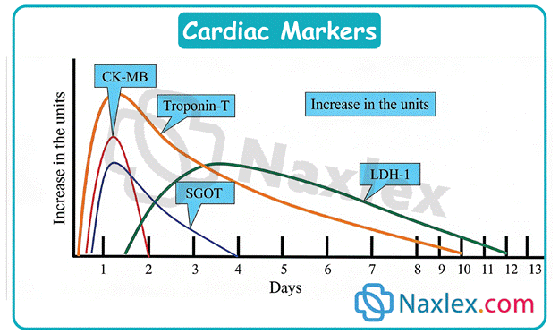
Indications
- Chest pain suggestive of acute coronary syndrome
- Suspected myocardial infarction
- Evaluation of unstable angina
- Assessment after PCI or thrombolytic therapy
- Detection of reinfarction
- Evaluation of heart failure (BNP/NT-proBNP)
Nursing Responsibilities
|
Before |
During |
After |
|
Document onset of symptoms |
Collect blood samples at prescribed intervals |
Trend serial laboratory values rather than relying on a single result |
|
Obtain baseline ECG |
Label and transport specimens promptly |
Monitor chest pain, dysrhythmias, vital signs, and oxygen saturation |
|
Explain the need for serial blood draws |
Verify correct specimen collection times |
Prepare patient for additional diagnostic tests or PCI if indicated |
Lipid Profile
Purpose: Evaluate cardiovascular risk and monitor response to lipid-lowering therapy.
Normal Values
|
Component |
Desired Level |
Clinical Significance |
|
Total Cholesterol |
<200 mg/dL |
Overall cholesterol level |
|
LDL |
<100 mg/dL (<70 mg/dL for very high-risk patients) |
Primary contributor to atherosclerotic plaque formation |
|
HDL |
>40 mg/dL (men); >50 mg/dL (women) |
Protective cholesterol that removes cholesterol from arteries |
|
Triglycerides |
<150 mg/dL |
Elevated levels increase cardiovascular and pancreatitis risk |
Indications
- Routine cardiovascular screening
- Diabetes mellitus
- Hypertension
- Family history of coronary artery disease
- Obesity
- Monitoring response to statins or other lipid-lowering medications
Nursing Responsibilities
|
Before Test |
After Test |
|
Fast 8–12 hours if ordered |
Review results with the patient |
|
Water is allowed |
Reinforce heart-healthy diet |
|
Avoid alcohol for 24 hours |
Encourage regular exercise |
|
Review medications that may affect results |
Encourage smoking cessation and medication adherence |
Lifestyle Education
- Reduce saturated and trans fats
- Increase fruits, vegetables, whole grains, soluble fiber, and omega-3 fatty acids
- Exercise at least 150 minutes per week
- Maintain healthy body weight
- Report unexplained muscle pain, weakness, or dark urine while taking statins
Echocardiogram
Purpose: Evaluate cardiac anatomy, chamber size, valve function, wall motion, ejection fraction, and blood flow.
Types
|
Type |
Description |
Advantages |
|
Transthoracic Echocardiogram (TTE) |
Ultrasound transducer placed on chest |
Noninvasive; no sedation required |
|
Transesophageal Echocardiogram (TEE) |
Ultrasound probe inserted into the esophagus |
Superior visualization of posterior cardiac structures, valves, atria, and thrombi |
|
Stress Echocardiogram |
Images obtained before and after exercise or pharmacologic stress |
Detects myocardial ischemia |
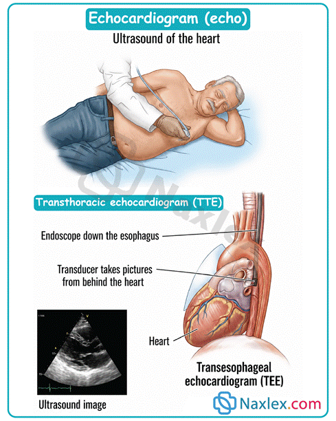
Indications
- Heart failure
- Cardiac murmurs
- Valvular heart disease
- Cardiomyopathy
- Pericardial effusion
- Infective endocarditis
- Congenital heart disease
- Evaluation of left ventricular ejection fraction
Nursing Responsibilities
|
Before |
During |
After |
|
TTE: No special preparation required |
Monitor ECG, oxygen saturation, and vital signs |
Resume normal activities after TTE |
|
TEE: NPO for 6–8 hours; remove dentures; obtain informed consent; establish IV access |
Monitor response to sedation and airway status |
Keep NPO until gag reflex returns; monitor swallowing, airway, oxygenation, and signs of esophageal injury |
Possible Complications
- Aspiration
- Respiratory depression
- Esophageal perforation
- Bleeding
- Dysrhythmias
- Sore throat following TEE
Normal Ejection Fraction (EF): 55–70%
Stress Testing
Purpose: Evaluate myocardial perfusion and identify ischemia during increased cardiac workload.
Types
|
Type |
Description |
|
Exercise Stress Test |
Performed on a treadmill or stationary bicycle |
|
Pharmacologic Stress Test |
Uses dobutamine, adenosine, regadenoson, or dipyridamole when exercise is not possible |
|
Nuclear Stress Test |
Radioactive tracer evaluates myocardial perfusion and identifies ischemic areas |
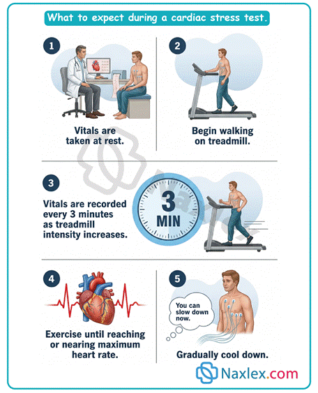
Indications
- Chest pain
- Suspected coronary artery disease
- Exercise tolerance evaluation
- Assessment of antianginal therapy
- Post-myocardial infarction evaluation
Contraindications
- Acute myocardial infarction
- Unstable angina
- Severe aortic stenosis
- Acute myocarditis
- Acute pericarditis
- Decompensated heart failure
Nursing Responsibilities
|
Before |
During |
After |
|
Avoid caffeine, smoking, alcohol, and heavy meals |
Continuous ECG and blood pressure monitoring |
Continue monitoring until vital signs return to baseline |
|
Hold beta blockers or nitrates if prescribed |
Observe for chest pain, dyspnea, dizziness, or dysrhythmias |
Document symptoms and provider interpretation |
|
Wear comfortable clothing and shoes |
Keep emergency equipment readily available |
Reinforce follow-up instructions |
Terminate the Test Immediately If
- Chest pain
- Significant ST elevation or depression
- Ventricular tachycardia
- Severe dyspnea
- Dizziness or syncope
- Severe hypertension
- Hypotension
Hemodynamic Monitoring
Purpose: Continuously assess cardiovascular pressures, cardiac function, and tissue perfusion in critically ill patients.
Hemodynamic Parameters
|
Parameter |
Normal Range |
Reflects |
|
Central Venous Pressure (CVP) |
2–8 mmHg |
Right ventricular preload and fluid volume |
|
Pulmonary Capillary Wedge Pressure (PCWP/PAWP) |
6–12 mmHg |
Left ventricular preload |
|
Cardiac Output (CO) |
4–8 L/min |
Amount of blood pumped per minute |
|
Cardiac Index (CI) |
2.5–4 L/min/m² |
Cardiac output adjusted for body size |
Monitoring Devices
|
Device |
Primary Use |
|
Central Venous Catheter |
Measures CVP and administers medications |
|
Pulmonary Artery (Swan-Ganz) Catheter |
Measures pulmonary pressures, wedge pressure, cardiac output, and mixed venous oxygen saturation |
|
Arterial Line |
Continuous blood pressure monitoring and frequent arterial blood sampling |
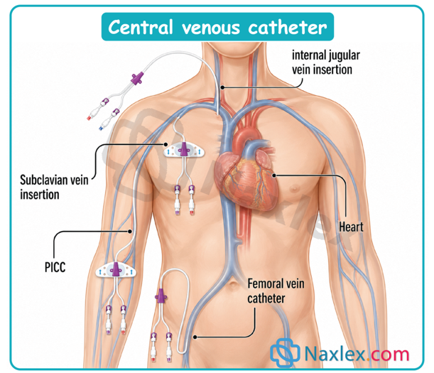
Indications
- Cardiogenic shock
- Septic shock
- Severe heart failure
- Major surgery
- Continuous vasoactive medication administration
- Unstable hemodynamic status
Nursing Responsibilities
- Level transducer to the phlebostatic axis
- Zero transducer every shift and after repositioning
- Maintain sterile technique
- Maintain pressure bag at approximately 300 mmHg
- Remove air bubbles from tubing
- Assess insertion site for infection or bleeding
- Monitor distal circulation and waveform quality
- Assess hemodynamic trends rather than isolated readings
Possible Complications
- Infection
- Air embolism
- Bleeding
- Dysrhythmias
- Thrombosis
- Pulmonary artery rupture
- Limb ischemia
- Catheter migration
Coronary Angiography
Purpose: Visualize coronary arteries, evaluate cardiac function, measure intracardiac pressures, and perform therapeutic interventions.
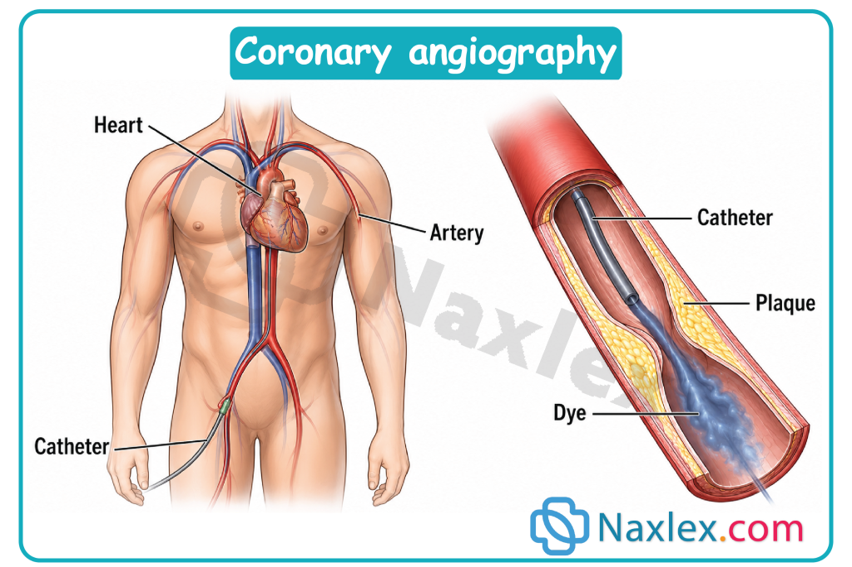
Common Uses
- Diagnose coronary artery disease
- Evaluate valvular disease
- Assess ventricular function
- Measure intracardiac pressures
- Balloon angioplasty (PCI)
- Coronary artery stent placement
Common Access Sites
- Radial artery
- Femoral artery
Nursing Responsibilities
|
Before Procedure |
After Procedure |
|
Obtain informed consent |
Monitor vital signs frequently |
|
NPO for 6–8 hours |
Assess puncture site for bleeding or hematoma |
|
Assess allergies to contrast media |
Monitor distal pulses, capillary refill, skin color, temperature, and sensation |
|
Review renal function (BUN, creatinine, eGFR) |
Encourage hydration if not contraindicated |
|
Mark peripheral pulses |
Monitor urine output and renal function |
|
Establish IV access |
Femoral access: Keep leg straight; Radial access: Monitor circulation to the hand |
Possible Complications
- Bleeding
- Hematoma
- Retroperitoneal hemorrhage
- Stroke
- Myocardial infarction
- Dysrhythmias
- Contrast-induced nephropathy
- Allergic reaction
- Arterial dissection
- Embolism
Patient Education
- Report chest pain immediately
- Report bleeding or swelling at the insertion site
- Report numbness, tingling, or coldness of the affected extremity
- Avoid strenuous activity and heavy lifting for several days
- Increase fluid intake if not contraindicated to promote contrast excretion
Vascular Access
Purpose: Provide reliable venous access for medications, fluids, nutrition, blood sampling, and hemodynamic monitoring.
Types of Vascular Access
|
Device |
Duration |
Common Uses |
|
Peripheral IV |
Short term |
Fluids, medications, blood products |
|
Midline Catheter |
Up to 4 weeks |
Antibiotics, fluids |
|
Peripherally Inserted Central Catheter (PICC) |
Weeks to months |
Long-term antibiotics, chemotherapy, TPN |
|
Non-tunneled Central Venous Catheter |
Short term |
ICU patients, vasoactive medications, hemodynamic monitoring |
|
Tunneled Catheter (Hickman/Broviac) |
Months to years |
Long-term therapy |
|
Implanted Port (Port-a-Cath) |
Months to years |
Chemotherapy and intermittent long-term therapy |
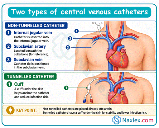
Indications
- Long-term intravenous therapy
- Chemotherapy
- Total parenteral nutrition
- Hemodynamic monitoring
- Difficult venous access
- Frequent blood sampling
- Dialysis
Possible Complications
- Central line-associated bloodstream infection (CLABSI)
- Air embolism
- Catheter occlusion
- Thrombosis
- Phlebitis
- Extravasation
- Catheter migration
- Pneumothorax
- Hemothorax
- Catheter fracture
Nursing Responsibilities
|
Insertion |
Ongoing Care |
|
Verify catheter tip placement before initial use |
Assess blood return and catheter patency |
|
Maintain sterile technique |
Flush according to agency policy |
|
Secure catheter appropriately |
Perform sterile dressing changes |
|
Document insertion details |
Disinfect catheter hub before access |
|
|
Monitor insertion site daily for redness, drainage, swelling, or tenderness |
|
|
Measure external catheter length routinely to detect migration |
Patient Education
- Keep dressing clean, dry, and intact
- Avoid pulling or kinking the catheter
- Report fever, redness, swelling, drainage, or pain immediately
- Avoid blood pressure measurements and venipuncture in the PICC arm whenever possible
- Implanted ports should only be accessed with a Huber needle
Summary
- Cardiovascular diagnostic tests provide essential information about cardiac structure, function, perfusion, electrical activity, and hemodynamic status, allowing early diagnosis and appropriate treatment of cardiovascular disease.
- Cardiac biomarkers, particularly troponin, are the primary laboratory indicators of myocardial injury, while CK-MB assists in detecting reinfarction and BNP is used to evaluate heart failure and ventricular overload.
- Lipid profiles assess long-term cardiovascular risk by measuring total cholesterol, LDL, HDL, and triglycerides, guiding both lifestyle interventions and pharmacologic therapy.
- Echocardiography offers noninvasive or minimally invasive visualization of cardiac anatomy and function, with transthoracic echocardiography serving as the initial imaging modality and transesophageal echocardiography providing enhanced visualization of posterior cardiac structures.
- Stress testing evaluates myocardial perfusion during increased cardiac workload and may be performed using exercise or pharmacologic agents when physical exercise is not feasible. Continuous monitoring is essential to identify ischemia or life-threatening complications.
- Invasive hemodynamic monitoring provides continuous assessment of cardiac performance through measurements such as central venous pressure, pulmonary artery pressures, pulmonary capillary wedge pressure, cardiac output, and cardiac index, supporting management of critically ill patients.
- Coronary angiography remains the gold standard for evaluating coronary artery disease and frequently serves as both a diagnostic and therapeutic procedure through percutaneous coronary intervention. Careful monitoring before and after the procedure is essential to detect bleeding, impaired perfusion, dysrhythmias, and contrast-related complications.
- Safe management of vascular access devices requires meticulous aseptic technique, routine assessment for patency and infection, appropriate flushing protocols, and early recognition of complications such as thrombosis, catheter occlusion, air embolism, and central line-associated bloodstream infection.
- Accurate interpretation of diagnostic findings must always be combined with comprehensive patient assessment, including vital signs, physical examination, symptom progression, and clinical history to guide nursing priorities and interdisciplinary collaboration.
- Effective nursing care throughout cardiovascular diagnostic testing emphasizes patient safety, evidence-based clinical judgment, early recognition of complications, therapeutic communication, patient education, and prompt intervention to optimize cardiovascular outcomes.
Naxlex
Videos
Login to View Video
Click here to loginTake Notes on Cardiovascular Diagnostic and Therapeutic Procedures
This filled cannot be empty
Join Naxlex Nursing for nursing questions & guides! Sign Up Now