



Please set your exam date
Diagnostic testing
Study Questions
Practice Exercise 4
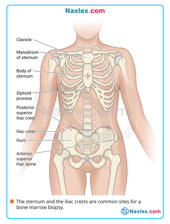
The nurse should take which of the following actions when assisting with a bone marrow biopsy?
Explanation
A bone marrow biopsy is an invasive diagnostic procedure that carries inherent risks of vascular damage and localized bleeding. Because the posterior superior iliac crest is highly vascular, close monitoring for hemorrhage and delayed subcutaneous hematomas is vital to prevent exsanguination or concealed retroperitoneal bleeding.
Rationale for correct answer:
C. Assessing the biopsy site for bleeding and hematoma formation for several days is a critical safety intervention. The posterior superior iliac crest is a deeply vascular area, and bleeding or a hidden hematoma can develop post-procedure. Continuous site checks prevent delayed hypovolemic complications.
Rationale for incorrect answers:
A. Assisting the client to a right side-lying position is not a standard universal post-procedure positioning requirement. The client should be placed in a supine position with a pressure dressing applied over the site for 30 to 60 minutes to maintain constant hemostasis through direct mechanical compression of the biopsy wound.
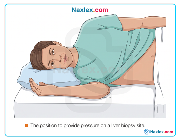
B. Observing for dyspnea, pallor, and coughing are clinical assessments targeted at identifying a pneumothorax or pulmonary embolism. These symptoms are typical complications following a liver biopsy or central venous access placement, rather than a standard iliac bone marrow aspiration.
D. Standing in front of the client to support the neck and knees describes the positioning assistance required for a lumbar puncture. For a bone marrow biopsy, the nurse places the client in a prone or lateral decubitus position to expose the posterior superior iliac crest safely.
Test-taking strategy
- Analyze the scenario and question: The nurse is assisting with a bone marrow biopsy and needs to identify the most appropriate nursing intervention from the options provided.
- Evaluate procedure-specific positioning: Review the anatomical site typically used for a bone marrow biopsy, which is the posterior superior iliac crest.
- Choice 1 is incorrect because the supine position is preferred initially to apply pressure to the posterior pelvic site.
- Choice 4 describes the fetal positioning required for a lumbar puncture, not an iliac crest biopsy.
- Differentiate post-procedure complications: Match the assessment parameters to the specific risks of the procedure.
- Choice 2 flags respiratory distress symptoms, which are complications associated with thoracic procedures or liver biopsies (due to diaphragmatic risk).
- Choice 3 targets bleeding and hematoma formation. Because bone marrow tissue is highly vascular and bleeding is the primary risk, checking the site for several days is the most logical and safe action. Rule out 1, 2, and D.
Take home points
- Hemorrhage and local hematoma formation are the primary risks following an invasive bone marrow aspiration or biopsy.
- Post-procedure positioning requires the client to lie supine for up to 60 minutes to apply natural pressure to the pelvic biopsy site.
- Severe local pain, radiating discomfort, or swelling at the biopsy site weeks later warrants immediate investigation for deep infection or hematoma.
- Proper positioning for a bone marrow biopsy involves exposing the posterior superior iliac crest via a prone or lateral recumbent position.
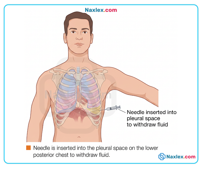
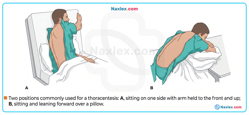
The nurse is preparing to assist a primary care provider who is going to perform a thoracentesis. The nurse’s role will include which action?
Explanation
A thoracentesis is an invasive procedure requiring the percutaneous needle aspiration of accumulated fluid or air from the pleural space. Proper mechanical positioning widens the intercostal spaces, facilitating safe needle insertion through the parietal pleura while reducing the risk of accidental laceration to the lung parenchyma or the intercostal neurovascular bundle.
Rationale for correct answer:
B. Seating the client upright with their arms supported on an overbed table optimizes anatomical access to the pleural cavity. This posture flexes the spine and expands the posterior intercostal spaces, lowering the diaphragm and allowing safe percutaneous access for fluid fluid removal while minimizing parenchymal injury risks.
Rationale for incorrect answers:
A. Placing the client supine in the Trendelenburg position shifts abdominal contents cranially against the diaphragm, restricting lung expansion. This position increases intrathoracic pressure and obscures access to the posterior pleural space, which is highly counterproductive for an effective lower intercostal fluid aspiration.
C. Delegating the measurement of vital signs to an unassigned assistive personnel during an invasive thoracentesis violates safety guidelines. The nurse must personally monitor the client's continuous respiratory status, oxygen saturation, and hemodynamic stability during the procedure to recognize immediate pleural shock or decompensation.
D. Administering an opioid analgesic immediately before the procedure is not a standard pre-thoracentesis nursing requirement. Local infiltration with lidocaine by the provider is typically sufficient, whereas heavy systemic opioids risk masking early symptoms of a developing pneumothorax or inducing unwanted respiratory depression.
Test-taking strategy
- Analyze the Scenario and Question: The nurse is assisting a provider with a thoracentesis. The question asks to identify the correct action within the nurse's scope of practice.
- Visualize the Anatomy: Thoracentesis targets fluid in the pleural space, which accumulates at the base of the lungs due to gravity. Access is easiest from the back.
- Choice 1 (Trendelenburg) positions the client upside down, making posterior thoracic access impossible and compressing the lungs. Ruled out.
- Choice 2 describes the classic upright, leaning-forward position that spreads the ribs and drops the fluid to the base for easy aspiration. This is correct.
- Evaluate Scope and Safety: Review delegation and assessment rules.
- Choice 3 is incorrect because an invasive procedure with a high risk of sudden decompensation requires registered nurse monitoring, not delegation to a UAP.
- Choice 4 is inappropriate because local anesthesia is standard, and opioids can suppress respiration or mask complications like an accidental lung puncture. Rule out 1, 3, and D.
Take home points
- The optimal position for a thoracentesis is sitting upright, leaning forward with arms resting on an overbed table to widen the intercostal spaces.
- Intra-procedural vital signs and respiratory assessments must be performed directly by the registered nurse due to the risk of sudden pneumothorax.
- Local anesthesia via subcutaneous lidocaine infiltration is the primary method of pain control during a percutaneous needle thoracentesis.
- If a client cannot sit up, a lateral decubitus position with the unaffected side down and the bed elevated may be utilized as an alternative.
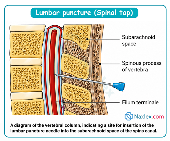
A nurse is assisting the provider with a lumbar puncture for a client who shows signs of meningism. Into which position should the nurse help place the client?
Explanation
A lumbar puncture requires opening the intervertebral spaces of the lumbar spine to allow safe needle insertion into the subarachnoid space. Maximizing spinal flexion separates the spinous processes of the third, fourth, or fifth lumbar vertebrae, which is vital to facilitate meningeal access, avoid local nerve root trauma, and prevent accidental dural laceration during subsequent percutaneous cerebrospinal fluid aspiration.
Rationale for correct answer:
A. Flexing the client's head toward the chest and drawing the knees up to the abdomen creates a lateral recumbent fetal position. This maximal curvature of the lumbar spine broadens the interspinous gaps, providing an unobstructed path for the spinal needle. Maintaining spinal flexion protects local tissues and ensures successful intrathecal access.
Rationale for incorrect answers:
B. Placing the client in the Trendelenburg position while lying in the Sims' position is an inappropriate and unsafe orientation for a lumbar puncture. Trendelenburg raises the pelvis above the head, which increases intracranial pressure and shifts cerebrospinal fluid mechanics unfavorably, complicating lumbar access and elevating herniation risks.
C. Raising the arms above the head and elevating the legs on pillows extends the spine rather than flexing it. Extension compresses the lumbar spinous processes together, completely blocking the needle entry pathway into the subarachnoid space. This posture makes intervertebral needle entry structurally impossible.
D. Positioning the client prone with the legs fully extended is not the standard position for a routine diagnostic lumbar puncture. While a prone position can be utilized under specialized fluoroscopic guidance in radiology, it lacks the protective vertebral separation achieved by the standard lateral fetal position at the bedside.
Test-taking strategy
- Analyze the scenario and question: The nurse is assisting with a lumbar puncture for a client showing signs of meningism. The question asks into which position the nurse should place the client.
- Visualize the spinal anatomy: Recall that to insert a needle into the subarachnoid space of the lumbar spine (usually L3-L4 or L4-L5), the bones of the spine must be separated as much as possible.
- Choice 3 (Extension) and Choice 4 (Prone extension) flatten or compress the lumbar spine, closing the intervertebral spaces and blocking access. These are ruled out.
- Apply postural biomechanics: Compare the remaining options.
- Choice 2 (Trendelenburg/Sims') shifts cerebrospinal fluid pressure dynamics toward the brain, which is dangerous in a patient with potential meningitis/meningism due to the risk of cerebral herniation.
- Choice 1 describes the classic lateral recumbent fetal position (chin to chest, knees to abdomen). This maximizes lumbar curvature and spreads the vertebrae apart, making it the correct option. Rule out 2, 3, and D.
Take home points
- The standard bedside position for a lumbar puncture is the lateral recumbent position with the head flexed and knees drawn to the abdomen.
- Maximum spinal flexion separates the lumbar spinous processes, widening the anatomical window for safe needle insertion into the subarachnoid space.
- After a lumbar puncture, the client should remain flat in a supine position for several hours to minimize the risk of a post-dural puncture headache.
- Extending the spine or using the Trendelenburg position is contraindicated because it closes the intervertebral spaces or unsafely alters intracranial pressure.
Comprehensive Questions
A nurse is contributing to the post-procedure care plan for a client who has just returned from cardiac catheterization with coronary angiography. An arterial closure device was applied at the access site. Which nursing intervention should be included in the client’s immediate post-procedure care?
Explanation
Arterial closure devices mechanically seal the femoral artery puncture site following cardiac catheterization to achieve rapid hemostasis. While these deployment systems significantly reduce necessary immobilization times compared to standard manual compression, maintaining absolute immobility for a targeted window is vital to secure the fibrin plug, prevent mechanical tract disruption, and block retroperitoneal hemorrhage during subsequent early ambulation recovery.
Rationale for correct answer:
B. Maintaining the client on bed rest for 2 hours is the correct protocol when an arterial closure device is utilized. These devices deploy an internal anchor and collagen plug to seal the femoral arteriotomy instantly. This allows safe ambulation within 2 to 4 hours, compared to the 6 hours required for manual compression.
Rationale for incorrect answers:
A. Restricting oral fluids for the first 4 hours after the procedure is contraindicated and dangerous. The nurse should actively encourage fluid intake to promote the osmotic diuresis and clearance of nephrotoxic iodinated contrast dye. Restricting fluids promotes stasis and increases the risk of contrast-induced nephropathy.
C. Raising the head of the bed to 45 degrees is a procedural error that risks vascular access failure. The client's affected groin must remain straight to prevent bending the femoral artery and disrupting the closure plug. The head of the bed must be kept below 30 degrees to avoid mechanical strain and bleeding.
D. Inserting an indwelling urinary catheter 1 hour after the procedure is an unnecessary invasive action that introduces an infection risk. Catheterization is reserved for clients experiencing acute urinary retention or those requiring strict hemodynamic monitoring, rather than being a routine intervention for post-catheterization care.
Test-taking strategy
- Analyze the scenario and question: A client returns from a cardiac catheterization with coronary angiography via the femoral artery. An arterial closure device was used. The question asks for an intervention to include in immediate post-procedure care.
- Understand closure device mechanics: Recall that arterial closure devices speed up hemostasis. Traditional manual compression requires 6 hours of strict bed rest. Closure devices reduce this time drastically, usually down to 2 hours of bed rest.
- Choice 2 matches this 2-hour immobilization window perfectly.
- Identify procedural contraindications: Critique choices 1, 3, and D.
- Choice 1 is incorrect because fluids should be encouraged, not restricted, to protect the kidneys from contrast.
- Choice 3 is incorrect because flexing the hip at 45 degrees puts pressure on the puncture site and can cause hematoma; the leg must remain flat (< 30 degrees).
- Choice 4 is an invasive error; catheters are not routinely placed post-cath. Rule out 1, 3, and D.
Take home points
- The use of an arterial closure device reduces mandatory post-cardiac catheterization bed rest from 6 hours down to approximately 2 hours.
- The head of the bed must be maintained at or below 30 degrees during bed rest to keep the groin straight and prevent access site bleeding.
- Clients must be encouraged to increase fluid intake immediately after the procedure to flush out nephrotoxic contrast dye.
- Frequent neurovascular assessments of the distal extremity are necessary to detect acute arterial occlusion or hematoma formation.
The nurse should take which of the following actions when assisting with a bone marrow biopsy?
Explanation
Preparing a client for diagnostic testing involves a holistic framework encompassing psychological, educational, and physical interventions. Systematically addressing pre-procedural protocols reduces client anxiety, minimizes bodily motion during invasive pathways, and ensures physical safety, which is vital to preserve anatomical alignment, secure valid diagnostic data, and prevent preventable procedural errors during the subsequent intravascular or percutaneous testing sequence.
Rationale for correct answers:
A. Ensuring appropriate preparation is vital to guarantee the validity of the diagnostic test results. This includes verifying pre-procedure fasting statuses, completing medication holds, and checking baseline coagulation panels prior to invasive access. Skipping these steps can cause immediate test cancellation or physical injury.
B. Providing individualized education helps reduce the client's anxiety and promotes active cooperation during the diagnostic test. Tailoring instructions to the client's cognitive and psychological state ensures they understand what to expect before, during, and after the test. Educational interventions maximize client compliance and procedural safety.
C. Placing the client in the correct position optimizes anatomical exposure and ensures safety during the procedure. Correct positioning prevents structural injuries and allows the practitioner to navigate needles or scopes into target cavities without obstruction. Maintaining postural alignment reduces the risk of accidental perforations.
E. Providing emotional support helps stabilize the client's autonomic nervous system, lowering stress-induced tachycardia or hypertensive spikes before a procedure. The nurse uses therapeutic communication and calming presence to mitigate fear of the unknown. Active coping support enhances the overall clinical experience.
Rationale for incorrect answers:
D. Initiating Standard Precautions is an intra-procedural and post-procedural infection control mandate rather than a client preparation action. While the nurse wears personal protective equipment, this action is a universal safety barrier designed to protect the clinician from bloodborne pathogens, rather than an act of preparing the client for testing.
F. Documenting the diagnostic outcomes is a post-procedural nursing action that occurs after the test has been fully executed and analyzed. The nurse cannot document results during the preparatory phase of care because the data does not yet exist. This makes it an evaluation step rather than a pre-test intervention.
Test-taking strategy
- Analyze the scenario and question: The question asks for actions included when preparing a client for diagnostic testing. The keyword is "preparing," which focuses exclusively on the pre-procedural phase of care.
- Filter actions by chronological phase: Evaluate each option based on whether it occurs before the diagnostic test begins.
- Choice 1 (Preparation), Choice 2 (Education), Choice 3 (Positioning), and Choice 5 (Emotional support) are all classic pre-procedural interventions that prepare the client physically and mentally. These are ruled in.
- Choice 6 (Documenting outcomes) occurs long after the test is completed and interpreted, ruling it out.
- Differentiate client care vs. infection control: Look closely at Choice 4 (Initiating Standard Precautions). Standard precautions are a continuous infection control framework for the provider during specimen handling, not an action directed at preparing the client's body or mind for a test. Rule out 4 and F.
Take home points
- Client preparation for diagnostic tests requires checking structural prerequisites like NPO status, medication restrictions, and informed consent.
- Individualized client education and continuous emotional support reduce anxiety, lowering stress-induced vital sign fluctuations during testing.
- Proper physical positioning expands anatomical entry windows and keeps the client stabilized to prevent procedural injuries or tissue damage.
- Documenting diagnostic outcomes is a retrospective post-procedural responsibility that occurs only after findings are reported by the lab or provider.
The nurse is preparing a staff education program on the Joint Commission’s 2009 hospitals’ national patient safety goals. The purpose of the Joint Commission is to do which of the following?
Explanation
Accreditation frameworks in healthcare focus on establishing rigorous clinical benchmarks to optimize operational safety. The Joint Commission implements evidence-based standards to systematically eliminate preventable medical hazards, optimize care execution, and reduce clinical sentinel events, making the reduction of healthcare errors and the preservation of patient safety the primary objective rather than commercial success or public relations optimization.
Rationale for correct answer:
B. The core purpose of The Joint Commission is to continuously improve patient safety and minimize clinical mistakes across healthcare delivery systems. Their National Patient Safety Goals are specifically targeted programs designed to address systemic vulnerabilities, such as misidentification or medication errors. Prioritizing these goals directly serves to decrease error and optimize client safety.
Rationale for incorrect answers:
A. Improving a hospital's public rating or competitive standing is a secondary, indirect byproduct of achieving accreditation rather than the organizational purpose. The Joint Commission functions as an independent, non-profit regulatory body focused strictly on quality metrics, making public marketing advantages irrelevant to their primary safety mandate.
C. Increasing hospital revenues or maximizing commercial profitability is not a focus of accreditation or patient safety guidelines. While preventing complications avoids financial penalties, the commission's standards are built around ethical clinical care, completely separating their operational criteria from hospital revenue or corporate profitability.
D. Elevating the public image or reputation of the nursing profession is a function of professional bodies like the American Nurses Association. The Joint Commission evaluates interdisciplinary hospital-wide safety cultures, focusing on collective operational systems rather than highlighting or increasing the status of an isolated discipline.
Test-taking strategy
- Analyze the scenario and question: The nurse is teaching a staff program on The Joint Commission's National Patient Safety Goals. The question asks to identify the fundamental purpose of this regulatory body.
- Identify the core mission of regulatory agencies: Recall that the National Patient Safety Goals (NPSGs) are created specifically to target vulnerabilities in patient care that lead to injury or death.
- Choice 1 (Hospital ratings) and Choice 4 (Image of nursing) focus on public relations and external perceptions rather than internal clinical safety. These are ruled out.
- Choice 3 (Hospital revenues) relates to financial and commercial gain, which contradicts the non-profit, safety-driven mission of the organization.
- Correlate terms to select safety objectives: Select Choice 2 (Decrease client error) because it directly matches the core clinical definition of "patient safety." Rule out 1, 3, and D.
Take home points
- The primary objective of The Joint Commission is to ensure patient safety and improve healthcare quality through rigorous accreditation standards.
- National Patient Safety Goals are evidence-based mandates updated regularly to target and mitigate high-risk areas prone to clinical errors.
- The focus of healthcare accreditation is purely clinical quality and risk reduction rather than improving financial margins or hospital marketing campaigns.
- Enhancing safety requires an interdisciplinary approach that evaluates institutional workflows rather than focusing on the prestige of an isolated profession.
The nurse understands that decreasing the length of time a client stands prior to venipuncture and the length of time the tourniquet is left on the client’s arm helps to prevent which of the following?
Explanation
Prolonged hydrostatic pressure and localized venous stasis during phlebotomy disrupt the fluid balance inside blood vessels. Restricting the duration of both pre-procedural standing and tourniquet compression prevents the excessive filtration of plasma water into the surrounding interstitial tissue, avoiding artificial solute elevation and altered diagnostic data during subsequent hematological or biochemical plasma profiling.
Rationale for correct answer:
A. Minimizing standing time and restricting tourniquet application to less than 60 seconds directly prevents hemoconcentration. Prolonged venous stagnation forces fluid out of the capillaries, causing an artificial concentration of large molecules like proteins and cells. Preventing hemoconcentration ensures that laboratory results reflect true baseline serum values.
Rationale for incorrect answers:
B. Altering tourniquet timing or standing durations does not prevent acute localized or systemic hemorrhage. Hemorrhage refers to excessive, uncontrolled bleeding caused by vascular wall rupture, severe coagulopathy, or improper post-puncture pressure, rather than standard pre-analytical fluid shifts occurring within an intact venous lumen.
C. Homeostasis represents the normal, healthy state of physiological equilibrium maintained continuously by the body's regulatory mechanisms. Decreasing tourniquet compression time is an intervention intended to preserve the accuracy of a diagnostic test rather than an action performed to block or prevent natural cellular homeostasis.
D. Hypoxemia represents a pathological decrease in the partial pressure of oxygen within arterial blood gas measurements. Leaving a venous tourniquet on an extremity causes temporary localized tissue ischemia and venous stasis, but it does not cause or prevent global systemic hypoxemia in the lungs or central arterial circulation.
Test-taking strategy
- Analyze the scenario and question: The nurse is reviewing phlebotomy techniques, specifically reducing the time a client stands and limiting tourniquet application duration. The question asks what complication these interventions help prevent.
- Understand phlebotomy fluid dynamics: Recall what happens when a tourniquet is left on too long (greater than 1 minute) or when prolonged standing causes venous pooling. Water escapes the vasculature, leaving large elements behind.
- Choice 3 (Homeostasis) is a positive, normal state of bodily balance, so a nurse wouldn't try to prevent it. Ruled out.
- Choice 2 (Hemorrhage) and Choice 4 (Hypoxemia) relate to gross blood loss and systemic arterial oxygen drops, which are not caused by brief venous stasis in an arm.
- Identify pre-analytical lab errors: Select Choice 1 (Hemoconcentration) because it is the specific, textbook term for the localized pooling of large molecules and blood cells due to prolonged venous obstruction. Rule out 2, 3, and D.
Take home points
- Tourniquet application must be strictly limited to less than 60 seconds during venipuncture to avoid pre-analytical errors.
- Hemoconcentration occurs when localized venous stasis forces fluid out of the vessel, falsely elevating parameters like hematocrit, calcium, and potassium.
- Instructing the client to avoid prolonged standing or pumping their fist immediately before a blood draw reduces hydrostatic fluid shifts.
- Hemorrhage is prevented post-procedure by applying direct mechanical pressure to the venipuncture site for at least 2 to 5 minutes.
A nurse has just obtained an arterial blood gas (ABG) sample from a client. Which of the following actions should the nurse take?
Explanation
Arterial blood gas analysis measures dissolved respiratory gases and cellular metabolites that remain highly volatile outside the human body. Because erythrocytes continue active anaerobic glycolysis post-collection, packing the syringe in an ice slurry immediately slows cellular metabolism, preserving true oxygen and carbon dioxide levels to prevent metabolic drift, erroneous partial pressure alterations, and inaccurate clinical interpretation during subsequent potentiometric blood gas quantification.
Rationale for correct answer:
A. Packing the arterial sample in ice maintains sample stability during transit to the laboratory. Lowering the temperature slows down cellular respiration and glycolysis within the trapped erythrocytes, preventing them from consuming oxygen and producing carbon dioxide. This thermal preservation ensures the laboratory measures accurate baseline arterial parameters.
Rationale for incorrect answers:
B. Maintaining pressure on the radial artery site for only 1 minute is insufficient to ensure complete hemostasis. Because arterial architecture functions under high hydrostatic pressures, a post-puncture site requires continuous manual compression for at least 5 minutes, or longer if the client takes anticoagulants, to prevent a large hematoma.
C. Transferring the blood sample to a separate test tube introduces ambient air exposure and invalidates the results. Arterial blood gas collections are drawn into a specialized, pre-heparinized, airtight syringe that must be capped immediately. Decanting the blood into an open tube alters volatile gas partial pressures.
D. Drawing a second arterial blood sample in 10 minutes is not a standard post-procedure action unless the client experiences acute clinical deterioration. Arterial punctures are invasive, painful, and carry risks of vascular spasm or thrombosis, making routine serial sticks without a fresh medical indication unwarranted.
Test-taking strategy
- Analyze the scenario and question: The nurse has just successfully drawn an arterial blood gas sample and must identify the correct immediate post-procedure handling action.
- Understand blood gas volatility: Recall that cells in the blood continue to live and consume oxygen inside the syringe. To prevent this from changing the pH, pO2, and pCO2 values, cellular metabolism must be paused or slowed down.
- Choice 2 is a dangerous understatement; 1 minute of pressure on an artery will cause severe bleeding (5 minutes is the minimum).
- Choice 3 destroys the sample because exposing arterial blood to atmospheric air ruins gas measurements.
- Identify preservation mechanisms: Select Choice 1 (Pack the sample in ice) because hypothermia reduces cellular metabolic consumption, keeping the blood gas values stable during the trip to the laboratory. Rule out 2, 3, and D.
Take home points
- Arterial blood gas specimens must be packed in ice immediately if analysis cannot be completed at the bedside within 10 to 15 minutes.
- Manual compression on an arterial puncture site must be maintained for a minimum of 5 continuous minutes to prevent hemorrhage or hematoma.
- ABG samples must never be transferred or exposed to air, as room air exposure falsely elevates oxygen and drops carbon dioxide values.
- The collection syringe must be pre-heparinized to prevent clotting, and all air bubbles must be expelled immediately prior to capping.
A nurse is preparing a urine specimen for laboratory analysis. If there is a delay in transporting the sample, which action should the nurse take?
Explanation
Delayed processing of an unpreserved urine specimen leads to rapid biochemical and microbiological decomposition at room temperature. Bacteria present in the sample multiply exponentially, catabolizing urea into ambient ammonia, which elevates the pH, causes crystalline precipitation, and triggers enzymatic degradation, necessitating strict thermal stabilization to arrest bacterial proliferation and maintain diagnostic validity before subsequent microscopic urinalysis.
Rationale for correct answer:
C. Placing the urine specimen in a plastic biohazard bag and refrigerating it is the correct measure when transport is delayed beyond 1 hour. Cold storage halts the replication of contaminating ambient bacteria and preserves the physical structure of cellular casts. This mechanical cooling guards the biochemical integrity of the sample.
Rationale for incorrect answers:
A. Notifying the prescribing practitioner about a simple logistics or transit delay is an inappropriate escalation of an operational nursing task. The nurse possesses the autonomy to preserve the specimen correctly via refrigeration or to re-collect the sample if it degrades, making an immediate practitioner page completely unnecessary.
B. Discarding the urine sample immediately upon a minor transport delay is a premature action that wastes clinical resources and delays diagnostic data. Re-collection is only required if the sample has remained at warm room temperatures for more than 1 to 2 hours without preservation, making automatic disposal incorrect.
D. Writing "stat" on the requisition form to force laboratory pickup does not fix an existing logistical delay or protect an uncooled sample. Falsifying clinical urgency or demanding immediate action can compromise truly critical patient samples within the hospital system while ignoring proper specimen cooling protocols.
Test-taking strategy
- Analyze the scenario and question: There is an unexpected delay in transporting a freshly collected urine sample to the laboratory. The nurse must choose the correct preservation intervention.
- Evaluate chemical and bacterial shifts: Recall that urine is an excellent culture medium at room temperature. Leaving it out causes bacteria to grow, which alters the pH and destroys cells or casts needed for an accurate reading.
- Choice 1 and Choice 4 are poor communication and management choices that fail to address the immediate physical state of the specimen.
- Choice 2 is wasteful and unnecessary if the sample can still be preserved.
- Apply cold storage principles: Select Choice 3 (Refrigerate the sample) because cooling is the standard, universal method to inhibit bacterial growth and preserve chemical analytes when immediate laboratory processing is unavailable. Rule out 1, 2, and D.
A nurse is preparing a staff education program about diagnostic testing. Which best defines culture and sensitivity?
Explanation
Infectious disease management relies on precise microbiological identification and targeted pharmacological profiling to eradicate pathogenic micro-organisms. Combining isolation techniques with resistance profiling guides clinicians away from empirical, broad-spectrum coverages toward narrow-spectrum therapies, which is vital to optimize antimicrobial stewardship, ensure targeted pathogen eradication, and prevent the global acceleration of multidrug resistance during subsequent bacterial or fungal infections.
Rationale for correct answer:
C. Culture and sensitivity testing definitively identifies both the specific invading pathogen and its susceptibility profile to targeted antimicrobials. The culture phase isolates and grows the microorganism, while the sensitivity phase exposes it to various agents to determine the minimum inhibitory concentration. This combined microbiological data guides safe antimicrobial selection.
Rationale for incorrect answers:
A. Defining a culture and sensitivity panel as a common screening test understates its specific diagnostic role. Screening tests are broad, non-specific assessments applied to asymptomatic populations to detect general disease risks, whereas a culture is a targeted, confirmatory diagnostic tool reserved for active infectious processes.
B. Explaining that it allows bacteria to grow so the exact organism can be identified describes only the culture portion of the test. This statement completely omits the sensitivity component, which determines which pharmacological agents will successfully eliminate the pathogen. It represents an incomplete and fragmented definition of the comprehensive assay.
D. Evaluating cell maturity, metabolic activity, and morphologic variations describes the parameters of a cytological examination or a peripheral blood smear. These cellular metrics are utilized to detect hematological malignancies or bone marrow disorders, rather than isolating and profiling pathogenic infectious microorganisms.
Test-taking strategy
- Analyze the scenario and question: The nurse is preparing an educational program on diagnostic testing. The question asks for the option that best and most completely defines a "culture and sensitivity" (C&S) test.
- Deconstruct the compound term: Break the test name into its two mandatory, sequential components: "Culture" (growing and identifying the organism) and "Sensitivity" (determining which drugs kill the organism).
- Choice 2 only describes the culture phase, making it incomplete. Ruled out.
- Choice 4 describes cytology or pathology slides, which is completely unrelated to infectious disease profiling.
- Evaluate for completeness: Select Choice 3 because it explicitly integrates both elements of the test name - identifying the nature of the invading organism (culture) and determining its susceptibility to antibiotics (sensitivity) - into a singular, clinically accurate definition. Rule out 1, 2, and D.
Take home points
- A culture and sensitivity test is a dual-phase diagnostic modality that isolates a pathogen and determines its specific antimicrobial vulnerability.
- The culture phase allows the microorganism to incubate and multiply so it can be precisely identified by genus and species.
- The sensitivity phase tests the isolated pathogen against common antibiotics to classify it as susceptible, intermediate, or resistant.
- Obtaining culture specimens before administering the first dose of an empirical antibiotic prevents false-negative growth results.
A nurse is reviewing diagnostic procedures that require the administration of a contrast medium to visualize internal body structures. Which of the following diagnostic studies are considered contrast-mediated procedures? Select all that apply
Explanation
Contrast-mediated radiologic imaging utilizes high-density substances to temporarily alter the radiation absorption characteristics of hollow organs or fluid-filled cavities. Introducing radiopaque contrast elements illuminates structural contours and intraluminal boundaries against surrounding soft tissues, which is vital to reveal mucosal lesions, track vascular patency, and isolate structural anomalies during subsequent fluoroscopic or radiographic examinations.
Rationale for correct answers:
A. A barium swallow requires the oral ingestion of a thick, radiopaque barium sulfate suspension to outline the upper gastrointestinal tract. As the heavy compound transits the esophagus and stomach, it blocks x-ray beams, creating high-contrast fluoroscopic images that reveal structural strictures or motility disorders.
B. A bronchography is an imaging procedure that relies on the direct instillation of an oil-based or water-soluble radiopaque contrast agent into the bronchial tree. The contrast substance coats the tracheobronchial mucosa, allowing detailed radiographic mapping of bronchial architecture to identify advanced bronchiectasis or localized airway obstructions.
C. An intravenous cholangiography utilizes an intravenously injected iodinated contrast medium that is selectively cleared and concentrated by the hepatobiliary system. The radiopaque dye flows directly into the hepatic, cystic, and common bile ducts, illuminating luminal narrowing, structural strictures, or radiolucent cholelithiasis on serial films.
E. A myelogram involves the percutaneous instillation of a water-soluble iodinated contrast medium directly into the spinal subarachnoid space via a lumbar puncture. The radiopaque fluid surrounds the spinal cord and nerve roots, providing sharp contrast to detect herniated discs, spinal canal stenosis, or intrathecal masses.
Rationale for incorrect answers:
D. Mammography is a non-contrast, low-dose screening x-ray technique used exclusively to evaluate breast tissue parenchyma. It relies entirely on the natural density differentials between normal fibroglandular tissue, adipose tissue, and abnormal microcalcifications or solid masses, making exogenous contrast media completely unnecessary.
F. Ultrasonography is a completely non-ionizing, non-contrast diagnostic modality that relies on high-frequency sound waves to generate real-time tissue images. Transducers record the acoustic impedance and echoes bouncing off internal organ interfaces, mapping solid and fluid-filled structures without any radioactive or chemical contrast media ingestion.
Test-taking strategy
- Analyze the scenario and question: The nurse is identifying diagnostic procedures that require the administration of an exogenous contrast medium (such as barium or iodine) to visualize internal body structures.
- Evaluate the imaging modality mechanics: Go through each option and determine whether it uses an injected, instilled, or ingested radiopaque substance to create a visible outline on an x-ray or fluoroscopy machine.
- Choice 1 uses barium to coat the GI tract (ruled in).
- Choice 2 instills contrast into the airways (ruled in).
- Choice 3 injects dye to illuminate the bile ducts (ruled in).
- Choice 5 injects dye into the spinal column (ruled in).
- Eliminate non-contrast screening modalities: Review the remaining choices for contrast needs.
- Choice 4 (Mammography) relies purely on compression and low-energy tissue x-rays.
- Choice 6 (Ultrasonography) uses sound waves, not contrast x-rays. Rule out 4 and F.
Take home points
- Contrast-mediated procedures introduce dense radiopaque substances into the body to sharply outline the internal lumens of organs and vessels.
- Barium swallows and bronchographies use contrast agents to coat mucosal surfaces within the gastrointestinal and respiratory systems, respectively.
- Intravenous cholangiograms and myelograms require targeted contrast injections to visualize hepatobiliary ducts and the spinal subarachnoid space.
- Screening modalities like mammography and ultrasound rely on inherent tissue densities or sound wave echoes without requiring contrast media.
A nurse is preparing to obtain a capillary blood glucose reading from a school-age child. Which action should the nurse plan to carry out?
Explanation
Capillary blood glucose testing requires strict chemical and physical isolation of the blood sample from localized environmental contaminants. Allowing the antiseptic solution to air-dry completely prevents chemical interaction with the lancet wound, which is vital to avoid sample hemolysis, prevent chemical interference with the reagent strip, and reduce localized burning pain during the subsequent percutaneous skin puncture.
Rationale for correct answer:
A. Allowing the antiseptic solution to dry completely prevents the alcohol from mixing with the blood sample. Wet alcohol on the skin causes cell lysis and dilutes the glucose concentration, leading to inaccurate diagnostic data. Ensuring the site is dry preserves the chemical integrity of the sample and reduces procedural discomfort.
Rationale for incorrect answers:
B. Applying a cool compress to the child's finger causes localized vasoconstriction and severely reduces capillary blood flow. This vascular contraction makes it difficult to obtain an adequate blood volume, forcing the nurse to squeeze the tissue forcefully. Forceful milking introduces interstitial fluid that alters glucose measurements.
C. Using the first drop of blood obtained after a skin puncture can yield an inaccurate, diluted reading. The initial drop is heavily contaminated with intracellular fluids, serous exudates, and chemical residues from the antiseptic wash. Wiping away the first drop guarantees that the second drop represents pure capillary blood.
D. Puncturing the center of the finger pad causes excessive pain and risks structural injury to the underlying tissue. The central pad of the digit contains a high density of sensitive tactile nerve endings and a thin epidermal layer. Avoiding the central pad protects the child from unnecessary neurological trauma.
Test-taking strategy
- Analyze the scenario and question: The nurse is preparing to obtain a capillary blood glucose reading from a school-age child. The question asks to identify the correct action to carry out.
- Evaluate chemical and physical site interferences: Review how pre-puncture skin preparation alters blood samples and patient comfort.
- Choice 1 highlights allowing the antiseptic to dry. This prevents hemolysis and guards against false glucose readings. This is correct.
- Eliminate physiological and procedural errors: Check the remaining choices against standard laboratory guidelines.
- Choice 2 is incorrect because cold compresses cause vasoconstriction; warming the hand is preferred to increase perfusion.
- Choice 3 is incorrect because the first drop of blood contains interstitial fluid and must be wiped away.
- Choice 4 targets the highly sensitive center of the finger pad instead of the less painful lateral sides. Rule out 2, 3, and D.
Take home points
- Antiseptic solutions must be allowed to air-dry completely prior to a lancet puncture to prevent hemolysis and burning sensations.
- Wiping away the first drop of blood with dry gauze removes interstitial fluid and chemical contaminants that skew glucose calculations.
- Lancet punctures should be performed exclusively on the lateral borders of the finger to minimize pain and protect nerve pathways.
- Warming the extremity with a warm, moist washcloth expands capillaries and enhances spontaneous blood flow at the puncture site.
A nurse is preparing to obtain a blood sample for a capillary blood glucose test. Which of the following sites should the nurse select?
Explanation
Anatomical site selection for capillary phlebotomy relies on balancing abundant microvascular networks with low nerve density profiles. Choosing a non-dominant, lateral digital surface maximizes capillary yield while safeguarding functional areas of the hand, which is vital to maintain perfusion density, minimize sensory pain paths, and ensure procedural safety during subsequent bedside monitoring diagnostic protocols.
Rationale for correct answer:
D. Selecting the side of the ring finger provides an optimal site with excellent capillary vascularity and lower nerve sensitivity. The lateral margins of the finger have a thicker epidermal layer and fewer pain receptors than the central pads. Utilizing the lateral surface ensures a high-volume sample with minimal neurological distress.
Rationale for incorrect answers:
A. Selecting the pad of the thumb is inappropriate due to its dense nerve networks and vital role in opposition and fine motor function. Puncturing this central surface causes intense pain and leaves a tender wound that interferes with daily grasping tasks. The thumb pad has a thick callus that complicates clean vascular access.
B. Choosing the pinna of the ear is not a standard or safe site for routine capillary blood glucose testing in an adult or child. The earlobe can be used in specific metabolic research settings, but the cartilage-dense pinna lacks sufficient capillary beds and carries a high risk of chondritis or localized tissue necrosis.
C. Selecting the pad of the big toe is an incorrect site for an ambulatory or older client due to safety and contamination risks. The feet are highly susceptible to microvascular insufficiency and environmental pathogens, meaning a toe puncture increases the risk of delayed healing and deep podiatric infections.
Test-taking strategy
- Analyze the scenario and question: The nurse is preparing to select an appropriate anatomical site for a capillary blood glucose test.
- Evaluate site specificity and safety: Review standard adult and pediatric peripheral puncture sites based on tissue thickness, nerve density, and infection risks.
- Choice 1 (Thumb pad) and Choice 3 (Big toe pad) involve highly sensitive, functional, or infection-prone weight-bearing surfaces that must be avoided.
- Choice 2 (Pinna) targets cartilage, which has poor vascularity and is prone to permanent structural infection.
- Identify the clinical standard: Select Choice 4 (Side of the ring finger) because the lateral aspects of the third and fourth fingers are the universal gold standard sites for capillary blood collection, offering excellent blood flow with the least amount of functional interference. Rule out 1, 2, and C.
Take home points
- The lateral borders of the ring and middle fingers are the preferred anatomical sites for adult capillary blood sampling.
- Central pads of the fingers and thumbs must be avoided due to dense sensory nerve endings and functional motor importance.
- Puncturing the feet or toes for blood glucose monitoring is contraindicated in older children and adults due to infection and neurovascular risks.
- In newborns and infants under 12 months of age, the lateral or medial plantar surfaces of the heel are utilized instead of the fingers.
A nurse is reinforcing teaching with a client about how to collect a stool specimen for occult blood testing. Which client statement demonstrates correct understanding?
Explanation
Guaiac-based fecal occult blood testing targets the pseudoperoxidase activity of heme molecules to detect occult gastrointestinal bleeding. Because common dietary items possess intrinsic chemical properties that can mimic or alter this chromogenic reaction, implementing strict pre-test dietary modifications is vital to eliminate peroxidase interference, avoid false-positive diagnostic outcomes, and ensure diagnostic specificity during subsequent colorectal cancer screening protocols.
Rationale for correct answer:
B. Stating the need to avoid red meat for 2 days before the test demonstrates an accurate understanding of the pre-procedural dietary restrictions. Red meat contains animal hemoglobin and myoglobin, which can trigger a false-positive blue reaction on the guaiac paper. Restricting beef or pork intake safeguards the accuracy of the test.
Rationale for incorrect answers:
A. Believing that consuming pasteurized dairy products will alter the test results represents a misunderstanding of the dietary guidelines. Milk, cheese, and yogurt do not contain hemoglobin or high peroxidase activity and do not interfere with the colorimetric reaction, making dairy consumption completely permissible during the screening window.
C. Thinking it is acceptable if urine mixes with the stool sample is incorrect because urine invalidates the test specimen. Urine can dilute the sample, wash away loose blood cells, or contain chemical contaminants and medications that alter the paper's reagent stability, making urine contamination a cause for specimen rejection.
D. Planning to collect one specimen each week for 4 weeks violates the established multi-sample screening protocol. The standard guaiac fecal occult blood test requires collecting consecutive samples from three separate, consecutive bowel movements within a few days, rather than an extended monthly longitudinal sampling sequence.
Test-taking strategy
- Analyze the scenario and question: The nurse is teaching a client how to collect a stool specimen for an occult blood test. The question asks which statement by the client indicates a correct understanding of the instructions.
- Identify sources of chemical interference: Recall that guaiac tests detect heme. Therefore, ingesting non-human blood or meat products within 48 to 72 hours of the test will cause a false-positive result.
- Choice 2 correctly states that red meat must be avoided for at least 2 days before testing, which directly aligns with this principle. This is correct.
- Eliminate procedural and dietary misconceptions: Review the remaining options.
- Choice 1 is incorrect because dairy products do not cause chemical interference.
- Choice 3 is a procedural error; urine must never mix with the stool sample because it alters the test chemistry and invalidates the specimen.
- Choice 4 describes an incorrect timing protocol; the test requires three consecutive daily stools, not weekly samples over a month. Rule out 1, 3, and D.
Take home points
- Clients must avoid eating red meat, including beef, lamb, and liver, for at least 48 to 72 hours prior to a guaiac fecal occult blood test.
- Mixing urine with the stool specimen invalidates the collection and requires harvesting a fresh, uncontaminated sample.
- The standard screening protocol requires collecting samples from three consecutive, separate bowel movements to optimize diagnostic accuracy.
- Raw vegetables containing high peroxidase activity, such as radishes, turnips, and horseradish, must be withheld before testing to prevent false-positives.
A client with leukemia has just undergone a bone marrow biopsy. Laboratory results reveal thrombocytopenia. Which nursing assessment is the highest priority following the procedure?
Explanation
Severe thrombocytopenia in a post-procedural client significantly compromises the coagulation cascade and primary hemostasis. Because the posterior superior iliac crest is a deeply vascular area, a deficiency in circulating platelets prevents adequate plug formation at the puncture site, necessitating vigilant site evaluation to detect concealed hemorrhage, localized hematoma formation, and impending hypovolemic shock during subsequent post-biopsy recovery phases.
Rationale for correct answer:
A. Inspecting the biopsy aspiration site is the highest priority nursing intervention to ensure early identification of bleeding. Thrombocytopenia drastically increases the risk of post-procedure hemorrhage due to the lack of functional platelets to form an initial plug. Frequently checking the dressing prevents severe hemorrhagic complications and localized hematoma pooling.
Rationale for incorrect answers:
B. Checking the client's body temperature is an important assessment for identifying infectious complications like neutropenic sepsis in a leukemia patient. However, infection presents a delayed risk rather than an immediate life-threatening hazard. This makes it secondary to monitoring for an acute post-puncture hemorrhage in the presence of severe thrombocytopenia.
C. Evaluating the client's skin turgor provides an assessment of overall cellular hydration and fluid volume status. While useful for long-term tracking of fluid balance in oncology patients, it is not a sensitive or specific indicator for detecting localized vascular bleeding or acute hypovolemia, making it a low priority intervention following an invasive bone marrow tap.
D. Measuring the client's urinary output is an essential method for evaluating ongoing renal perfusion and systemic hemodynamic stability. While fluid output will eventually drop during profound hemorrhagic shock, it is a delayed physiological indicator that follows direct visualization of active bleeding at the primary wound site, rendering it secondary to physical wound inspection.
Test-taking strategy
- Analyze the scenario and question: The client has leukemia, is thrombocytopenic, and has just undergone an invasive bone marrow biopsy. The question asks for the highest priority nursing assessment following the procedure.
- Correlate pathology with procedural risk: Recognize that thrombocytopenia means a low platelet count, which directly impairs the blood's ability to clot. An invasive bone marrow biopsy creates a direct puncture into a highly vascular bone cavity (usually the iliac crest). Therefore, the client's absolute greatest risk is immediate bleeding.
- Choice 2 (Temperature) addresses infection risks from leukemia, which is a real concern but develops over days rather than minutes.
- Choice 3 (Skin turgor) addresses general dehydration, which is not an acute risk here.
- Apply prioritization principles (risk reduction): Compare Choice 1 (Inspect the site) and Choice 4 (Measure urine output). Inspecting the surgical site allows the nurse to detect active bleeding immediately at its source, whereas monitoring urinary output is a delayed method of tracking systemic volume depletion. Thus, site inspection is the most immediate safety action. Rule out 2, 3, and D.
Take home points
- Thrombocytopenia severely compromises hemostasis, making localized hemorrhage the most critical immediate risk following an invasive bone marrow biopsy.
- Direct physical inspection of the posterior superior iliac crest dressing is the primary nursing action to detect post-procedural bleeding early.
- While monitoring for infection via temperature checks is necessary for leukemia clients, it represents a delayed risk compared to acute blood loss.
- Post-procedure care for thrombocytopenic clients requires applying direct manual pressure to the puncture site for an extended period to prevent hematomas.
A nurse is providing pre-procedure education to a client who is scheduled for a barium enema. Which statement made by the client suggests a need for additional teaching?
Explanation
Pre-procedural preparation for a contrast-enhanced lower gastrointestinal examination requires complete evacuation of the large intestine to ensure optimal visualization. Ingesting solid foods or a standard breakfast prior to the scan obscures the mucosal wall with fecal artifact, creating diagnostic debris that can mask structural luminal lesions, mimic polyps, and cause test invalidation during subsequent fluoroscopic x-ray imaging protocols.
Rationale for correct answer:
A. Planning to eat breakfast indicates that the client misinterprets the pre-procedural dietary instructions and requires additional education. A barium enema requires a strict clear liquid diet for 24 hours prior to the test and complete fasting on the morning of the procedure. Consuming a breakfast solidifies stool, which obscures the bowel lumen and invalidates the x-ray images.
Rationale for incorrect answers:
B. Stating that a laxative may be given after the test demonstrates a correct understanding of post-procedure care. Barium sulfate can dry and impact within the colon, leading to severe constipation or bowel obstruction. Administering a post-test laxative or fluid bolus is standard to facilitate rapid evacuation of the chalky contrast material.
C. Recognizing that the contrast material will be administered rectally confirms an accurate understanding of the route of delivery. A barium enema is a retrograde lower gastrointestinal study where the contrast liquid is instilled directly through a rectal tube to distend the large intestine. The client's rectal statement matches correct procedural methodology.
D. Understanding that they must change positions and hold the contrast fluid indicates proper knowledge of the intra-procedural expectations. The client must rotate into supine, prone, and lateral angles to allow the heavy barium to coat all segments of the colon during x-ray capture. Retaining the fluid ensures clear mucosal visualization.
Test-taking strategy
- Analyze the scenario and question: The nurse is teaching a client about an upcoming barium enema. The question asks which statement indicates a need for additional teaching, meaning the correct answer will be a false or dangerous client statement.
- Evaluate pre-procedure dietary rules: Recall that any endoscopic or contrast-mediated visualization of the gastrointestinal tract requires a clean, empty bowel. If the colon contains stool or digested food, the barium cannot coat the walls smoothly, and the x-rays will be unreadable.
- Choice 1 features the client stating they plan to eat breakfast the morning of the test. This violates the mandatory NPO (nothing by mouth) status required for lower GI imaging and will cause the test to be canceled. This is the correct choice.
- Verify correct procedural concepts: Review the remaining options to ensure they reflect accurate clinical standards.
- Choice 2 is a true and necessary post-procedure step (barium causes impaction, so laxatives flush it out).
- Choice 3 correctly identifies the rectal route of contrast administration.
- Choice 4 accurately describes the physical requirements of changing positions to distribute the contrast medium. Rule out 2, 3, and D.
Take home points
- Clients undergoing a barium enema must remain NPO after midnight and maintain a clear liquid diet the day before to ensure complete bowel clearance.
- Ingesting solid foods on the morning of a lower GI contrast study creates fecal artifacts that obscure the intestinal lumen and invalidate the x-ray films.
- Post-procedure administration of laxatives and aggressive hydration is essential to facilitate the prompt expulsion of hardened barium from the colon.
- The white or chalky appearance of stools for 24 to 72 hours following a barium study is an expected finding that confirms contrast clearance.
A nurse is reinforcing discharge instructions for a client who recently underwent a cystoscopy to investigate blood in the urine. Which information should the nurse include?
Explanation
Cystoscopy involves the direct insertion of a rigid or flexible endoscope through the urethra to visually inspect the bladder urothelium. Because passing the instrument causes localized mechanical trauma and mucosal irritation, informing the client about transient post-procedural symptoms is vital to alleviate unnecessary anxiety, encourage regular hydration, and distinguish between normal physiologic recovery and acute infectious complications during subsequent home care management.
Rationale for correct answer:
C. Informing the client that mild burning and pink-tinged urine are expected findings ensures proper expectation management post-procedure. The cystoscope physically irritates the sensitive urethral mucosa, leading to small amounts of localized capillary bleeding and stinging during initial voiding. Validating these temporary symptoms prevents premature panic or unnecessary emergency visits.
Rationale for incorrect answers:
A. Advising the client to resume all usual heavy activities immediately after returning home ignores basic tissue healing principles. The traumatized urinary tract requires a brief period of physiological rest to minimize pelvic congestion and prevent secondary delayed bleeding. Clients should avoid strenuous lifting or vigorous exercise for the first 24 hours.
B. Restricting fluid intake for the next 24 hours is highly counterproductive and increases the risk of urinary tract infection and clot retention. The nurse must instruct the client to increase oral hydration to dilute the urine, flush out residual bacteria, and minimize localized burning. Restricting fluids promotes stasis and worsening dysuria.
D. Using strong or heavily scented soaps when performing perineal hygiene can severely exacerbate chemical irritation of the freshly traumatized urethral meatus. Perfumes and additives disrupt the protective flora and trigger localized contact dermatitis, compounding post-cystoscopy discomfort. Clients should use only mild, unscented soap and warm water.
Test-taking strategy
- Analyze the scenario and question: The nurse is reinforcing discharge instructions for a client who just underwent a cystoscopy. The question asks for the correct information to include in the discharge teaching.
- Understand procedural trauma: Recall that a cystoscopy involves passing a camera scope up the urethra. This causes immediate, localized mechanical friction, meaning minor inflammation, micro-bleeding, and dysuria are universal temporary side effects.
- Choice 3 directly identifies mild burning and a small amount of blood in the urine as normal, expected post-procedure findings, making it the correct option.
- Eliminate unsafe and illogical interventions: Evaluate the remaining options against safe standard nursing practice.
- Choice 1 is too broad and aggressive; strenuous activity must be avoided initially to prevent bleeding.
- Choice 2 tells the client to restrict fluids, which is dangerous because concentrated urine worsens burning and increases the risk of blood clots and urinary tract infections; fluids should be increased.
- Choice 4 suggests heavily scented soaps, which cause severe chemical irritation to an already raw urethral opening. Rule out 1, 2, and D.
Take home points
- Mild burning upon urination and pink-tinged urine are common, self-limiting findings due to instrumental trauma of the urethral mucosa.
- Clients must be encouraged to increase their oral fluid intake post-procedure to flush the bladder and minimize the severity of dysuria.
- Strenuous physical exertion, heavy lifting, and sexual intercourse should be avoided for 24 to 48 hours to minimize bleeding risks.
- Severe pelvic pain, inability to void, bright red clots, or a high fever are abnormal findings that require immediate medical evaluation.
A nurse is monitoring a client following a coronary angiography. Which of the following findings should the nurse report to the provider?
Explanation
Coronary angiography utilizes iodinated contrast media that undergoes exclusive clearance via glomerular filtration. Because these hyperosmolar dyes cause direct renal vasoconstriction and tubular toxicity, an elevated post-procedure blood urea nitrogen signals impending nephrotoxicity, demanding aggressive hydration to prevent contrast-induced nephropathy and subsequent acute renal failure.
Rationale for correct answer:
A. A blood urea nitrogen of 32 mg/dL is abnormally elevated and requires immediate provider notification. The normal reference range for BUN is 10 to 20 mg/dL. An elevated level indicates compromised renal function, placing the client at high risk for acute kidney injury secondary to the nephrotoxic contrast medium.
Rationale for incorrect answers:
B. A sinus rhythm of 97 beats per minute represents a stable, although slightly elevated, heart rate within expected physiological limits. Normal adult heart rate ranges between 60 and 100 beats per minute. Because the rhythm lacks arrhythmias or acute ischemic changes, it does not warrant an immediate emergency report.
C. A respiratory rate of 12 breaths per minute is a normal finding that indicates adequate ventilation and pulmonary stability. The standard respiratory reference range for a healthy adult is 12 to 20 breaths per minute. This stable baseline rules out respiratory depression or acute distress post-angiography, making reporting unnecessary.
D. A partial thromboplastin time of 26 seconds is within the normal reference interval for an unanticoagulated client. Standard baseline PTT values range from 25 to 35 seconds. Because this indicates a normal intrinsic coagulation pathway with no acute risk of spontaneous hemorrhage, it requires no provider notification.
Test-taking strategy
- Analyze the scenario and question: The nurse is monitoring a client after a coronary angiography. The question asks for a finding that must be reported to the provider, which means looking for an abnormal value or complication.
- Correlate procedure with specific organ risks: Recall that coronary angiography involves injecting iodinated contrast dye into the coronary arteries. The main system at risk from this dye is the renal system (contrast-induced nephropathy).
- Choice 2 (HR 97), Choice 3 (RR 12), and Choice 4 (PTT 26) are all within normal physiological parameters.
- Identify laboratory abnormalities: Select Choice 1 because a BUN of 32 mg/dL is significantly higher than the normal limit of 20 mg/dL. This indicates renal insufficiency, meaning the client is struggling to clear the contrast dye and needs urgent medical adjustments. Rule out 2, 3, and D.
Take home points
- Iodinated contrast dye used during coronary angiography is highly nephrotoxic and requires close monitoring of renal laboratory values.
- Blood urea nitrogen levels above 20 mg/dL post-procedure indicate renal impairment and the risk of contrast-induced nephropathy.
- Vital signs and coagulation parameters must be kept within normal biological limits to prevent acute cardiopulmonary complications.
- Aggressive post-procedure hydration is the primary nursing strategy to flush contrast media out of the renal tubules.
Practice Exercise 1
The nurse would call the primary care provider immediately for which laboratory result?
Explanation
Anemia is a pathological decrease in total circulating red blood cell mass, reducing the oxygen-carrying capacity of blood. A critically low hematocrit represents severe hemodilution or blood loss, risking cellular hypoxia, tissue ischemia, and high-output heart failure if left untreated.
Rationale for correct answer:
B. The client's hematocrit is 22%, which is severely low and requires an immediate provider notification. Normal hematocrit for adult females ranges from 37% to 47%. A value of 22% indicates severe anemia or active hemorrhage, requiring urgent intervention like blood transfusions to prevent decompensation.
Rationale for incorrect answers:
A. The hemoglobin value is 16 g/dL, which falls within the expected physiological limits. The standard reference interval for an adult male hemoglobin is 14 to 18 g/dL. Because this value demonstrates adequate oxygen-carrying capacity, it does not represent an acute abnormality requiring provider notification.
C. The white blood cell count is 9 x 10³/mL3, which reflects normal immune system homeostasis. The standard reference range for an adult leukocyte count is 5 to 10 x 103/mLC. This finding rules out acute leukocytosis or leukopenia, meaning no immediate telephone call is warranted.
D. The platelet count is 300 x 103/mL3, which indicates normal clotting capability and bone marrow function. The standard laboratory reference range for thrombocytes is 150 to 400 x 103/mLC. Because this value falls squarely within limits, there is no risk of spontaneous hemorrhage or acute thrombosis.
Test-taking strategy
- Analyze the scenario and question: The question asks for a laboratory result that requires immediate notification of the primary care provider. This indicates the correct option must be a critical, life-threatening panic value.
- Apply laboratory reference values: Evaluate each hematological parameter against standard adult biological ranges to detect abnormalities.
- Choice 1 (Hgb 16 g/dL) and Choice 3 (WBC 9 x 10³/mL3) are within safe physiological limits.
- Choice 4 (Platelets 300 x 103/mL3) is normal, ruling out clotting disorders.
- Identify critical thresholds: Recognize that a hematocrit of 22% (Choice 2) is significantly below the transfusion threshold (typically hemoglobin < 7 g/dL or hematocrit < 21-24% depending on comorbidities). Rule out 1, 3, and 4 based on stability.
Take home points
- A hematocrit value below 25% in an adult female indicates severe anemia or acute blood loss requiring immediate medical evaluation.
- Normal adult female hematocrit ranges from 37% to 47%, while male hemoglobin ranges from 14 to 18 g/dL.
- Platelet and white blood cell counts must be evaluated concurrently to rule out pancytopenia or systemic infection.
- Immediate provider notification is reserved for critical panic values that put the client at risk for imminent hemodynamic instability.
The nurse learns that the client has a history of liver disease during an assessment. Which diagnostic tests might be indicated for this client? Select all that apply
Explanation
Hepatic insufficiency disrupts crucial metabolic, detoxification, and protein synthesis pathways within the body. When hepatocyte integrity is compromised, intercellular enzymes leak into circulation, and the loss of functional parenchymal mass impairs the clearing of metabolic toxins and the endogenous synthesis of structural lipid biomarkers, necessitating comprehensive serological profiling to evaluate systemic insufficiency and prevent hepatic encephalopathy.
Rationale for correct answers:
A. Alanine aminotransferase is a highly specific intracellular enzyme concentrated primarily within hepatocytes. When hepatocellular injury or inflammation occurs, this enzyme leaks directly into the bloodstream, elevating serum transaminase levels. Measuring it allows clinicians to quantify active cellular necrosis or hepatic inflammation.
C. Cholesterol synthesis occurs predominantly within hepatocytes, which regulate global lipid homeostasis and lipoprotein metabolism. Advanced parenchymal liver disease severely impairs this biosynthetic pathway, leading to abnormally low circulating lipid profiles. Tracking these levels assists in monitoring the organ's structural synthetic capacity.
D. Ammonia is a toxic byproduct of protein catabolism that is normally converted into urea by functional hepatocytes via the urea cycle. Impaired hepatic clearance or portosystemic shunting causes serum accumulation of this neurotoxin, directly crossing the blood-brain barrier to trigger astrocytes. Monitoring its levels helps manage hepatic encephalopathy and cognitive deterioration.
Rationale for incorrect answers:
B. Myoglobin is an iron-binding, oxygen-storing protein found exclusively within cardiac and skeletal muscle tissue. Serum elevations serve as an early biomarker for acute myocardial infarction or skeletal rhabdomyolysis, rather than indicating primary biliary or hepatic tissue pathology.
C. Brain natriuretic peptide is a neurohormone synthesized and secreted primarily by ventricular myocytes in response to high intracardiac pressures and stretching. It serves as a definitive biomarker for diagnosing and staging congestive heart failure, but it holds no diagnostic value for monitoring isolated hepatic disease.
Test-taking strategy
- Analyze the scenario and question: The client has a history of liver disease. The question asks to select all diagnostic tests that are indicated for monitoring or evaluating this specific organ system.
- Identify hepatobiliary biomarkers: Recall which laboratory parameters assess hepatocyte damage, hepatic synthetic function, and detoxification capacity.
- Choice 1 (ALT) is a direct marker of liver cell injury and is ruled in.
- Choice 3 (Cholesterol) reflects the synthetic function of the liver and is ruled in.
- Choice 4 (Ammonia) measures the detoxification capability of the liver and is ruled in.
- Eliminate unrelated organ biomarkers: Evaluate the remaining options for tissue specificity.
- Choice 2 (Myoglobin) is specific to muscle damage and is ruled out.
- Choice 5 (BNP) is specific to ventricular stretch in heart failure and is ruled out. Rule out 2 and E.
Take home points
- Alanine aminotransferase is a highly specific enzyme used to detect and monitor acute hepatocellular injury and inflammation.
- Serum ammonia levels accumulate when the liver can no longer convert protein waste products into urea, increasing the risk for hepatic encephalopathy.
- The liver is the primary site for cholesterol synthesis, and dropping lipid profiles can reflect worsening hepatic synthetic failure.
- Biomarkers like myoglobin and brain natriuretic peptide are specific to musculoskeletal and cardiovascular pathologies, respectively, rather than hepatic disease.
The nurse requests a laboratory blood test to determine how well a client has controlled her diabetes during the past 3 months. Which blood test will provide this information?
Explanation
Chronic hyperglycemia leads to the non-enzymatic attachment of circulating glucose to the hemoglobin molecules inside red blood cells. Because erythrocytes possess a fixed lifespan, quantifying this specific modification provides an accurate reflection of average systemic glycemic control over a multi-month period without being influenced by acute diurnal fluctuations or temporary dietary compliance.
Rationale for correct answer:
C. Glycosylated hemoglobin reflects the average blood glucose concentration over the preceding 2 to 3 months. Glucose binds irreversibly to the hemoglobin molecule for the lifetime of the erythrocyte, which averages 120 days. This makes it the definitive standard for assessing long-term therapeutic adherence.
Rationale for incorrect answers:
A. Fasting blood glucose measures the concentration of circulating monosaccharides at a single, isolated point in time after an 8-hour fast. While useful for acute screening or daily titrations, it is easily influenced by recent dietary intake and cannot evaluate long-term glycemic stability.
B. A capillary blood specimen provides an immediate, real-time measurement of blood glucose using a bedside glucometer. It reflects the exact circulating glucose level at that specific minute, making it ideal for managing acute hypoglycemia but useless for evaluating a multi-month historical trend.
D. Gamma-glutamyl transferase is a microsomal enzyme found primarily in the liver, biliary tract, and pancreas. It is utilized clinically to detect cholestasis, biliary obstruction, or chronic alcohol ingestion, but it plays no role in tracking carbohydrate metabolism or therapeutic diabetes control.
Test-taking strategy
- Analyze the scenario and question: The nurse needs a laboratory blood test to determine how well a client has controlled her diabetes over the past 3 months.
- Evaluate timing parameters: Look for a biomarker that has a long half-life or represents historical data rather than immediate, real-time measurements.
- Choice 1 (Fasting blood glucose) and Choice 2 (Capillary blood specimen) capture transient, instantaneous glucose levels that fluctuate hourly. These are ruled out.
- Correlate physiology with diagnostic windows: Recall that red blood cells circulate for approximately 120 days (3 to 4 months) and that glucose binds to them permanently via glycation.
- Choice 3 (Glycosylated hemoglobin / HbA1c) directly correlates with this 3-month physiological window, making it the correct option.
- Choice 4 (GGT) is a liver enzyme completely unrelated to glucose tracking. Rule out 1, 2, and D.
Take home points
- Glycosylated hemoglobin provides an accurate assessment of glycemic control over a 90 to 120-day window based on erythrocyte lifespan.
- Bedside capillary blood glucose and fasting plasma glucose tests only reflect real-time, immediate circulating sugar levels that fluctuate throughout the day.
- Regular monitoring of HbA1c allows healthcare providers to evaluate long-term treatment efficacy and adjust anti-diabetic regimens appropriately.
- Gamma-glutamyl transferase is an enzyme marker for hepatobiliary disorders and alcohol consumption, holding no relevance for diabetes management.
A nurse is preparing to measure a client’s capillary blood glucose using a glucometer. Which action should the nurse implement?
Explanation
Capillary blood glucose testing requires optimizing peripheral perfusion to obtain an adequate sample volume without causing tissue trauma. Positioning the puncture site below the level of the heart utilizes gravity to increase local hydrostatic pressure, which is vital to maximize arterial inflow, promote spontaneous blood flow, and prevent sample dilution from excessive tissue squeezing during subsequent percutaneous lancet punctures.
Rationale for correct answer:
A. Positioning the client's finger in a dependent posture enhances local capillary blood flow through the effects of gravity. This vascular engorgement allows a sufficient blood drop to form spontaneously after the lancet puncture. Maximizing perfusion minimizes the need to forcefully squeeze or milk the tissue, which alters glucose readings.
Rationale for incorrect answers:
B. Applying sterile gloves before performing a capillary blood glucose measurement is an unnecessary and inefficient use of clinical resources. Bedside glucometry is a clean, minimally invasive procedure rather than a surgical operation. Wearing standard, clean examination gloves is sufficient to maintain proper bloodborne pathogen precautions.
C. Choosing the central tip of the finger for the lancet puncture causes unnecessary pain and increases the risk of tissue injury. The central pad of the digit contains a high density of sensitive nerve endings and a thin epidermal layer. Puncturing the lateral aspect of the finger provides excellent vascularity with significantly less neurological discomfort.
D. Using the first drop of blood obtained immediately after the skin puncture can lead to an inaccurate diagnostic reading. The initial drop typically contains intracellular fluids, serous exudates, and traces of alcohol skin prep that dilute the glucose concentration. Wiping away the first drop ensures the second drop reflects pure capillary blood.
Test-taking strategy
- Analyze the scenario and question: The nurse is preparing to measure a client's capillary blood glucose level using a bedside glucometer. The question asks which action the nurse should implement to ensure an accurate and safe procedure.
- Evaluate peripheral hemodynamics: Review options that influence blood flow and sample collection.
- Choice 1 suggests placing the finger in a dependent position. Gravity increases blood pooling in the fingertips, which yields an abundant sample without forcing the nurse to squeeze the finger. Squeezing causes hemolysis and fluid shifts. This is correct.
- Identify procedural errors: Critically evaluate choices 2, 3, and 4 against standard nursing guidelines.
- Choice 2 overstates infection control needs; clean gloves are the standard for glucose checks, not sterile gloves.
- Choice 3 targets the center of the finger pad, which is highly sensitive and painful; the fleshy lateral sides should be used instead.
- Choice 4 selects the first drop of blood, which is contaminated with interstitial fluid and skin cleansers. The first drop must always be wiped away. Rule out 2, 3, and D.
Take home points
- Holding the selected finger in a dependent position prior to puncture uses gravity to engorge capillaries and ensure an adequate blood volume.
- The first drop of blood must always be wiped away with dry gauze because it contains interstitial fluid and chemical residues that skew results.
- Lancet punctures must be performed on the lateral or side aspects of the fingertip to minimize pain and protect dense nerve pathways.
- Capillary blood glucose testing is a clean procedure requiring standard non-sterile examination gloves rather than surgical sterile gloves.
Practice Exercise 2
A 78-year-old male client needs to complete a 24-hour urine specimen. In planning his care, the nurse realizes that which measure is most important?
Explanation
A 24-hour urine collection measures specific metabolic products and clearance rates over an exact diurnal period. Ensuring complete collection without accidental loss of any specimen is vital to prevent falsified parameters, inaccurate diagnostic conclusions, and mandatory test replication due to missed voiding.
Rationale for correct answer:
C. Posting a sign in the client's restroom is the most critical action to prevent inadvertent specimen disposal. If any urine is accidentally discarded during the 24-hour timeframe, the entire collection is invalidated and must be completely restarted. Clear communication via visual alerts ensures that all healthcare providers and the client maintain compliance with the strict protocol.
Rationale for incorrect answers:
A. The first voiding at the start of the 24-hour collection period must be completely discarded rather than saved. Discarding the initial specimen empties the bladder of residual urine accumulated before the exact start time, ensuring the collection captures only metabolic components produced within the true duration of the scheduled test.
B. A 24-hour urine collection does not require sterile individual collection containers for each subsequent voiding. The specimen is typically collected into a large, clean, non-sterile container provided by the laboratory, as the test measures chemical analytes and total volume rather than identifying active bacterial growth or microorganisms.
D. While keeping the urine cold is standard to prevent bacterial proliferation and decomposition, it is secondary to ensuring no urine is lost. Refrigeration or keeping the container on ice preserves chemical stability of the components, but it is useless if the collection is ruined by someone discarding an unrecorded void.
Test-taking strategy
- Analyze the scenario and question: The client requires a 24-hour urine specimen collection. The question asks for the most important measure in planning care, which requires identifying the action that preserves test validity and prevents the need to restart the entire process.
- Apply concepts of test invalidation: Recall that the primary reason 24-hour urine collections fail is the accidental discarding of a single voiding by the client or staff.
- Choice 1 is a procedural error because the first void must be discarded.
- Choice 2 is incorrect because sterile technique is reserved for urine cultures.
- Prioritize communication and error prevention: Compare the remaining options: refrigeration (Choice 4) and posting a sign (Choice 3). While refrigeration maintains chemical stability, failing to collect all urine completely invalidates the test immediately. Therefore, posting a sign is the highest priority intervention to guarantee protocol adherence. Rule out 1, 2, and D.
Take home points
- Accidental omission or disposal of even a single voiding invalidates a 24-hour urine collection and requires restarting the entire process.
- Clear visual signage in the client's bathroom is the primary nursing intervention to prevent accidental disposal of specimens by staff or visitors.
- The 24-hour collection sequence must begin by having the client empty their bladder, discarding that initial specimen, and recording that exact time as the start.
- Preserving the collection container on ice or in a designated refrigerator maintains the integrity of the chemical compounds being analyzed.
The client is supposed to have a fecal occult blood test done on a stool sample. The nurse is going to use the Hemoccult test. Which of the following indicates that the nurse is using the correct procedure? Select all that apply
Explanation
Guaiac-based fecal occult blood testing relies on the pseudoperoxidase activity of hemoglobin to detect hidden gastrointestinal bleeding. Applying separate aliquots from distinct regions of the fecal mass maximizes the probability of capturing intermittent bleeding lesions, while avoiding chemical interference from exogenous reducing agents prevents false-negative results during the subsequent chromogenic oxidation reaction that confirms microscopic hemorrhage.
Rationale for correct answers:
B. Collecting specimens from two distinct areas of the fecal matter increases diagnostic sensitivity for localized gastrointestinal pathology. Bleeding from colorectal polyps or malignant lesions is often irregular and not uniformly distributed throughout the stool. Sampling separate locations ensures favorable detection of localized occult blood drops.
C. Assessing for a blue color change on the guaiac paper confirms a positive reaction for occult blood. The structural iron in hemoglobin acts as a catalyst when developer solution is added, oxidizing the alpha-guaiaconic acid reagent into a quinone structure. This chemical transition turns the testing window blue, signaling gastrointestinal hemorrhage.
E. Screening the client for recent high-dose ascorbic acid ingestion prevents false-negative diagnostic outcomes. Vitamin C is a potent antioxidant that directly interferes with the chromogenic oxidation reaction on the guaiac paper, inhibiting the color change despite the presence of blood. Validating ascorbic intake preserves test accuracy.
Rationale for incorrect answers:
A. Mixing the developer reagent directly with the stool sample before card application violates the standard manufacturing protocol. The raw stool specimen must be smeared as a thin film directly onto the designated paper windows first, allowing it to dry slightly before the stabilizing reagent drops are applied to the reverse side.
D. Requesting a colleague to verify a pink color reaction reflects a misunderstanding of guaiac-based colorimetric results. A positive reaction specifically produces a distinct blue or cyan hue rather than a pink pigmentation. Chasing a pink indicator would cause an incorrect interpretation of a completely negative test window.
Test-taking strategy
- Analyze the scenario and question: The nurse is performing a guaiac-based fecal occult blood test (Hemoccult). The question asks to select all actions that indicate the correct clinical procedure is being followed.
- Recall the chemical mechanism: Remember that the Hemoccult test uses guaiac paper and a developer solution to detect the peroxidase activity of hemoglobin, resulting in a blue color change.
- Choice 3 correctly identifies the blue color change and is ruled in.
- Choice 4 mentions a pink color, which is incorrect and ruled out.
- Evaluate sampling and interferences: Review procedural steps for sample collection and patient preparation.
- Choice 1 is a procedural error because the reagent is dropped onto the back of the card after the stool is applied, not mixed beforehand.
- Choice 2 is correct because sampling different areas accounts for intermittent or localized tumor bleeding and is ruled in.
- Choice 5 is correct because Vitamin C can cause false-negatives by inhibiting the oxidation reaction, making it an essential pre-test screening question. Rule out 1 and D.
Take home points
- A positive guaiac-based fecal occult blood test is definitively indicated by a distinct blue color change within the specified reaction timeframe.
- Stool samples must be obtained from different areas of the specimen to maximize the detection of intermittent or localized colorectal bleeding.
- High-dose Vitamin C can cause false-negative results, while red meat and NSAIDs can cause false-positive reactions on a guaiac card.
- The developer reagent solution must never be mixed with the stool sample; it is applied to the opposite side of the card after the smear is placed.
A nurse is teaching a male client how to obtain a clean-catch midstream urine specimen for culture and sensitivity testing. Which of the following actions should the nurse instruct the client to perform? Place the steps in the correct order.
Explanation
A clean-catch midstream urine specimen isolates bladder urine by mechanically clearing external urethral pathogens prior to voiding. Executing a strict, sequential cleansing and collection process minimizes contamination from the penile skin or distal urethra, which is vital to ensure an accurate bacterial count, prevent false-positive diagnostic outcomes, and direct appropriate targeted therapy during subsequent microbiological culture and susceptibility analysis.
Rationale for correct answer:
The correct sequence optimizes asepsis during sample collection. Proper hand hygiene (1) must precede handling any sterile supplies. Opening the kit and placing the lid face down (3) preserves internal sterility. Retracting the foreskin and cleansing the glans three separate times (2) physically decontaminates the urethral path. Initiating voiding into the toilet clears out distal urethral flora before capturing a 30 to 60 cc midstream sample (4) into the cup. Securing the lid tightly (5) prevents environmental contamination, and labeling the container inside a biohazard bag (6) ensures safe specimen transport to the laboratory.
Test-taking strategy
- Analyze the scenario and question: The question requires the nurse to order the correct sequential steps for a male client obtaining a clean-catch midstream urine specimen.
- Apply principles of asepsis and logic: Establish the chronological order based on infection control standards.
- Step 1 (Wash hands) must always occur first before touching any medical equipment.
- Step 3 (Open the kit and position the lid) prepares the sterile collection vessel before the client's hands become contaminated by body fluids.
- Sequence cleansing and collection: Order the physical preparation of the anatomy and the capture of the sample.
- Step 2 (Retract foreskin and cleanse the glans three times) must occur immediately prior to voiding to eliminate localized skin flora.
- Step 4 (Void into toilet, then pass the cup into the stream) captures the true midstream bladder sample while flushing away transient urethral exit bacteria.
- Finalize specimen security: Close the procedure by locking in the sample.
- Step 5 (Secure lid tightly) prevents leaks and airborne contamination.
- Step 6 (Label and place in a biohazard bag) satisfies final documentation and safety requirements before laboratory transport.
Take home points
- Hand hygiene and sterile kit preparation must occur before any anatomical cleansing or voiding begins.
- The male glans must be wiped completely clean three separate times using a fresh antiseptic towelette for each circular stroke.
- The first portion of the urine stream must be discarded into the toilet to flush out normal flora inhabiting the distal urethra.
- The sterile specimen container must be capped immediately post-void and placed into a biohazard bag to maintain strict diagnostic integrity.
The nurse needs to collect a sputum specimen to identify the presence of tuberculosis (TB). Which nursing action(s) is/are indicated for this type of specimen? Select all that apply
Explanation
Sputum collection for Mycobacterium tuberculosis requires isolating deep pulmonary secretions rather than superficial oral or pharyngeal fluids. Because the acid-fast bacilli pool in the respiratory tree during prolonged sleep, adhering to strict timing, avoiding cross-contamination, and securing multiple serial samples is vital to ensure adequate bacterial load detection and prevent diagnostic false-negatives during microscopic acid-fast smear examination and molecular amplification testing.
Rationale for correct answers:
B. Sending the specimen immediately to the laboratory prevents the overgrowth of contaminating oral flora that can compromise test results. Rapid processing ensures the viability of the sample for acid-fast bacilli smear and culture analysis. Delaying delivery can lead to specimen degradation, rendering the diagnostic laboratory sample unusable.
D. Offering mouth care before collection minimizes oral contamination by removing food particles, saliva, and superficial oral bacteria without introducing antiseptics. Providing mouth care after the procedure cleanses the oral cavity of lingering, foul-tasting sputum secretions. This intervention maintains mucosal hygiene and enhances client comfort.
E. Collecting sputum specimens for 3 consecutive days maximizes the diagnostic sensitivity needed to isolate the pathogen. Mycobacterium tuberculosis is shed intermittently into the respiratory tract, meaning a single sample might miss the active bacteria. Securing serial morning specimens drastically improves bacterial recovery and diagnostic accuracy.
Rationale for incorrect answers:
A. Collecting the specimen in the evening yields a lower diagnostic yield compared to early morning collections. Sputum secretions pool deeply in the bronchi and alveoli overnight during sleep, making a morning sample the most concentrated and rich in bacillary volume. An evening collection consists mostly of dilute saliva.
C. Asking the client to spit into the container results in a contaminated saliva sample rather than deep pulmonary secretions. The client must perform a deep, diaphragmatic cough to clear material directly from the lower bronchioles into the sterile cup. Mere spitting will fail to harvest the necessary endobronchial specimen.
Test-taking strategy
- Analyze the scenario and question: The nurse is collecting a sputum specimen specifically for tuberculosis identification. The question asks to select all indicated nursing actions for this specific type of specimen collection.
- Evaluate timing and frequency guidelines: Recall that Mycobacterium tuberculosis is an acid-fast bacillus shed intermittently, and secretions pool during sleep.
- Choice 1 is incorrect because morning is the gold standard for pooling.
- Choice 5 is correct because a 3-day consecutive series is required to catch intermittent shedding and is ruled in.
- Assess collection and contamination controls: Differentiate between saliva and true sputum, and evaluate preservation needs.
- Choice 3 is a procedural error because spitting only yields saliva; deep coughing is required.
- Choice 4 is correct because rinsing with water before clears debris without killing bacteria, and post-care helps clean the mouth.
- Choice 2 is correct because immediate transport maintains specimen integrity before ambient flora overgrows. Rule out 1 and C.
Take home points
- Sputum specimens for tuberculosis must be collected on 3 consecutive days, preferably first thing in the morning, to account for intermittent bacterial shedding.
- Clients must perform deep diaphragmatic coughing to obtain true endobronchial secretions rather than clearing simple saliva from the oral cavity.
- Performing oral hygiene with plain water before collection reduces food and normal flora contamination without killing the target mycobacteria.
- Sputum samples must be transported to the laboratory immediately to prevent specimen degradation and ensure accurate acid-fast bacilli cultures.
Practice Exercise 3
The client has a urinary health problem. Which procedure is performed using indirect visualization?
Explanation
Indirect visualization of the urinary tract relies on radiographic imaging and contrast media rather than direct endoscopic optics. Intravenous pyelography uses an injected radiopaque contrast medium to examine structural anatomy, filtering capacity, and transit pathology within the kidneys, ureters, and bladder under continuous fluoroscopy.
Rationale for correct answer:
A. Intravenous pyelography is an indirect visualization procedure that utilizes intravenous contrast and serial x-ray films to view the urinary tract. The provider reviews radiographs to visualize the structures rather than looking directly into the organs with a scope. This makes it an imaging modality rather than direct endoscopy.
Rationale for incorrect answers:
B. A kidneys, ureter, and bladder x-ray is a basic screening film that does not provide structural visualization of internal lumens or mucosal surfaces. It is a plain abdominal radiograph used primarily to detect radiopaque calculi or gross organ boundaries, rather than a diagnostic procedure for intraluminal visualization.
C. Retrograde pyelography involves the direct insertion of catheters into the ureters via cystoscopy to inject contrast material up toward the kidneys. Because it requires concurrent endoscopic guidance to place the catheters, it combines elements of direct access with radiographic imaging rather than acting as a purely non-invasive indirect method.
D. Cystoscopy is a direct visualization procedure where a specialized endoscope is inserted directly through the urethra into the urinary bladder. The clinician views the urothelium, urethral sphincter, and bladder wall structures in real time via an optical lens system, classifying it as endoscopy rather than indirect visualization.
Test-taking strategy
- Analyze the scenario and question: The client has a urinary health problem, and the question asks which diagnostic procedure represents an indirect visualization technique.
- Differentiate direct vs. indirect modalities: Direct visualization requires an endoscope to look directly at the tissue structures, whereas indirect visualization utilizes radiography, contrast, or scanning technologies to create images.
- Choice 4 (Cystoscopy) is a classic direct visualization technique using a lighted scope and is ruled out.
- Choice 2 (KUB) is a plain x-ray film that lacks the contrast required to visualize internal pathways or structures.
- Evaluate contrast-enhanced imaging: Compare Choice 1 (Intravenous pyelography) and Choice 3 (Retrograde pyelography). Retrograde pyelography requires cystoscopy (direct access) to introduce contrast. Intravenous pyelography relies entirely on intravenous injection and subsequent radiographic tracking as the kidneys excrete the dye, making it the definitive indirect visualization procedure. Rule out 2, 3, and D.
Take home points
- Indirect visualization procedures rely on contrast media and radiographic films to assess the structure and function of the urinary tract.
- Intravenous pyelography provides an indirect structural assessment by tracking the clearance and transit of intravenously administered iodine contrast.
- Direct visualization procedures, such as cystoscopy, involve the physical insertion of an optical scope into the body cavities to inspect tissue integrity.
- Plain film radiographs like a KUB are baseline screening tools used to identify stones or gas patterns rather than detailed luminal anatomy.
The nurse is aware that which noninvasive procedure provides information about the physiology or function of an organ?
Explanation
Functional metabolic imaging provides distinct cellular-level insights compared to structural anatomic scans. Positron emission tomography evaluates tissue radiotracer uptake to measure cellular glycolysis, perfusion, and metabolic pathways, revealing true tissue pathophysiology before structural changes manifest on traditional anatomic imaging cross-sections.
Rationale for correct answer:
D. Positron emission tomography is a functional imaging modality that tracks the metabolic activity of tissue. By utilizing radiopharmaceuticals like fluorodeoxyglucose, it captures real-time metabolism and organ function noninvasively. This distinguishes it from modalities that focus purely on structural anatomy details.
Rationale for incorrect answers:
A. Angiography is an invasive diagnostic procedure requiring arterial catheterization and fluoroscopy to visualize the vascular lumen. It is utilized to detect structural anomalies like stenosis or aneurysms, which rules it out as a completely noninvasive functional scanning technique.
B. Computerized tomography is a noninvasive structural imaging modality that utilizes cross-sectional x-ray beams. It provides exceptional anatomical detail regarding organ boundaries, tissue density, and structural masses, but it does not measure cellular physiology or dynamic organ function directly.
C. Magnetic resonance imaging is a noninvasive modality that uses strong magnetic fields and radiofrequency pulses to map soft tissues. It provides superior contrast for anatomical pathology and structural boundaries, but standard MRI scans do not evaluate real-time cellular metabolic function or physiology.
Test-taking strategy
- Analyze the scenario and question: The question asks for a noninvasive procedure that specifically provides information regarding the physiology or function of an organ.
- Differentiate structure vs. function: Eliminate options that primarily yield structural, anatomical data rather than dynamic metabolic or physiological activity.
- Choice 1 (Angiography) is ruled out immediately because it is an invasive procedure requiring arterial puncture and catheter insertion.
- Choice 2 (CT) and Choice 3 (MRI) are noninvasive but are fundamentally structural imaging techniques designed to show anatomy, lesions, and structural defects.
- Identify cellular metabolic modalities: Recognize that Choice 4 (PET) measures the emission of positrons from an injected radioactive tracer to evaluate metabolic processes. Because it assesses dynamic metabolic functioning rather than static anatomy, it is the correct choice. Rule out 1, 2, and C.
Take home points
- Positron emission tomography assesses organ function and cellular metabolism by measuring the uptake of radioactive glucose tracers.
- Computerized tomography and magnetic resonance imaging primarily offer detailed structural and anatomical images rather than dynamic physiological function.
- Angiography is an invasive vascular procedure that requires contrast injection and catheter placement to visualize blood vessel architecture.
- Functional diagnostic imaging can detect early cellular-level pathological changes before macroscopic structural abnormalities appear on standard x-rays.
A. A nurse is preparing a client for a scheduled magnetic resonance imaging (MRI) scan. Which client statements should prompt the nurse to immediately notify the health care provider? Select all that apply
Explanation
Magnetic resonance imaging exposes the human body to powerful, static magnetic fields and shifting radiofrequency pulses. Ferromagnetic objects or implanted microelectronic arrays within these fields experience extreme kinetic torques, thermal currents, and functional programming erasures, making strict pre-scan clearance protocols vital to detect implanted hardware, manage extreme claustrophobia, and prevent lethal malfunction or localized tissue thermal injury during subsequent diagnostic imaging sequences.
Rationale for correct answers:
B. Stating that they have an implanted pacemaker represents an absolute safety contraindication that requires immediate provider notification. The strong magnetic fields can instantly deactivate, reprogram, or physically dislodge the internal generator and pacemaker leads. This risk of sudden cardiac arrest requires checking for MRI-conditional models.
C. Disclosing severe anxiety and an inability to remain still identifies a critical barrier to test completion. An MRI requires the client to remain completely immobile inside a narrow, loud bore for up to 60 minutes to prevent movement artifacts from ruining the scan. The provider must be notified to secure an order for an anxiolytic or alternative sedation.
Rationale for incorrect answers:
A. Having a history of type 2 diabetes mellitus managed with long-term insulin therapy is not a contraindication for an MRI scan. While the client cannot wear an external, non-detachable continuous glucose monitor or automated insulin pump into the magnet room, the underlying metabolic condition itself poses zero risk during the imaging sequence.
D. Verifying a previous mitral valve replacement with a prosthetic valve does not automatically bar the client from an MRI or require an immediate alert. Most modern mechanical or bioprosthetic cardiac valves are composed of non-ferromagnetic materials that are safe for scanning, although the nurse must verify the specific implant manufacturer model documentation before room entry.
E. Recovering from a hip fracture repaired with a joint replacement does not represent an immediate medical danger or contraindication. Orthopedic hardware like pins, plates, and total joint prostheses are firmly anchored into bone and typically fabricated from titanium or stainless steel, which resist magnetic torque but can create localized imaging artifacts on nearby tissue cross-sections.
Test-taking strategy
- Analyze the scenario and question: The nurse is preparing a client for an MRI scan. The question asks to select all client statements that require an immediate notification to the healthcare provider due to safety hazards or procedural barriers.
- Identify electromagnetic traps: Recall that the primary hazard of an MRI is its powerful magnet, which acts on ferromagnetic metals and electronic implants.
- Choice 2 flags an implanted pacemaker. Older models will experience component failure, heating, or tearing of the heart tissue. This is a life-threatening contraindication and must be reported immediately.
- Evaluate practical and behavioral barriers: Consider what is physically required of a client during an MRI scan.
- Choice 3 identifies extreme anxiety and an inability to lie still. Movement destroys image quality, and severe claustrophobia can cause panic attacks inside the tight bore. The provider needs to know this to order a sedative. This is ruled in.
- Screen stable implants and conditions: Review the remaining options for true magnet risks.
- Choice 1 is a metabolic condition with no magnetic properties.
- Choice 4 and Choice 5 describe older surgical repairs. Modern heart valves and orthopedic joint replacements are typically titanium or non-ferromagnetic alloys and are safe, making an immediate provider alert unnecessary compared to an electronic pacemaker. Rule out 1, 4, and E.
Take home points
- Implanted electronic devices like pacemakers and implantable cardioverter-defibrillators represent major, life-threatening hazards during an MRI scan.
- Clients experiencing severe claustrophobia or anxiety must be identified early so that the provider can prescribe prophylactic anxiolytic medications.
- Most modern orthopedic hardware and prosthetic heart valves are made of non-ferromagnetic metals that are safe to enter the magnetic field.
- All external wearable medical technology, including continuous glucose monitors, nicotine patches, and insulin pumps, must be detached before room entry.
A nurse is providing pre-procedure instructions to a client with type 2 diabetes mellitus who is scheduled for a colonoscopy. The nurse advises the client to discontinue metformin for 48 hours before and after the procedure. Which rationale best explains this recommendation?
Explanation
Metformin is an oral biguanide antidiabetic agent that undergoes exclusive renal clearance and can accumulate during states of reduced kidney perfusion. While a standard colonoscopy does not routinely utilize iodinated contrast, clinical protocol requires withholding the drug whenever a procedure carries an unpredictable risk of dehydration or contrast-induced acute kidney injury, which is vital to prevent metformin accumulation, avert profound systemic tissue hypoxia, and block the fatal development of lactic acidosis during subsequent perioperative care sequences.
Rationale for correct answer:
D. Decreasing the risk of lactic acidosis if contrast media is used is the definitive physiological rationale for this multi-day restriction. Metformin suppresses hepatic gluconeogenesis and can cause a buildup of lactic acid if renal clearance drops. If the provider unexpectedly utilizes intravenous contrast during concurrent imaging, the combination can trigger fatal lactic accumulation and acute nephrotoxicity.
Rationale for incorrect answers:
A. Stating that the hold is to reduce the risk of fasting-induced hypoglycemia represents an incorrect understanding of metformin's pharmacodynamics. Unlike insulin or sulfonylureas, metformin increases insulin sensitivity and does not cause spontaneous carbohydrate depletion or hypoglycemia during fasting, rendering this specific rationale clinically invalid.
B. Explaining that withholding the drug prevents stress-induced hyperglycemia is logically flawed and contradictory. Discontinuing an anti-diabetic medication removes glycemic control, which would increase rather than decrease the risk of rising blood sugars. Stress-induced spikes are managed with short-acting insulin rather than stopping metformin.
C. Claiming the recommendation directly minimizes the risk of kidney injury from bowel preparation fluid loss misinterprets the causal sequence. While aggressive bowel preps cause dehydration, metformin itself does not damage the renal tubules; rather, existing dehydration traps the drug, causing systemic acidosis rather than primary nephron injury.
Test-taking strategy
- Analyze the scenario and question: A client with Type 2 diabetes is scheduled for a colonoscopy. The nurse instructs the client to hold metformin for 48 hours before and after the test. The question asks for the rationale that best explains this safety recommendation.
- Correlate drug risks with contrast media: Recall the major, black-box safety warning associated with metformin: lactic acidosis. Metformin is excreted by the kidneys. If a client receives iodinated contrast dye (which can happen if a colonoscopy converts to a CT scan or uses fluoroscopy) or becomes severely dehydrated, renal function drops. This causes metformin to build up, leading to lethal lactic acidosis.
- Choice 4 directly highlights this critical safety link (decreasing lactic acidosis risk if contrast is used), making it the best answer.
- Differentiate biguanide mechanics from other antidiabetics: Critique choices 1, 2, and C.
- Choice 1 is incorrect because metformin does not cause hypoglycemia when fasting (it does not increase insulin secretion).
- Choice 2 is backward; stopping a diabetes drug allows hyperglycemia to occur.
- Choice 3 misstates the action; metformin doesn't cause kidney injury from fluid loss; dehydration causes metformin retention. Rule out 1, 2, and C.
Take home points
- Metformin must be withheld for 48 hours before and after contrast-mediated procedures or major surgeries to avoid the risk of lactic acidosis.
- Iodinated contrast dye can cause acute kidney injury, leading to metformin accumulation and a subsequent dangerous drop in blood pH.
- Unlike sulfonylureas or insulin, metformin operates by reducing hepatic glucose production and does not cause acute hypoglycemia during fasting windows.
- Resuming metformin post-procedure is strictly contingent on verifying that the client's renal function and serum creatinine have returned to normal baseline values.
Exams on Diagnostic testing
Custom Exams
Login to Create a Quiz
Click here to loginLessons
Naxlex
Just Now
Notes Highlighting is available once you sign in. Login Here.
Aspiration/Biopsy
Aspiration is the withdrawal of fluid that has abnormally collected (e.g., pleural cavity, abdominal cavity) or the obtaining of a specimen (e.g., cerebrospinal fluid).
A biopsy is the removal and examination of tissue. Biopsies are usually performed to determine a diagnosis or to detect malignancy.
Both aspiration and biopsy are invasive procedures and require strict sterile technique.
10.1 Lumbar puncture (Spinal tap)
- Purpose: Withdraw cerebrospinal fluid (CSF) from the subarachnoid space for diagnostics or pressure readings via a manometer.
- Anatomical Site: Between L3–L4 or L4–L5 (avoids the spinal cord and major nerve roots).
Clinical Workflow
- Pre-procedure: Explain that they must remain still for ~15 minutes; they will feel a local anesthetic pinprick and insertion pressure. Have the client empty their bladder and bowels before starting to optimize comfort.
- Positioning: Lateral (side-lying) with head bent toward the chest and knees flexed up to the abdomen (arches the back to widen intervertebral spaces).

- During: Stand in front of the client to help them stay still. Monitor color, respirations, and pulse. Label specimen tubes in exact sequence and wear gloves.
- Post-procedure:
- Place a small sterile dressing over the site.
- Position client flat in dorsal recumbent with only one head pillow for 1 to 12 hours (per provider orders) to prevent spinal headaches.
- Encourage frequent oral fluids to help restore CSF volume.
- Monitor neurologic status and check for numbness, tingling, or radiating leg pain.
Nursing insights:
LIFESPAN CONSIDERATIONS: Lumbar Puncture
CHILDREN
- Briefly demonstrate the procedure on a doll or stuffed animal. Allow time to answer questions.
- One member of the health care team should stay in close physical contact with the child, maintain eye contact, and talk to and reassure the child during the procedure.
OLDER ADULTS
- Some clients need help maintaining the flexed position due to arthritis, weakness, or tremors.
- Provide an extra blanket to keep the client warm during the procedure. Older adults have a decreased metabolism and less subcutaneous fat.
- If the client has a hearing loss, speak slowly, distinctly, and loud enough, especially when unable to make eye contact.
10.2 Abdominal paracentesis
- Purpose: To drain excess peritoneal fluid (ascites) to relieve abdominal organ pressure or collect fluid samples. Normal ascitic fluid is serous, clear, and light yellow.
- Anatomical Site: Midway between the umbilicus and the symphysis pubis on the abdominal midline.

Clinical Workflow
- Pre-procedure: Have the client void immediately before the procedure to empty the bladder and prevent accidental puncture.
- Positioning: Sitting position up in bed, in a chair, or at the edge of the bed supported by pillows.
- During: Observe closely for distress and signs of hypovolemic shock due to rapid fluid shifts (pallor, dyspnea, diaphoresis, dropping blood pressure, anxiety).
- Post-procedure:
- Apply a small sterile dressing.
- Monitor vital signs, urine output, and site drainage every 15 minutes for the first 2 hours, then hourly for 4 hours.
- Measure abdominal girth at the level of the umbilicus before and after.
- For male clients: Check specifically for scrotal edema.
Critical Safety Limit: Drain fluid very slowly and cap the maximum volume at 1,500 mL at one time to avoid hypovolemic shock. Older adults have an even lower tolerance for rapid fluid loss.
10.3 Thoracentesis
- Purpose: Remove excess fluid or air from the pleural cavity to ease breathing, or to introduce intrapleural chemotherapy.
- Anatomical Site: Tailored by chest X-ray, percussion, and palpation. Typically, the lower posterior chest is selected to remove fluid, while the upper anterior chest is used to remove air.

Clinical Workflow
- Pre-procedure: Advise the client that they will feel pressure. Instruct them absolutely not to cough during needle insertion to avoid lung puncture.
- Positioning: Sitting up with arms raised above the head or leaning forward over a bedside pillow table. This spreads the ribs and enlarges the intercostal spaces.

- During: Monitor for respiratory distress (dyspnea, pallor, coughing). Ensure the stopcock attached to the aspirating needle stays closed when not actively draining so air cannot enter the pleural space. Keep collection containers below lung level.
- Post-procedure:
- Apply a sterile dressing. Do not remove more than 1,000 mL of fluid within the first 30 minutes.
- Assess breath sounds, respiratory rate/depth, chest pain, and changes in cough or sputum.
- Position the client on their unaffected side with the head of the bed elevated 30° for at least 30 minutes to facilitate lung expansion.
Nursing insights:
LIFESPAN CONSIDERATIONS: Thoracentesis
OLDER ADULTS
- Some older clients will need help maintaining the proper position due to arthritis, tremors, or weakness.
- Provide support with pillows during the procedure.
- Absence of body fat in older adults can help the primary care provider locate the intercostal spaces.
- Provide an extra blanket to keep your client warm during the procedure. Older adults have a decreased metabolism and less subcutaneous fat.
10.4 Bone marrow biopsy
- Purpose: Remove a sample of red bone marrow to diagnose blood disorders like leukemia or pernicious anemia.
- Anatomical Site: Sternum, iliac crests, anterior/posterior iliac spines. The posterior superior iliac crest is preferred.

Clinical Workflow
- Pre-procedure: Warn the client they may hear a "crunching" sound as the needle passes through the bone cortex and feel brief pain during marrow aspiration (1–2 mL collected). Administer sedatives as ordered.
- Positioning: Supine for a sternal biopsy; prone or side-lying for an iliac crest biopsy.
- During: Monitor for pallor, sweating (diaphoresis), or faintness from pain or bleeding.
- Post-procedure:
- Apply a small dressing. Many protocols require direct pressure for 5–10 minutes to prevent bleeding.
- Assess the site closely for bleeding and hematoma formation for several days.
- Provide prescribed analgesics for bone tenderness.
Nursing insights
LIFESPAN CONSIDERATIONS: Bone Marrow Biopsy
CHILDREN
- Young clients need emotional support due to the pain and pressure associated with this procedure.
- Young clients may require gentle restraint to prevent movement during the procedure.
OLDER ADULTS
- Older adults with osteoporosis will experience less needle pressure.
- Ask the client to empty the bladder for comfort before the procedure.
- Provide pillows and blankets to help older adults remain comfortable during the procedure.
10.5 Liver biopsy
- Purpose: Aspirate a small sample of liver tissue at the bedside.
- Anatomical Site: Through the intercostal space between the two right lower ribs or subcostally (below the right rib cage).

Clinical Workflow
- Pre-procedure: Verify prothrombin time and platelet counts well in advance (abnormal clotting speeds make this highly contraindicated). Ensure the client fasts for at least 2 hours. Administer ordered sedatives or Vitamin K (given days prior to minimize hemorrhage risk).
- Positioning: Supine with the upper right quadrant exposed.
- During:
- Crucial breathing command: Instruct the client to take a few deep breaths, exhale completely, and hold their breath for up to 10 seconds during needle insertion and withdrawal. This immobilizes the chest wall and holds the diaphragm at its highest position, avoiding lung or liver laceration.
- Post-procedure:
- Apply immediate pressure and a small dressing.
- Positioning: Turn the client onto a right side-lying position with a small pillow or folded towel directly under the biopsy site for several hours to compress the liver against the chest wall and prevent bleeding.

-
- Monitor vital signs every 15 minutes for the first hour, then hourly for 24 hours.
- Assess for severe abdominal pain, which can indicate bile peritonitis (bile leaking into the abdominal cavity).
Nursing insights
LIFESPAN CONSIDERATIONS: Liver Biopsy
OLDER ADULTS
- Observe for skin irritation from tape applied to the sterile dressing. Older adults often have fragile skin.
- Ask the client to empty the bladder before the procedure. Older adults may need to void more often and in smaller amounts.
10.6 Core charting requirements for all procedures
For every procedure, the nurse must explicitly document:
- Date, time, and the performing provider's name.
- Fluid/tissue characteristics: Exact volume, color, and clarity/character of any fluid drained.
- Total number of specimens obtained and confirmation of immediate, correctly labeled transport to the lab.
- Specific measurements (e.g., pre/post abdominal girth for paracentesis, CSF pressure readings for LP).
- Detailed nursing interventions and client tolerance (vital signs, pain levels, and neuro/respiratory checks).
Summary
- Diagnostic testing involves three phases.
- Client preparation is the focus during the pretest phase.
- During the intratest phase, the nurse performs or assists with the diagnostic test and collects the specimen.
- Providing nursing care of the client and follow-up activities and observations are the role of the nurse during the post-test phase.
- Nursing responsibilities associated with specimen collection include
- providing client comfort, privacy, and safety
- explaining the purpose of and procedure for the specimen collection
- using correct procedure for obtaining the specimen
- noting relevant information on the laboratory requisition slip
- transporting the specimen promptly
- reporting abnormal findings.
- Visualization procedures include indirect visualization or noninvasive procedures such as lung scan, echocardiogram, electrocardiography, x-ray, CT, and MRI.
- In contrast, direct visualization or invasive techniques visualize body organs and system functions. Examples of invasive procedures include colonoscopy, barium enema, intravenous pyelography, and angiography.
Objectives
To:
- Describe the nurse’s role for each of the phases involved in diagnostic testing.
- List common blood tests.
- Discuss the nursing responsibilities for specimen collection.
- Explain the rationale for the collection of each type of specimen.
- Compare and contrast CT, MRI, and nuclear imaging studies.
- Describe the nurse’s role in caring for clients undergoing aspiration/biopsy procedures.
- Demonstrate appropriate documentation and reporting of diagnostic testing information.
Introduction
Diagnostic and laboratory tests are tools that provide information about clients.
- Tests may be used for basic screening as part of a wellness check.
- Frequently tests are used to help confirm a diagnosis, monitor an illness, and provide valuable information about the client’s response to treatment.
Nurses require knowledge of the most common laboratory and diagnostic tests because one primary role of the nurse is to teach the client and family or significant other how to prepare for the test and the care that may be required following the test.
Diagnostic Testing Phases
Diagnostic testing involves three phases: pretest, intratest, and post-test.
Pretest:
The major focus of the pretest phase is client preparation. A thorough assessment and data collection (e.g., biologic, psychological, sociologic, cultural, and spiritual) assist the nurse in determining communication and teaching strategies.
Examples of questions:
- What type of sample will be needed and how will it be collected?
- Does the client need to stop oral intake for a certain number of hours prior to the test?
- Does the test include administration of dye (contrast media) and, if so, is it injected or swallowed?
- Are fluids restricted or forced?
- Are medications given or withheld?
- How long is the test?
- Is a consent form required?
Intratest:
This phase focuses on specimen collection and performing or assisting with certain diagnostic testing. The nurse ensures correct labeling, storage, and transportation of the specimen to avoid invalid test results.
Post-Test:
The focus of this phase is on nursing care of the client and follow-up activities and observations. The nurse also reports the results to appropriate health team members.
Blood Tests
A phlebotomist, a person from a laboratory who performs venipuncture, usually collects the blood specimen for the tests ordered by the primary care provider.
- Complete Blood Count
The CBC is a basic screening test and one of the most frequently ordered blood tests
|
Component |
Normal Findings (Adult) |
Possible Causes: Increased |
Possible Causes: Decreased |
|
RED BLOOD CELL (RBC) COUNT The number of RBCs per cubic millimeter (mm3 )
|
Men: 4.6–6.0 million/ mm3 Women: 4.0–5.0 million/ mm3
|
Dehydration, Polycythemia vera, High altitude, cardiovascular disease |
Blood loss, Anemias, Overhydration, Leukemias, Chronic renal failure, Pregnancy |
|
HEMOGLOBIN (HGB)
Composed of a pigment (heme), which contains iron, and a protein (globin). |
Men: 13.5–18 g/dL
Women: 12–15 g/dL |
Polycythemia, Dehydration, Chronic obstructive pulmonary disease, Heart failure |
Blood loss, Anemias, Kidney diseases, Cancers |
|
HEMATOCRIT (HCT) The hematocrit or packed cell volume (Hct, PCV, or crit) is a fast way to determine the percentage of RBCs in the plasma. Reported as a percentage because it is the concentration of RBCs in the blood. |
Men: 40–54% Women: 36–46% |
Dehydration, Burns, Hypovolemia |
Acute blood loss, Pregnancy, Dietary deficiencies, Anemias |
|
RBC INDICES: Mean corpuscular volume (MCV) The mean or average size of the individual RBC. |
Men: 80–98 μm3 Women: 78–102 μm3 |
Chronic liver disease, Pernicious anemia |
Microcytic iron deficiency anemia, Lead poisoning |
|
Mean corpuscular hemoglobin (MCH) Amount of Hgb present in one cell. |
25–35 pg |
Macrocytic anemias |
Radiation |
|
Mean corpuscular hemoglobin concentration (MCHC) The proportion of each cell occupied by Hgb. |
31–37% |
Rarely seen |
Microcytic, hypochromic anemia |
|
WHITE BLOOD CELL (WBC) COUNT Count of the total number of WBCs in a cubic millimeter of blood. |
4,500–11,000 mm3 |
Acute infections, Tissue necrosis (e.g., myocardial infarction), Collagen diseases |
Viral infections, Hematopoietic diseases, Rheumatoid arthritis |
|
DIFFERENTIAL COUNT The proportion of each of the five types of WBCs in a sample of 100 WBCs. Neutrophils |
50–70% |
Acute infections |
Viral diseases, Leukemias, Aplastic and iron deficiency anemia |
|
Lymphocytes |
25–35% |
Viral infection, Chronic infections, Lymphocytic leukemia |
Cancers, Leukemia, Multiple sclerosis, Renal failure |
|
Monocytes |
4–6% |
Viral diseases, Parasitic diseases, Collagen diseases, Cancer |
Lymphocytic leukemia, Aplastic anemia |
|
Eosinophils |
1–3% |
Allergic reactions Phlebitis Thrombophlebitis Parasitic infestations |
Stress (burns, shock) Adrenocortical hyperfunction |
|
Basophils |
0.4–1.0% |
Leukemia Inflammatory process |
Hypersensitivity reaction Stress Pregnancy |
|
PLATELET COUNT Platelets are basic elements in the blood that promote coagulation. |
150,000–400,000/mm3 |
Infections Polycythemia vera Acute blood loss Splenectomy |
Idiopathic thrombocytopenic purpura Cancer, Systemic lupus erythematosus (SLE) Some types of anemias |
- Serum Electrolytes
The most commonly ordered serum tests are for sodium, potassium, chloride, and bicarbonate ions.
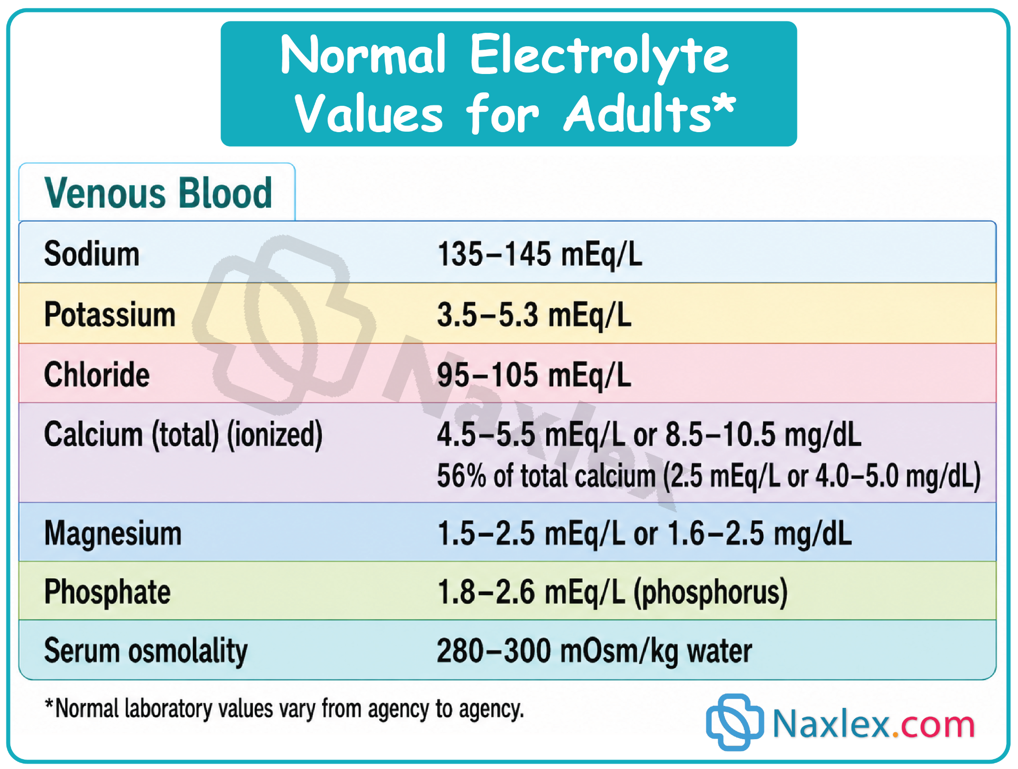
Blood levels of two metabolically produced substances, urea and creatinine, are routinely used to evaluate renal function. The kidneys, through filtration and tubular secretion, normally eliminate both.
- Urea, the end product of protein metabolism, is measured as blood urea nitrogen (BUN).
- Creatinine is produced in relatively constant quantities by the muscles and is excreted by the kidneys. Thus the amount of creatinine in the blood relates to renal excretory function.
- Serum Osmolality
Serum osmolality is a measure of the solute concentration of the blood. The particles included are sodium ions, glucose, and urea (BUN).
Serum osmolality can be estimated by doubling the serum sodium, because sodium and its associated chloride ions are the major determinants of serum osmolality. Normal values are 280 to 300 mOsm/kg.
- Drug Monitoring
Therapeutic drug monitoring is often conducted when a client is taking a medication with a narrow therapeutic range (e.g., digoxin, theophylline, aminoglycosides).
Peak and trough levels to determine if the blood serum levels of a specific drug are at a therapeutic level and not a subtherapeutic or toxic level.
- Arterial Blood Gases
Specimens of arterial blood from the radial, brachial, or femoral arteries.
- Blood Chemistry
These are tests that may be performed on blood serum (the liquid portion of the blood).
Include determining certain enzymes that may be present (including lactic dehydrogenase [LDH], creatine kinase [CK], aspartate aminotransferase [AST], and alanine aminotransferase [ALT]), serum glucose, hemoglobin A1C (HbA1C) test, hormones such as thyroid hormone, and other substances such as cholesterol and triglycerides.
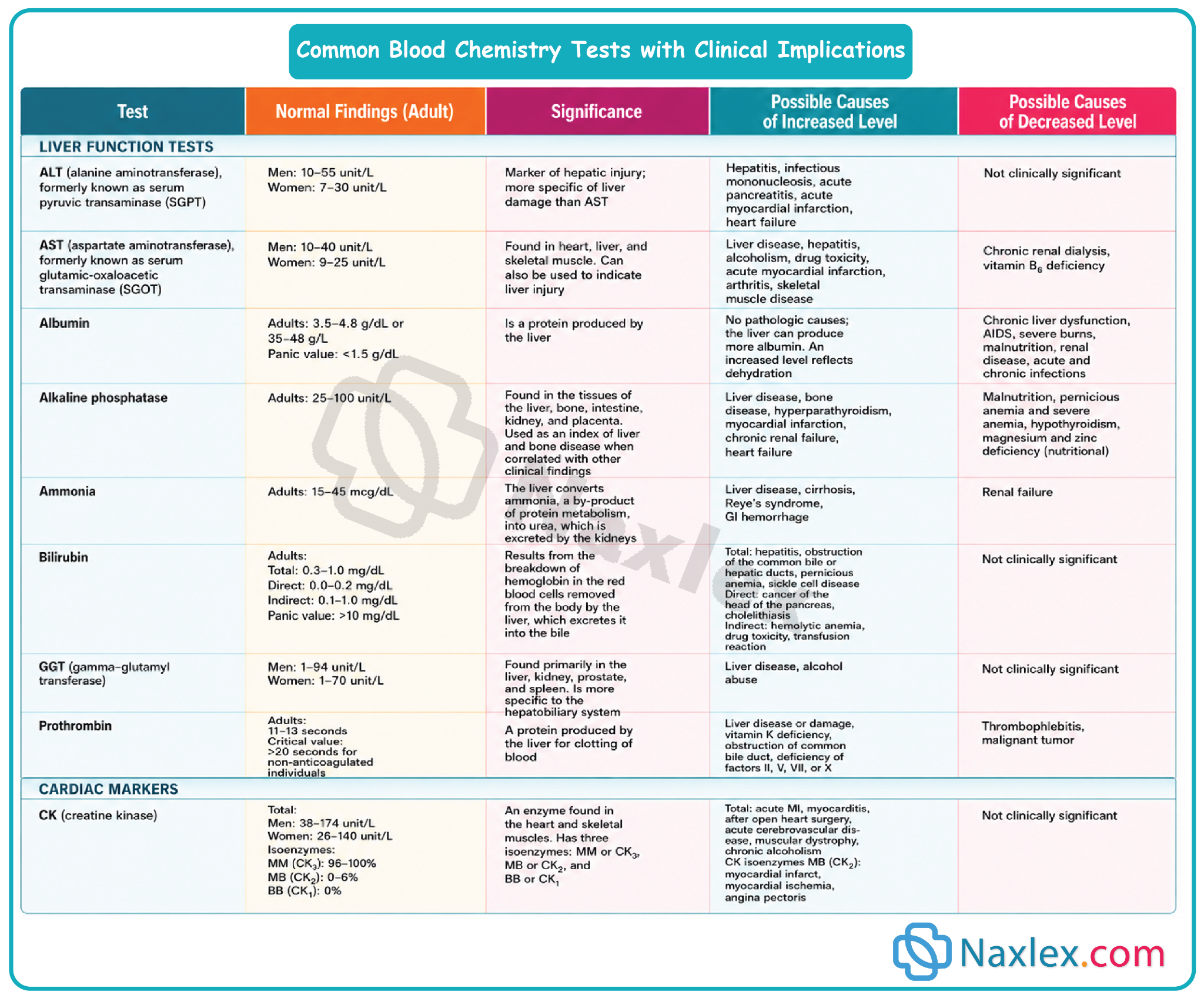
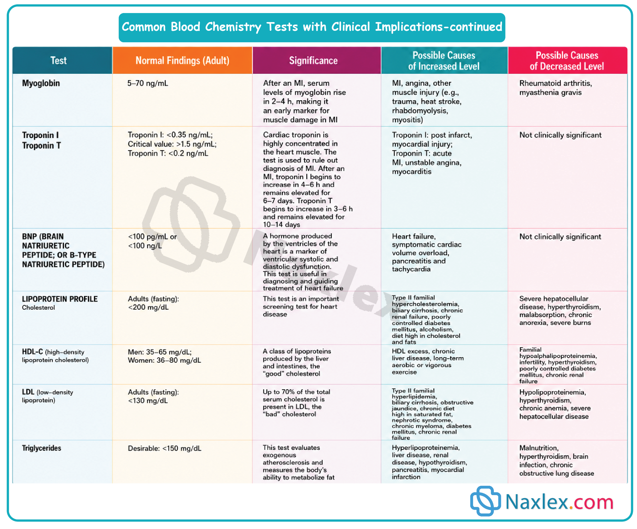
- Metabolic screening:
Newborns are routinely screened for congenital metabolic conditions- Tests for phenylketonuria (PKU), congenital hypothyroidism, sickle cell disease and galactosemia.
- Capillary Blood Glucose:
A capillary blood specimen is taken to measure the current blood glucose level when frequent tests are required or when a venipuncture cannot be performed.
Steps for obtaining a capillary blood specimen to measure blood glucose:
1. Preparation & Setup
- Review the specific manufacturer instructions for your glucose meter.
- Assemble all necessary equipment right at the client's bedside.
2. Implementation Procedure
Patient Intro & Hygiene
- Verify the client's identity, introduce yourself, and explain the procedure, its purpose, and how the results will impact their care.
- Perform hand hygiene, apply gloves, and ensure client privacy.
Site & Equipment Prep
- Turn on and prepare the meter (insert the test strip or calibrate/confirm the code if using an older model).
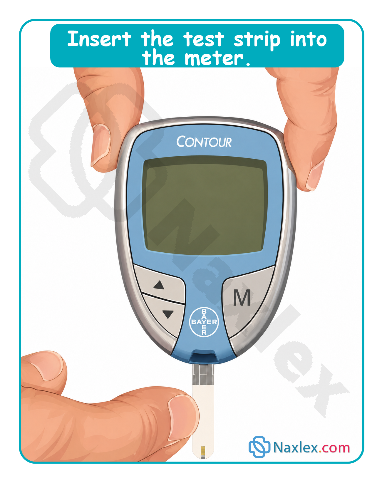
- Select the site: Choose the side of an adult's finger (avoiding bone) or an earlobe.
- Increase blood flow: Hold the finger below heart level, wrap it in a warm cloth, or gently rub the earlobe.
- Clean: Clean with soap and water or an antiseptic swab, allowing it to dry completely so alcohol doesn't alter the accuracy or cause stinging.
Sampling & Reading
- Puncture: Place the lancet perpendicular to the skin and pierce it using a quick darting motion.
- Collect: Gently squeeze for a drop of blood. Hold the strip to the drop to let it absorb or wick the blood without smearing it.
- Post-puncture: Have the client apply pressure to the site with gauze.
- Analyze: Leave the blood on the strip for the manufacturer's designated time. The meter will automatically display the glucose reading.
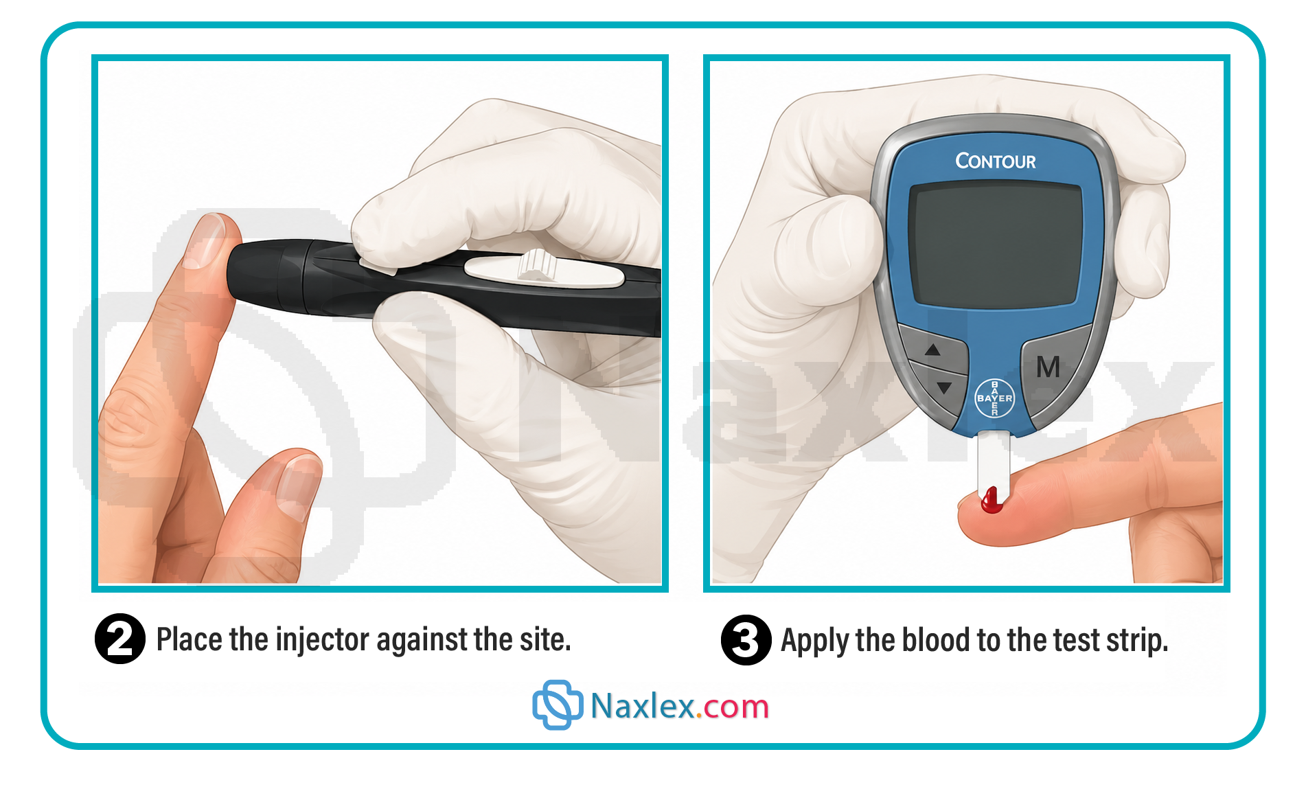
Cleanup & Documentation
- Dispose of the strip and gauze in a biohazard container, place the lancet in a sharps container, remove gloves, and wash hands.
- Document the results, testing method, and patient understanding on the client's record or diabetic flow sheet.
- Check for sliding scale insulin orders and administer medication as prescribed.
3. Evaluation
- Analyze Results: Compare the reading against normal ranges, previous results, and the client's current health status.
- Report & Follow-Up: Report abnormal results to the provider (confirming via venipuncture if required by agency policy).
- Patient Education: Assess the client's skin puncture site, evaluate their motivation and ability to self-test, and prepare them for home monitoring (frequency, record-keeping, and insulin administration).
Specimen Collection and Testing
Laboratory examination of specimens such as urine, blood, stool, sputum, and wound drainage provides important adjunct information for diagnosing health care problems and also provides a measure of the responses to therapy.
Nurses often assume the responsibility for specimen collection:
- Provide client comfort, privacy, and safety.
- Explain the purpose of the specimen collection and the procedure for obtaining the specimen.
- Use the correct procedure for obtaining a specimen or ensure that the client or staff follows the correct procedure.
- Note relevant information on the laboratory requisition slip, for example, medications the client is taking that may affect the results.
- Transport the specimen to the laboratory promptly. Fresh specimens provide more accurate results.
- Report abnormal laboratory findings to the health care provider in a timely manner consistent with the severity of the abnormal results.
6.1 Stool specimen collection and testing
1. Purposes of Fecal Analysis
|
Test Type |
Purpose / Clinical Significance |
Special Requirements |
|
Occult Blood (Guaiac) |
Detects hidden blood from ulcers, inflammatory disease, or tumors. |
Small smear on a card; sensitive to hemoglobin peroxidase. |
|
Dietary / Secretion |
Steatorrhea (excess fat) - absorption issue. Decreased bile - liver/gallbladder obstruction. |
Must collect the total quantity of a single bowel movement, not a small sample. |
|
Ova & Parasites (O&P) |
Identifies parasites and eggs. Requires 3 specimens over consecutive days. |
Must transport immediately while still warm. |
|
Bacteria / Viruses |
Cultures small amounts of feces to pinpoint infections. |
Must use sterile container/swab and aseptic technique. Note current antibiotics on lab form. |
2. Collection Guidelines & Patient Teaching
Patient Instructions (Crucial for Accuracy)
- Location: Defecate into a clean bedpan or bedside commode.
- No contamination: Void urine before the test. Do not contaminate the sample with urine or menstrual discharge.
- No toilet paper: Do not throw toilet tissue into the collection pan (chemicals alter lab results).
- Preparation: Remove toilet cleaners and flush twice before home collection.
Nursing Actions & Technique
- Safety & Hygiene: Wear clean gloves. Use 1–2 clean tongue blades to transfer the specimen; wrap sticks in paper towels and discard in regular waste (do not flush).
- Sample Size: Send ~2.5 cm (1 inch) of formed stool, or 15–30 mL of liquid stool.
- Target Selection: Intentionally include areas with visible pus, mucus, or blood.
- Timing & storage: Send to the lab immediately. If delayed, check container directions; some samples require refrigeration (never store in a fridge with food/meds).
- Delegation to UAP: Random collection and occult blood testing can be delegated. The nurse must perform sterile stool cultures. Instruct UAP to save positive cards for the nurse to verify.
3. Fecal Occult Blood Testing (FOBT)
Guaiac Smear (Hemoccult)
- Mechanism: Detects the enzyme peroxidase in hemoglobin.
- Interpretation: A blue color indicates a positive result (blood is present).
Nursing Alert: Color-blind nurses should not read these results.
- Interferences (The 3-to-7 Day Rule):
- False-Positives: Caused by red meat, raw vegetables/fruits (radishes, turnips, melons), and irritating medications (NSAIDs, aspirin, steroids, iron, anticoagulants). Restrict foods rich in Vit C for 3 days and meds for 7 days before testing.
- False-Negatives: Caused by taking >250 mg/day of Vitamin C up to 3 days before the test.
Fecal Immunochemical Test (FIT)
- Advantages over Guaiac: Higher sensitivity/specificity for colorectal cancer, no dietary or medication restrictions, and only requires 2 samples (instead of 3).
- Methods: Available as a liquid-based method (stored in a buffer solution) or a dry-slide method (collected via a long-handled brush).
Stool DNA Testing (Cologuard)
- A non-invasive screening tool for colorectal cancer that measures both human hemoglobin and specific cancer-related gene mutations.
4. Documentation Checklist
- Date, time, and specific collection method.
- Stool Characteristics: Color, odor, consistency, amount, and abnormal findings (mucus, blood).
- FOBT results (if performed).
- Patient tolerance (anal bleeding, discomfort during/after defecation) and perianal skin status.
- Patient/family education and ability to self-test.
Nursing insights:
LIFESPAN CONSIDERATIONS: Stool Specimen
INFANTS
- To collect a stool specimen for an infant, the stool is scraped from the diaper, being careful not to contaminate the stool with urine.
CHILDREN
- A child who is toilet trained should be able to provide a fecal specimen, but may prefer being assisted by a parent.
- When explaining the procedure to the child, use words appropriate for the child’s age rather than medical terms.
- Ask the parent what words the family normally uses to describe a bowel movement.
- A specimen for pinworms is collected by the parent early in the morning, after sleep and before the child has a bowel movement. Scotch tape is attached to a tongue blade and the sticky side is laid flat against the perineum and anus to pick up any eggs or small worms. The tongue blade is then examined under a microscope.
OLDER ADULTS
- Older adults may need assistance if serial stool specimens are required.
6.2 Urine specimens
Kidney function is directly dependent on cardiac output. Any systemic alteration that reduces cardiac output will inherently compromise renal perfusion and decrease urine output.
The nurse is responsible for collecting urine specimens for a number of tests:
- clean voided urine specimens for routine urinalysis
- clean-catch or midstream urine specimens for urine culture
- timed urine specimens for a variety of tests that depend on the client’s specific health problem
1. Routine & Culture Collection Methods
Clean Voided Specimen (Routine Urinalysis)
Used for general screening and non-culture testing.
- Optimal sample: First morning void. It has a higher, more uniform solute concentration and a more acidic pH.
- Volume required: A minimum of 10 mL is generally required for a standard urinalysis (UA).
- Contamination controls: Must be strictly free of fecal matter. Instruct female clients to discard toilet tissue in the toilet or waste bag, never in the collection pan (tissue fibers degrade lab analysis).
- Delegation: Collection can be delegated to Unlicensed Assistive Personnel (UAP) with clear instructions regarding client assistance or bedpan use.
Clean-Catch / Midstream Specimen (Culture & Sensitivity)
Ordered to identify specific uropathogens causing a Urinary Tract Infection (UTI).
-
- Perineal pre-wash:
Wash the external genitalia with soap and water to clear transient surface bacteria, then assess for any allergies to antiseptic cleansers.
-
- Targeted antiseptic cleansing:
Clean the urinary meatus using specific anatomical protocols, using each provided towelette exactly once.
- Female: Spread labia minora; cleanse front to back.
- Male: Retract foreskin if uncircumcised; cleanse in a circular motion from the meatus outward and down the shaft.
-
- Initiate voiding (initial flush):
Instruct the client to start urinating into the toilet or commode first. This initial stream clears out normal flora residing in the distal urethra and meatus.
-
- Midstream collection:
Without stopping the stream, pass the sterile specimen cup into the path of the running urine. Avoid letting the cup touch the penis, perineum, or labia.
2. Indwelling catheter specimen collection
Sterile urine specimens must be obtained from a closed drainage system using the built-in sampling port. Never collect a sample from the dependent drainage bag, as stagnant urine undergoes biochemical changes and bacterial overgrowth.
Structural Requirements
- Needleless Systems (Modern Standard): Utilize a Luer-Lok syringe inserted at a 90-degree angle perpendicular to the needleless port. Twist and lock to aspirate. The port self-seals upon removal.
- Traditional Rubber Catheters: Can only be performed on self-sealing rubber tubing (never on plastic, silicone, or Silastic). Insert a sterile needle at a 30- to 45-degree angle directly into the designated patch/port to facilitate self-sealing.
Procedure Sequence
- Apply clean gloves.
- Clamp the tubing: If fresh urine is not present in the tubing, clamp it at least 8 cm (3 inches) below the sampling port for up to 30 minutes to allow fresh urine to pool.
- Disinfect: Scrub the sampling port with a disinfectant swab. Ensure the site is distal to the balloon inflation tube to prevent accidental puncture of the balloon channel.
- Aspirate sample: Unclamp the tubing and withdraw the required volume:
- 3 mL for a sterile urine culture.
- 30 mL for a routine urinalysis.
- Transfer & clean: Transfer to a sterile container without touching the rim. Wipe the exterior of the container with disinfectant if contaminated.
- Transit: Unclamp the catheter line and send the sample to the lab immediately (or refrigerate).
3. Timed urine specimen
Some urine examinations require collection of all urine produced and voided over a specific period of time, ranging from 1 to 2 hours to 24 hours.
Timed specimens generally either are refrigerated or contain a preservative to prevent bacterial growth or decomposition of urine components. Each voiding of urine is collected in a small, clean container and then emptied immediately into the large refrigerated bottle or carton.
Timed urine specimens tests are performed for the following purposes:
- To assess the ability of the kidney to concentrate and dilute urine.
- To determine disorders of glucose metabolism, for example, diabetes mellitus.
- To determine levels of specific constituents, for example, albumin, amylase, creatinine, urobilinogen, or certain hormones (e.g., e striol or corticosteroids), in the urine.
Point-of-Care Urine Testing (Reagent Strips/Dipsticks)
Nurses or trained UAP can perform bedside reagent strip testing. Ensure strips are not outdated, follow exact manufacturer timing blocks for color reactions, and have UAP save positive samples for nursing verification.
|
Testing Parameter |
Clinical Range / Values |
Clinical Significance & Pathophysiology |
|
Specific Gravity |
1.010 to 1.025
|
Measures concentration/solute density.
• High (>1.025): Dehydration/fluid volume deficit, or excess solutes (glucose). • Low (<1.010): Fluid volume excess, high fluid intake, or renal inability to concentrate urine. |
|
Urinary pH |
Average: 6 (Slightly acidic; range 4.5–8.0) |
Reflects systemic acid-base regulation. • Metabolic Acidosis: Expected renal compensation drops pH (excreting H+ ions). • Metabolic Alkalosis: Expected renal compensation elevates pH. |
|
Glucose |
Negligible / Negative |
Screens for diabetes mellitus or gestational glucose intolerance. Inadequate for active glycemic monitoring; only indicated if blood glucose testing is refused or impossible. |
|
Ketones |
Negative |
Products of fatty acid catabolism. • Positive findings: Poorly controlled Type 1 diabetes (indicated during illness, fever, or BG consistently over 300 mg/dL), starvation, fasting, alcoholism, or strict high-protein diets. |
|
Protein |
Negative |
Plasma proteins are normally too large to pass the glomerular filtration barrier. • Positive findings: Indicates damage/increased permeability of the glomerular membrane (e.g., glomerulonephritis). |
|
Occult Blood |
Negative |
Highly sensitive indicator of micro/macro-hematuria. • > Clinical Alert: Indicates structural damage to the kidney parenchyma or the urinary tract lining. |
|
Osmolality |
50 to 1,200 mOsm/kg
|
Note: This is a laboratory test, not a bedside dipstick. Measures exact particle concentration (urea, creatinine, uric acid). • Elevated: Fluid volume deficit. • Decreased: Fluid volume excess. |
Documentation Checklist
- Exact date, time, and collection method (e.g., midstream vs. needleless catheter aspiration).
- Physical Characteristics: Color, clarity, presence of sediment, and distinct odors.
- Client tolerance, dysuria, or specific difficulties voiding.
- Lab Requisition Notations: Explicitly note if the client is currently on antibiotic therapy or is menstruating, as both alter diagnostic accuracy.
6.3 Sputum specimens
Sputum is a mucous secretion from the deep lower airways (lungs, bronchi, trachea). It must be strictly differentiated from saliva, which is clear oral secretions. Healthy individuals do not produce sputum.
Clinical indications & rationale
- Culture & Sensitivity (C&S): Identifies specific lower airway pathogens and determines targeted antimicrobial therapies.
- Cytology: Evaluates cellular structure to identify lung cancer and specific malignant cell types. Requires 3 serial early-morning specimens.
- Acid-Fast Bacillus (AFB): Screens for Mycobacterium tuberculosis (TB). Requires 3 consecutive daily early-morning collections, often using dedicated glass containers.
- Therapeutic Evaluation: Used to track if current respiratory therapies are resolving an infection.
Procedural mechanics & collection timing
- Optimal Timing: Early morning upon awakening (captures pooled nocturnal secretions) or immediately following postural drainage treatments.
- Volume Target: 1 to 2 teaspoons (4 to 10 mL) of thick expectorated sputum.
Step 1: Pre-Collection Oral Care:
Provide the client with fresh water to rinse the mouth. This clears residual food and transient oral microorganisms without altering lower airway flora.
Step 2: Airborne & droplet isolation setup:
If tuberculosis is suspected, perform collection in a negative-airflow isolation room, under UV lighting, or outdoors. If unavailable, wear a fitted respirator mask (capable of filtering droplet nuclei) along with standard gloves and PPE.
Step 3: Deep breathing & expectorated puncture:
Instruct the client to take deep breaths to expand the lungs, then perform a deep, productive cough to expectorate mucus directly into a sterile container. Prevent sputum from touching the container's exterior.
Step 4: Post-procedure hygiene & transit:
Offer mouthwash to alleviate an unpleasant taste. If the container's exterior is contaminated, wipe it down with disinfectant. Send the labeled sample to the lab immediately or refrigerate it to prevent normal contaminant overgrowth.
Nursing Scope & Delegation:
Expectorated collection can be delegated to UAP with directions on positioning and timing. However, if a client cannot cough, the nurse must perform an invasive pharyngeal suctioning procedure using a sterile sputum trap.
6.4 Throat culture
A throat culture samples the mucosa of the oropharynx and tonsillar pillars to isolate pathogenic microorganisms. This is an invasive procedure requiring nurse implementation to protect client safety and navigate airway reflexes.
Minimizing the gag reflex
The gag reflex is triggered by touching the pharyngeal walls or soft palate, causing the constrictor muscles of the pharynx to contract. To suppress this response and expose the target tissue, guide the patient through these steps:
- Positioning: Sit upright (if tolerated) to facilitate optimal visual alignment.
- Anatomical exposure: Have the client open their mouth wide and fully extend their tongue forward.
- Vocal relaxation: Instruct the client to say "ah." This vocalization lowers the back of the tongue, elevates the soft palate, and relaxes the pharyngeal constrictor muscles.
Collection technique
- Apply clean gloves.
- If the posterior wall is obscured, utilize a localized light source and gently depress the tongue with a clean tongue blade.
- Insert the sterile swab swiftly. Firmly run it across the tonsillar pillars and posterior pharynx, targeting areas that are explicitly reddened, inflamed, or contain purulent exudate.
- Remove the swab quickly without touching the teeth, gums, or buccal mucosa to prevent contamination. Secure it into the transport medium.
Documentation & Lab Requisition Checklist
- Exact date, time, and specific collection method utilized (e.g., expectorated vs. suctioned trap).
- Sputum Metrics: Record total volume, color (e.g., rust, green, clear), consistency (thick, tenacious, watery, mucoid), distinct odors, and any signs of hemoptysis (blood streaks).
- Auxiliary therapies used to assist collection (e.g., chest physiotherapy, postural drainage).
- Client comfort and tolerance of the procedure.
- Lab Requisition Entry: Explicitly note current or newly initiated antibiotic therapies on the laboratory requisition form, as this directly affects culture growth interpretation.
Nursing insights:
LIFESPAN CONSIDERATIONS: Sputum and Throat Specimens
INFANTS
- When taking a throat swab, avoid occluding an infant’s nose because infants normally breathe only through the nose.
CHILDREN
- Have a parent stand the young child between the parent’s legs with the child’s back to the parent and the parent’s arms gently but firmly around the child. As the parent tips the child’s head back, ask the child to open wide and stick the tongue out.
- Assure the child that the procedure will be over quickly and may “tickle” but should not hurt.
OLDER ADULTS
- Older adults may need encouragement to cough because a decreased cough reflex occurs with aging.
- Allow time for older adults to rest and recover between coughs when obtaining a sputum specimen.
Visualization Procedures
Diagnostic visualization splits into two primary modalities:
- Direct visualization (Invasive): Inside-the-body viewing via an inserted scope. Requires specialized consent, infection control, and post-procedural monitoring (e.g., perforation, bleeding, return of gag reflex).
- Indirect visualization (Noninvasive): Exterior imaging using structural or functional scanning modalities (X-ray, ultrasound, CT, MRI, Nuclear medicine).
1. Gastrointestinal (GI) System
Direct Visualization (Invasive)
Uses specialized scopes to view the luminal mucosal lining. All require post-procedural monitoring for complications like perforation or hemorrhage.
- Anoscopy: Visualizes the anal canal.
- Proctoscopy: Visualizes the rectum.
- Proctosigmoidoscopy: Visualizes the rectum and sigmoid colon.
- Colonoscopy: Visualizes the entire large intestine.
Indirect Visualization (Noninvasive)
Roentgenography (X-Ray & Fluoroscopy): Uses structural imaging to detect strictures, obstructions, tumors, ulcers, inflammatory disease, or hiatal hernias. Enhanced via radiopaque contrast (Barium Sulfate). Continuous flow is monitored in real-time via fluoroscopy.
|
Procedure |
Technique |
Nursing Implications |
|
Barium Swallow |
Patient drinks barium sulfate to image the upper GI tract/small bowel. |
Pre-op preparation; Post-op fluid management to avoid barium impaction (chalky white stools are expected). |
|
Barium Enema |
Barium enema is administered rectally to image the lower GI tract. |
Clear liquid diet/laxatives pre-procedure; evacuative clearance post-procedure. |
2. Urinary System
Direct Visualization (Invasive)
- Cystoscopy: Direct mucosal examination of the bladder, ureteral orifices, and urethra using a lighted cystoscope inserted transurethrally. Requires sterile technique.
Indirect Visualization (Noninvasive)
- KUB: Plain structural X-ray of the Kidneys, Ureters, and Bladder without contrast.
- Renal Ultrasonography: High-frequency reflected sound waves used to evaluate renal structures noninvasively.
- Intravenous Pyelography (IVP): Radiographic assessment of the urinary tract where contrast medium is injected intravenously and cleared renally.
- Retrograde Pyelography: Contrast medium is instilled directly into the renal pelvis via transurethral catheterization of the bladder and ureters.
3. Cardiopulmonary System
Cardiovascular Assessments
- Electrocardiography (ECG): Graphic recording of cardiac electrical activity using skin-surface electrodes.
- Echocardiogram: Noninvasive cardiac ultrasound assessing structural integrity and left ventricular ejection fraction. Nursing Note: Reassure the patient it is painless, though the conductive gel feels cold.
- Angiography: Invasive structural mapping of vascular patency using injected radiopaque dye under fluoroscopy.
- Coronary Angiography: Assesses the extent of coronary artery disease (CAD).
- Pulmonary Angiography: Evaluates the pulmonary vasculature, primarily to confirm a pulmonary embolism (PE).
- Other Sites: Carotid, cerebral, renal, and lower extremity vessels.
Respiratory assessments
- Chest X-Ray (CXR): Basic noninvasive structural imaging to diagnose and track pulmonary disease. Nursing Note: Ensure all jewelry and waist-up clothing are removed.
- Lung Scan (V/Q Scan): Nuclear imaging measuring gas and blood distribution. Used primarily to identify mismatch anomalies like pulmonary emboli.
- Perfusion Scan (Q Scan): Measures pulmonary blood flow via IV radioisotopes.
- Ventilation Scan (V Scan): Evaluates airway distribution (e.g., in emphysema) via inhaled radioactive gas.
- Nursing Note: Patient stays under a mask or exhales into room air. Scan takes 20 to 40 minutes. No radiation isolation precautions are needed (the dose is nominal).
Direct airway visualization (Invasive)
- Laryngoscopy & Bronchoscopy: Sterile visualization of the larynx and tracheobronchial tree. Used for diagnostic examination or tissue biopsy.
- Nursing & Safety Protocols:
- Informed consent is mandatory.
- Administer local anesthetic spray or gargle to the pharynx to completely abolish the gag reflex.
- Strictly NPO post-procedure until a positive gag reflex returns to prevent aspiration.
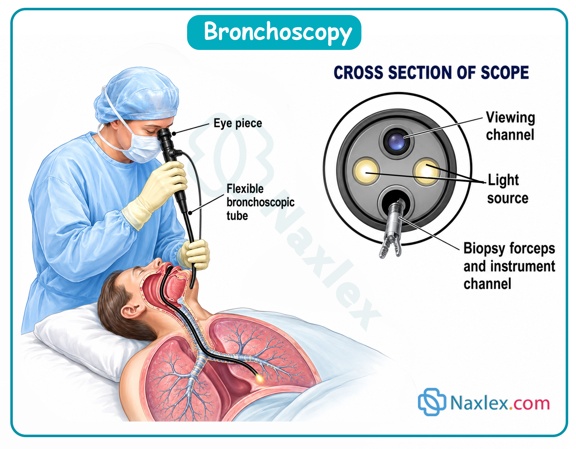
4. Advanced Advanced Cross-Sectional Imaging
Computed Tomography (CT)
- Mechanism: Multi-angle, high-sensitivity X-ray imaging creating three-dimensional structural reconstructions. Very sensitive to minor differences in tissue density.
Magnetic Resonance Imaging (MRI)
- Mechanism: Noninvasive scanning leveraging high-energy magnetic fields to align atomic nuclei. Avoids ionizing radiation. Provides superior soft-tissue contrast compared to CT.
MRI Safety Screen Checklist
- Implanted medical devices: Strictly contraindicated for ferromagnetically active implants (e.g., older pacemakers, specific metal hip prostheses).
- Removable metal: Remove all rings, watches, phones, and body jewelry. Exception: Titanium, niobium, and true surgical stainless steel are non-ferromagnetic and safe.
- Transdermal Patches: Remove all patches containing foil or metallic backings prior to scanning to prevent local thermal skin burns. Advise the patient to reapply a fresh patch post-procedure.
- Tattoos & Permanent Makeup: Screen for body tattoos (especially red pigments). Remind patients they may feel localized warming or mild edema and should report unusual sensations immediately to the operator.
- Environment/Comfort: High-magnet closed tunnels can trigger severe claustrophobia. Utilize the internal two-way communication system for constant monitoring. Provide earplugs to attenuate the loud acoustic noises during the 60–90 minute scan.
5. Nuclear Imaging
Nuclear medicine tracks organ physiological dynamics using targeted radiopharmaceuticals. Tissue uptake patterns showcase metabolic activity:
- Normal tissue- uniform, equal gray distribution
- Hyperfunctioning tissue- increased uptake; darker images (hot spots)
- Hypo functioning tissue- decreased uptake; lighter images (cold spots)
Positron Emission Tomography (PET): A functional nuclear study using an inhaled or injected radioisotope to dynamically track organ perfusion, cellular metabolic activity, and malignant tumor growth patterns.

Naxlex
Videos
Login to View Video
Click here to loginTake Notes on Diagnostic testing
This filled cannot be empty
Join Naxlex Nursing for nursing questions & guides! Sign Up Now