





 Video Library


Angina and Myocardial Infarction
Study Questions
Practice Exercise 1
A nurse is teaching a client about the differences between stable angina and myocardial infarction (MI). Which of the following statements by the client indicates an understanding of the teaching?
Explanation
Myocardial infarction represents irreversible myocardial necrosis caused by prolonged ischemia resulting from complete coronary artery occlusion. This acute coronary syndrome triggers ventricular remodeling and electrical instability, often presenting with subendocardial injury, elevated cardiac troponins, and potentially fatal ventricular arrhythmias requiring prompt reperfusion therapy.
Rationale for correct answer
4. The pain from stable angina is typically relieved after resting for 5 minutes because myocardial oxygen demand decreases. In contrast, myocardial infarction pain persists despite rest or nitroglycerin administration. Rest restores the balance between myocardial oxygen supply and demand in angina.
Rationale for incorrect answers
1. Chest pain lasting for 45 minutes typically indicates myocardial infarction rather than stable angina. Stable angina episodes generally last less than 15 minutes. Prolonged ischemic duration causes irreversible necrosis of myocardial tissue, distinguishing infarction from transient ischemic episodes.
2. Nausea and diaphoresis are classic associated symptoms of myocardial infarction rather than stable angina. Autonomic nervous system activation during extensive myocardial ischemia triggers these systemic responses. Stable angina usually presents with predictable substernal chest discomfort without autonomic symptoms.
3. Chest pain from stable angina is relieved by taking sublingual nitroglycerin or resting, not morphine. Morphine is reserved for acute myocardial infarction pain refractory to nitrates. Nitroglycerin induces vasodilation, decreasing cardiac workload and myocardial oxygen consumption effectively.
Test-taking strategy
- Analyze the Scenario and Question: The client is learning about the pathophysiological and clinical differences between stable angina and myocardial infarction, requiring the identification of correct diagnostic indicators regarding symptom duration, associated features, and treatment responses.
- Apply Pathophysiological Principles:
- Stable angina involves transient myocardial ischemia caused by fixed atherosclerotic plaques, characterized by predictable pain relieved by rest or nitrates within 5 minutes.
- Myocardial infarction involves prolonged ischemia leading to tissue necrosis, where pain lasts longer than 20 to 30 minutes and is unresponsive to rest.
- Rule Out Incorrect Choices:
- Rule out choice 1 because chest pain lasting 45 minutes exceeds the typical transient duration of stable angina and signifies myocardial infarction.
- Rule out choice 2 because autonomic symptoms like nausea and diaphoresis are characteristic of myocardial infarction, not stable angina.
- Rule out choice 3 because stable angina responds to rest or sublingual nitroglycerin, whereas morphine is utilized for acute myocardial infarction.
- Select the Correct Finding: Rule in choice 4 because relief of pain after resting for 5 minutes accurately describes the transient nature of stable angina compared to myocardial infarction.
Take home points
- Stable angina causes predictable chest discomfort that resolves rapidly with rest or sublingual nitrates.
- Myocardial infarction involves prolonged coronary occlusion resulting in irreversible myocardial necrosis and unremitting pain.
- Autonomic manifestations such as diaphoresis and nausea frequently accompany acute coronary events like myocardial infarction.
- Prompt differentiation between ischemic pain syndromes dictates appropriate pharmacological interventions and emergency reperfusion protocols.
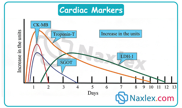
A nurse is caring for a client who has a suspected myocardial infarction. The client reports that the chest pain began 2 hours ago. Which of the following cardiac enzymes is the earliest marker of cardiac muscle injury?
Explanation
Myocardial infarction results from acute coronary artery occlusion causing irreversible cellular death and necrosis of cardiac muscle tissue. This ischemic injury compromises membrane integrity, leading to the intracellular release of specific structural proteins and enzymes into the bloodstream, which serve as essential diagnostic biomarkers for detecting acute coronary syndromes.
Rationale for correct answer
2. Myoglobin is a low molecular weight heme protein found in cardiac and skeletal muscle. It is released rapidly into the circulation following muscle injury, making it the earliest detectable serum marker of myocardial necrosis, typically rising within one to three hours after symptom onset.
Rationale for incorrect answers
1. Creatine kinase myocardial band is a specific isoenzyme found predominantly in myocardial tissue. While it rises following acute myocardial injury, its serum levels typically begin to elevate between three to six hours after symptom onset, rendering it less rapid than myoglobin as the earliest marker.
3. Troponin I is a highly sensitive and specific regulatory protein of the contractile apparatus in cardiac muscle. Although it remains the gold standard biomarker for diagnosing myocardial infarction, its initial elevation occurs four to six hours following the onset of ischemic injury.
4. Troponin T is a structural protein involved in the contraction of cardiac muscle. Similar to troponin I, serum levels of troponin T begin to rise approximately four to six hours after myocardial damage occurs, meaning it is not the earliest biochemical indicator available following injury.
Test-taking strategy
- Analyze the Scenario and Question: The client presents with a suspected myocardial infarction occurring two hours ago, requiring the identification of the earliest cardiac biomarker released into the bloodstream following initial muscle injury.
- Apply Pathophysiological Principles:
- Different cardiac biomarkers have distinct release kinetics based on their molecular weight, intracellular localization, and the extent of cellular membrane disruption during ischemia.
- Smaller cytoplasmic proteins diffuse out of damaged myocytes significantly faster than larger structural regulatory proteins or enzymatic complexes.
- Rule Out Incorrect Choices:
- Rule out choice 1 because creatine kinase myocardial band appears in the serum later than myoglobin, typically taking three to six hours to rise.
- Rule out choice 3 because troponin I elevates four to six hours post-injury, missing the earliest window of detection.
- Rule out choice 4 because troponin T follows a similar delayed timeline of four to six hours compared to rapid-release markers.
- Select the Correct Finding: Rule in choice 2 because myoglobin is a low molecular weight protein that leaks into the circulation within one to three hours, establishing it as the earliest marker of injury.
Take home points
- Myoglobin serves as the earliest serological indicator of myocardial injury due to its rapid release from damaged muscle cells.
- Cardiac troponins provide superior diagnostic specificity and sensitivity for myocardial infarction despite having a slightly delayed initial rise.
- Biomarker testing timelines dictate clinical decision-making during the initial evaluation of acute chest pain syndromes.
- Serial cardiac enzyme measurements are essential for confirming acute myocardial infarction when early markers show ambiguous initial results.
A nurse is caring for a client who is experiencing an acute myocardial infarction. The client asks why they are being given aspirin. Which of the following is the most appropriate response by the nurse?
Explanation
Myocardial infarction represents irreversible myocardial necrosis caused by prolonged ischemia resulting from complete coronary artery occlusion. This acute coronary syndrome triggers ventricular remodeling and electrical instability, often presenting with subendocardial injury, elevated cardiac troponins, and potentially fatal ventricular arrhythmias requiring prompt reperfusion therapy.
Rationale for correct answer
3. Aspirin inhibits the synthesis of thromboxane A2, a potent inducer of platelet aggregation. By preventing platelets from binding together, aspirin effectively stops the expansion of the existing thrombus, which is the primary mechanism obstructing blood flow during an acute myocardial infarction.
Rationale for incorrect answers
1. Aspirin does not possess direct vasodilatory properties to dilate coronary arteries. Coronary vasodilation is achieved through medications such as nitrates, which reduce preload and afterload, rather than antiplatelet agents, which specifically focus on preventing the formation of obstructive clots.
2. Although aspirin has antipyretic and anti-inflammatory effects, these are not the therapeutic goals during an acute myocardial infarction. The primary objective in this life-threatening emergency is to achieve immediate antiplatelet therapy to arrest the propagation of coronary artery thrombosis.
4. Aspirin is not utilized as a primary analgesic for acute chest pain. While it reduces systemic inflammation, it lacks the potent analgesic properties of opioids like morphine, which are required to relieve the severe, crushing ischemic pain associated with acute myocardial infarction.
Test-taking strategy
- Analyze the Scenario/Question: The client is undergoing an acute myocardial infarction and requires an explanation of the therapeutic mechanism of aspirin, specifically focusing on its role in cardiovascular emergency management versus its common pharmacological uses.
- Apply Pathophysiological Principles:
- Myocardial infarction is triggered by plaque rupture and subsequent thrombus formation.
- Antiplatelet therapy is essential to stop the cycle of platelet aggregation, thereby preventing the thrombus from enlarging and further occluding the affected coronary artery.
- Rule Out Incorrect Choices:
- Rule out choice 1 because aspirin is an antiplatelet agent, not a vasodilator used for increasing coronary blood flow.
- Rule out choice 2 because antipyretic and anti-inflammatory effects are not clinically relevant to preventing acute myocardial damage.
- Rule out choice 4 because aspirin does not provide sufficient analgesic relief for the intense pain experienced during an infarction.
- Select the Correct Finding: Rule in choice 3 because it accurately describes the pharmacological inhibition of platelet aggregation and thrombus formation, which is the cornerstone of emergency coronary care.
Take home points
- Aspirin is a cornerstone of initial myocardial infarction treatment due to its potent antiplatelet efficacy.
- The primary mechanism of aspirin in this context is the inhibition of thromboxane A2 synthesis.
- Immediate administration of aspirin during suspected infarction significantly improves mortality by limiting thrombus expansion.
- Nurses must distinguish between the antiplatelet, analgesic, and anti-inflammatory roles of aspirin in different clinical contexts.
A nurse is assessing a client for risk factors associated with coronary artery disease. Which of the following client history findings should the nurse identify as a nonmodifiable risk factor?
Explanation
Coronary artery disease involves the progressive accumulation of atherosclerotic plaque within coronary arteries, significantly reducing myocardial perfusion and oxygen delivery. This complex pathology is influenced by a combination of both inherent biological traits and acquired behavioral or environmental factors, making a comprehensive assessment of nonmodifiable and modifiable determinants essential for effective long-term risk stratification.
Rationale for correct answer
3. Male gender is a nonmodifiable risk factor for coronary artery disease. Statistically, men are at higher risk for developing cardiovascular conditions at a younger age compared to pre-menopausal women, due to genetic and hormonal factors influencing atherogenesis and endothelial function.
Rationale for incorrect answers
1. Tobacco use is a modifiable risk factor that profoundly damages the vascular endothelium. Smoking promotes systemic inflammation and oxidative stress, leading to the rapid progression of arterial plaque, yet this risk can be mitigated through smoking cessation and lifestyle interventions.
2. Hypertension is a modifiable risk factor characterized by elevated systemic vascular resistance. Chronic high blood pressure exerts mechanical stress on arterial walls, accelerating atherosclerosis, but this condition can be managed through pharmacological therapy and strict dietary sodium restrictions.
4. Metabolic disorders, such as diabetes mellitus and hyperlipidemia, are considered modifiable risk factors for coronary artery disease. These conditions alter lipid metabolism and glucose homeostasis, but they are treatable through consistent glycemic control and aggressive cholesterol management strategies.
Test-taking strategy
- Analyze the Scenario/Question: The client is being evaluated for coronary artery disease risk factors, requiring the nurse to differentiate between biological determinants that cannot be altered and behavioral or physiological conditions that can be managed therapeutically.
- Apply Pathophysiological Principles:
- Nonmodifiable factors include age, family history, and gender, which represent fixed variables that predispose an individual to disease.
- Modifiable factors involve lifestyle choices and treatable chronic diseases that can be controlled to decrease the risk of arterial plaque progression.
- Rule Out Incorrect Choices:
- Rule out choice 1 because tobacco use is a behavioral choice that can be stopped to reduce cardiovascular risk.
- Rule out choice 2 because hypertension is a clinical condition that responds to medical management and life changes.
- Rule out choice 4 because metabolic disorders involve physiological variables that can be modified through treatment.
- Select the Correct Finding: Rule in choice 3 because male gender is an inherent, unchangeable biological variable influencing disease development.
Take home points
- Nonmodifiable risk factors include age, gender, and genetics, which cannot be changed by the client.
- Modifiable risk factors encompass lifestyle behaviors like smoking, diet, and physical inactivity.
- Chronic medical conditions such as hypertension and diabetes are modifiable through sustained pharmacological and lifestyle control.
- Identifying nonmodifiable factors helps nurses focus interventions on the aggressive management of all modifiable risks.
A nurse is caring for a client who has unstable angina. The client asks the nurse how this differs from stable angina. Which of the following is the correct explanation?
Explanation
Unstable angina represents a dangerous acute coronary syndrome characterized by myocardial ischemia resulting from complex coronary plaque progression. Unlike stable manifestations, this state features unpredictable symptoms, indicating an imminent risk for myocardial infarction or total vessel occlusion, thus requiring urgent evaluation and aggressive medical intervention to restore adequate tissue perfusion.
Rationale for correct answer
3. Unstable angina manifests with an unpredictable pattern that intensifies in frequency, duration, and symptom severity. Unlike stable forms, this ischemic pain occurs at rest or with minimal exertion, indicating a worsening underlying coronary artery obstruction and potential thrombosis.
Rationale for incorrect answers
1. Unstable angina is not exclusively associated with rest, nor is it defined by coronary artery spasms. Prinzmetal angina is the specific variant caused by vasospasm, whereas unstable angina stems from progressive plaque instability and increasing myocardial oxygen demand.
2. Stable angina typically responds to rest or sublingual nitroglycerin, while unstable angina often remains refractory to these standard interventions. Morphine is indicated for severe pain associated with myocardial infarction, not for the routine management of anginal episodes or conditions.
4. Unstable angina significantly differs from stable angina because it represents a progressive deterioration of coronary flow. Stable angina involves predictable episodes of short duration, whereas unstable episodes are longer, more painful, and carry a higher risk of irreversible necrosis.
Test-taking strategy
- Analyze the Scenario/Question: The client is inquiring about the pathophysiology and clinical characteristics of unstable angina compared to stable angina, requiring an understanding of symptom frequency, intensity, and responsiveness to traditional pharmacological management.
- Apply Pathophysiological Principles:
- Stable angina is predictable, triggered by exertion, and relieved by rest or nitrates.
- Unstable angina is characterized by worsening symptoms, occurring at rest or with decreasing activity levels, signaling a high risk for cardiac events.
- Rule Out Incorrect Choices:
- Rule out choice 1 because it inaccurately describes vasospastic angina rather than the progressive nature of unstable angina.
- Rule out choice 2 because it confuses the therapeutic roles of nitroglycerin and morphine for angina versus infarction.
- Rule out choice 4 because unstable angina implies a worsening clinical status, not a shorter or identical duration.
- Select the Correct Finding: Rule in choice 3 because it correctly identifies the progressive, worsening nature of unstable angina symptoms.
Take home points
- Unstable angina is an acute coronary syndrome signaling high risk for myocardial infarction.
- Symptoms of unstable angina are unpredictable and often occur during periods of rest.
- Stable angina follows a predictable pattern triggered by exertion and resolves with rest.
- Distinguishing between stable and unstable angina is critical for determining appropriate urgency and treatment.
A nurse is performing an admission assessment on a client who has a suspected myocardial infarction. Which of the following findings should the nurse expect?
Explanation
Myocardial infarction manifests as acute cardiac tissue ischemia caused by obstructed coronary blood flow. This systemic stress response involves intense sympathetic nervous system activation, leading to significant hemodynamic instability and a profound psychological response, which clinicians must identify to ensure rapid intervention and cardiac output restoration.
Rationale for correct answer
3. A feeling of impending doom is a hallmark psychological symptom reported by clients experiencing an acute myocardial infarction. The massive surge of catecholamines triggered by cardiac distress creates intense anxiety, reflecting the body's acute awareness of physiological crisis and potential mortality.
Rationale for incorrect answers
1. Clients experiencing myocardial infarction typically present with cool, clammy, and diaphoretic skin. Peripheral vasoconstriction occurs due to sympathetic activation to shunt blood toward vital organs, which contradicts the presence of warm, flushed skin that might suggest systemic vasodilation.
2. Tachycardia, not bradycardia, is the expected assessment finding during an acute myocardial infarction. The sympathetic nervous system increases the heart rate to compensate for decreased myocardial contractility and maintain adequate systemic perfusion in the setting of coronary artery blockage.
4. Decreased level of consciousness is often observed rather than an increased level. Diminished cerebral perfusion resulting from acute cardiac failure or cardiogenic shock frequently causes confusion, restlessness, or lethargy, as the brain receives inadequate oxygenated blood during this acute event.
Test-taking strategy
- Analyze the Scenario/Question: The client is presenting with a suspected myocardial infarction, requiring the nurse to identify the classic clinical manifestations that reflect acute physiological distress and systemic autonomic responses associated with this life-threatening condition.
- Apply Pathophysiological Principles:
- Myocardial infarction triggers a powerful sympathetic nervous system response, manifesting in physical signs like diaphoresis and tachycardia.
- Patients often experience psychological distress, such as an overwhelming sense of doom, due to sudden, severe physiological trauma and hypoxia.
- Rule Out Incorrect Choices:
- Rule out choice 1 because vasoconstriction leads to cool, clammy skin, not warm, flushed skin.
- Rule out choice 2 because the sympathetic response increases heart rate, causing tachycardia rather than bradycardia.
- Rule out choice 4 because cerebral hypoperfusion results in altered or decreased consciousness, not an increase.
- Select the Correct Finding: Rule in choice 3 because the feeling of impending doom is a well-documented, classic psychiatric symptom of acute myocardial ischemia.
Take home points
- Acute myocardial infarction causes a surge in catecholamines, resulting in tachycardia and diaphoresis.
- Impending doom is a classic psychological presentation of cardiac distress and warrants immediate attention.
- Hypoperfusion during myocardial infarction commonly leads to cerebral impairment, not alertness.
- Clinical assessment must focus on signs of hemodynamic compromise and the systemic sympathetic stress response.
A nurse is reviewing the diagnostic results for a client who has angina. Which of the following ECG changes should the nurse identify as an indicator of ischemia?
Explanation
Myocardial ischemia occurs when coronary blood flow is insufficient to meet myocardial metabolic demands, leading to electrical and metabolic abnormalities. On an electrocardiogram, these changes reflect impaired repolarization processes in the myocardium, which serve as crucial diagnostic markers for identifying transient ischemia versus permanent myocardial necrosis in clinical practice.
Rationale for correct answer
3. ST-segment depression and T-wave inversion are classic electrocardiographic indicators of myocardial ischemia. These findings represent alterations in the repolarization sequence of ventricular cells when blood flow is significantly reduced, signaling that the myocardium is suffering from acute oxygen supply-demand imbalances.
Rationale for incorrect answers
1. ST-segment elevation is the hallmark sign of a full-thickness myocardial injury, known as a STEMI. This indicates complete coronary occlusion and transmural myocardial damage, which differentiates it from the transient or reversible state of simple ischemia observed in angina.
2. An abnormal Q-wave is a definitive diagnostic indicator of prior myocardial infarction or tissue necrosis. The presence of these waves indicates that the affected area of the myocardium is no longer electrically active, which represents irreversible scarring rather than active, reversible ischemia.
4. Complete heart block is an electrical conduction rhythm disorder involving the total interruption of signal transmission between the atria and the ventricles. While ischemia can sometimes lead to heart blocks, this condition is not a specific diagnostic indicator of myocardial ischemia.
Test-taking strategy
- Analyze the Scenario/Question: The client is being evaluated for angina, requiring the nurse to identify the specific electrocardiographic patterns that signify reversible myocardial ischemia rather than injury or infarction.
- Apply Pathophysiological Principles:
- Ischemia generally results in ST-segment depression or T-wave inversion due to altered electrical activity in the subendocardial layers.
- Injury and Infarction involve ST-segment elevation or pathological Q-waves, reflecting deeper levels of myocardial damage.
- Rule Out Incorrect Choices:
- Rule out choice 1 because ST-elevation typically denotes an acute STEMI, indicating injury rather than just ischemia.
- Rule out choice 2 because abnormal Q-waves signify old, completed tissue necrosis rather than current ischemia.
- Rule out choice 4 because complete heart block represents an electrical conduction problem rather than a standard ischemic change.
- Select the Correct Finding: Rule in choice 3 because it describes the classic electrical changes that occur when cardiac muscle is deprived of adequate oxygen.
Take home points
- ST-segment depression and T-wave inversion are the standard ECG indicators of myocardial ischemia.
- ST-segment elevation is specific to acute myocardial injury, requiring immediate reperfusion intervention.
- Pathological Q-waves indicate permanent myocardial damage or previous infarction occurring over time.
- Diagnostic ECG interpretation is vital for differentiating reversible ischemic states from acute myocardial necrosis.
A nurse is educating a client who has angina about the use of sublingual nitroglycerin. Which of the following instructions should the nurse include?
Explanation
Nitroglycerin acts as a potent vasodilator by releasing nitric oxide, which relaxes vascular smooth muscle throughout the coronary arteries and systemic vasculature. This mechanism significantly reduces myocardial oxygen demand by decreasing preload and afterload, though it frequently induces predictable adverse effects due to systemic vascular relaxation that nurses must effectively manage during patient education.
Rationale for correct answer
2. A headache is an extremely common side effect of nitroglycerin therapy. The drug causes significant systemic vasodilation, which increases blood flow to the cerebral vessels and raises intracranial pressure, resulting in a throbbing sensation that typically subsides as the patient develops medication tolerance.
Rationale for incorrect answers
1. Clients should take a second tablet if pain persists after 5 minutes, not 10. The protocol mandates calling emergency services if pain remains unrelieved after the first dose, as delaying intervention significantly increases the risk for permanent myocardial injury.
3. Sublingual nitroglycerin must be dissolved under the tongue to ensure rapid mucosal absorption. Swallowing the tablet with water introduces the medication to the first-pass effect in the liver, which would render the drug ineffective for the rapid management of anginal pain.
4. Nitroglycerin must be taken immediately upon the onset of chest pain to prevent progressive ischemic damage. Waiting 30 minutes is dangerous because prolonged ischemia leads to irreversible necrosis of cardiac muscle, transforming stable angina into a severe, life-threatening myocardial infarction event.
Test-taking strategy
- Analyze the Scenario/Question: The client requires education on the safe administration and expected side effects of sublingual nitroglycerin, focusing on proper dosing intervals, administration technique, and monitoring for common pharmacological adverse reactions.
- Apply Pathophysiological Principles:
- Nitroglycerin is a rapid-acting vasodilator intended for immediate rescue from myocardial ischemia.
- Proper administration is crucial to avoid systemic absorption issues or delays that could worsen the patient's underlying cardiac condition.
- Rule Out Incorrect Choices:
- Rule out choice 1 because the standard protocol requires shorter intervals for repeated dosing to avoid prolonged ischemia.
- Rule out choice 3 because swallowing the medication prevents the necessary rapid absorption required for therapeutic vascular relief.
- Rule out choice 4 because waiting 30 minutes is medically unsafe and permits further myocardial tissue death.
- Select the Correct Finding: Rule in choice 2 because it correctly identifies a frequent side effect resulting from the drug's potent vasodilatory action on cerebral blood vessels.
Take home points
- Sublingual nitroglycerin must dissolve under the tongue for rapid, effective systemic absorption.
- Persistent chest pain after one dose necessitates immediate emergency medical intervention.
- Headaches are an expected result of nitroglycerin-induced systemic and cerebral vasodilation.
- Clients must take nitroglycerin immediately at the first sign of angina to preserve cardiac muscle.
A nurse is caring for a client who has an acute myocardial infarction. Which of the following findings should the nurse recognize as an indication of cardiogenic shock?
Explanation
Cardiogenic shock is a life-threatening state of myocardial pump failure resulting in inadequate tissue perfusion despite adequate intravascular volume. This severe condition triggers a massive sympathetic response and profound hemodynamic collapse, necessitating rapid identification of clinical markers like systemic hypotension and peripheral hypoperfusion to prevent multi-organ dysfunction and irreversible cellular necrosis.
Rationale for correct answer
3. Tachycardia and hypotension are clinical hallmarks of cardiogenic shock. The failing heart struggles to maintain cardiac output, leading to decreased blood pressure, while the sympathetic nervous system accelerates the heart rate to compensate for impaired systolic function and restore tissue delivery.
Rationale for incorrect answers
1. Decreased urinary output is a classic sign of cardiogenic shock due to diminished renal perfusion. As the heart fails, the body prioritizes blood flow to the brain and heart, causing oliguria and elevated blood urea nitrogen, which contradicts the presence of increased renal filtration.
2. Hypertension is inconsistent with the hemodynamics of cardiogenic shock, which is defined by profound hypotension. The significant loss of myocardial contractility causes systolic pressure to fall, preventing the heart from generating sufficient pressure to overcome systemic vascular resistance and maintain perfusion.
4. Bradycardia and warm skin indicate conditions like neurogenic shock, not cardiogenic shock. Cardiogenic shock typically presents with tachycardia and cool, clammy skin due to vasoconstriction, as the body attempts to compensate for failing ventricular performance through intense peripheral sympathetic stimulation.
Test-taking strategy
- Analyze the Scenario/Question: The client is presenting with symptoms of cardiogenic shock following an acute myocardial infarction, requiring the nurse to identify the hemodynamic and physical findings that reflect severe cardiac pump failure and resultant systemic hypoperfusion.
- Apply Pathophysiological Principles:
- Cardiogenic shock involves an inability of the left ventricle to pump blood, leading to a sudden drop in cardiac output and systemic blood pressure.
- The body compensates for this failure via the sympathetic nervous system, resulting in rapid heart rate and peripheral vasoconstriction.
- Rule Out Incorrect Choices:
- Rule out choice 1 because shock results in decreased, not increased, renal perfusion and urine output.
- Rule out choice 2 because cardiogenic shock is fundamentally characterized by low blood pressure, not high blood pressure.
- Rule out choice 4 because cardiogenic shock involves a compensatory tachycardia, and vasoconstriction causes cool, clammy skin rather than warmth.
- Select the Correct Finding: Rule in choice 3 because it accurately describes the physiological compensation for low cardiac output and the symptomatic hypotension of pump failure.
Take home points
- Cardiogenic shock occurs when myocardial infarction impairs the heart's ability to maintain adequate systemic perfusion.
- Classic clinical signs include hypotension, tachycardia, oliguria, and cool, clammy peripheral skin.
- Hemodynamic monitoring often reveals low cardiac index and elevated filling pressures due to ventricular dysfunction.
- Rapid identification of shock is critical for initiating inotropic support and urgent revascularization therapy.
A nurse is preparing a client for a cardiac catheterization. Which of the following is the priority nursing action?
Explanation
Cardiac catheterization is an invasive diagnostic procedure requiring the administration of radiopaque contrast medium to visualize coronary anatomy. Identifying potential hypersensitivity reactions is critical because contrast dye, often containing iodine, can trigger severe anaphylactic responses, making preoperative screening the most essential safety step to prevent life-threatening complications during the intervention.
Rationale for correct answer
3. Assessing for an allergy to iodine or shellfish is the priority nursing action because the contrast medium used during angiography contains iodine-based substances. A severe allergic reaction can lead to acute anaphylaxis, which necessitates immediate medical management to maintain the client's airway and hemodynamic stability.
Rationale for incorrect answers
1. Ensuring the client remains NPO is a standard preoperative requirement to prevent aspiration during sedation. While this is necessary for client safety, it is not the most critical physiological priority compared to identifying a potentially fatal systemic allergic reaction to the procedural contrast dye.
2. Teaching the client about a PCA pump is a component of postoperative care planning. This action does not directly impact the immediate safety or survival of the client during the procedure and is secondary to identifying contraindications that could cause immediate harm during catheterization.
4. Obtaining written informed consent is a legal and ethical requirement that must be completed before any invasive procedure. Although this is essential for the medical record, it does not prioritize the physical safety of the client in the same manner as screening for a lethal allergic reaction.
Test-taking strategy
- Analyze the Scenario/Question: The client is scheduled for a cardiac catheterization, requiring the nurse to identify the priority action that most directly prevents a life-threatening physiological complication during or immediately following the procedure.
- Apply Prioritization Principles:
- Prioritize actions that address immediate life-threatening safety threats or physiological risks, such as anaphylaxis, over legal requirements or routine preoperative preparation steps.
- Maslow's Hierarchy of Needs places physical safety and the prevention of physical harm as a physiological priority.
- Rule Out Incorrect Choices:
- Rule out choice 1 because, while important for aspiration risk, it is less immediately life-threatening than an anaphylactic reaction.
- Rule out choice 2 because it is a routine educational task for post-procedural pain management rather than a safety priority.
- Rule out choice 4 because obtaining consent is a procedural safeguard rather than a direct clinical intervention for physical safety.
- Select the Correct Finding: Rule in choice 3 because the risk of anaphylaxis from contrast media is a direct, preventable, and potentially fatal clinical risk.
Take home points
- Pre-procedural allergy screening is mandatory when contrast media will be administered to prevent anaphylaxis.
- Contrast media for cardiac catheterization is typically iodine-based, necessitating queries regarding shellfish or iodine sensitivities.
- Patient safety priorities always focus on preventing immediate physiological harm during or after invasive procedures.
- Nurses must balance administrative requirements with essential clinical assessments that ensure client survival and well-being.
Practice Exercise 2
A nurse is caring for a client who is having a thallium scan to assess for myocardial ischemia. Which of the following instructions should the nurse include in the client's pre-procedure education?
Explanation
Thallium scans are diagnostic imaging procedures that utilize radioactive isotopes to evaluate myocardial perfusion and detect regions of myocardial ischemia. Accurate results depend heavily on standardized preparation protocols that prevent exogenous interference with cardiac blood flow and heart rate responses, ensuring the isotope effectively highlights viable versus infarcted or ischemic myocardial tissue during stress testing.
Rationale for correct answer
2. Avoiding tobacco and caffeine for 4 hours before the test is essential. Caffeine is a adenosine receptor antagonist that can mask or prevent the detection of ischemic regions during the scan. Smoking induces vasoconstriction, which also interferes with the assessment of cardiac perfusion.
Rationale for incorrect answers
1. There is no clinical requirement to maintain a liquid diet for 24 hours prior to a thallium scan. Patients are generally advised to eat a light meal before the procedure to avoid hypoglycemia, although specific fasting times are usually limited to several hours rather than an entire day.
3. Remaining NPO for 12 hours is excessive and could lead to hypoglycemia or patient discomfort. Most protocols require only a few hours of fasting to ensure patient comfort and prevent potential gastrointestinal complications during the physical exertion portion of the stress protocol.
4. Expecting a warm sensation is typical for iodine-based contrast dye used in coronary angiography, not radioactive isotopes used in thallium scans. Thallium isotopes do not produce immediate thermal sensations upon injection, and providing this information misleads the client regarding the specific nature of the imaging agent.
Test-taking strategy
- Analyze the Scenario/Question: The client is scheduled for a thallium scan, requiring the nurse to explain specific dietary and behavioral restrictions that are critical for achieving accurate, interpretable diagnostic results while avoiding unnecessary patient fasting or misinformation.
- Apply Pathophysiological Principles:
- Diagnostic imaging protocols must minimize physiological variables, such as caffeine and nicotine, which alter coronary artery diameter and heart rate responsiveness.
- Excessive fasting or incorrect expectations regarding procedural sensations can cause physical distress or interfere with the diagnostic efficacy of the radioactive isotope.
- Rule Out Incorrect Choices:
- Rule out choice 1 because a 24-hour liquid diet is unnecessary and contradicts standard pre-procedural nutritional support.
- Rule out choice 3 because a 12-hour fast is unnecessarily long and risks physiological complications like hypoglycemia.
- Rule out choice 4 because it incorrectly attributes sensations from contrast dye to the radioisotope injection process.
- Select the Correct Finding: Rule in choice 2 because it correctly addresses the substances that significantly interfere with cardiac physiological responses during stress imaging.
Take home points
- Caffeine must be avoided before myocardial perfusion scans due to its antagonist effects on cardiac imaging agents.
- Nicotine intake must be restricted to prevent coronary vasoconstriction, which alters blood flow distribution.
- Pre-procedural fasting should be limited to the minimum time required to prevent aspiration during exercise.
- Client education regarding diagnostic procedures must be specific to the type of agent being utilized for imaging.
A nurse is analyzing the laboratory results for a client who had chest pain 2 hours ago. Which of the following cardiac enzymes would the nurse expect to be elevated, considering the time elapsed since the onset of pain?
Explanation
Myocardial infarction involves the rapid release of intracellular proteins following irreversible myocardial necrosis. Because these biomarkers have different kinetic profiles, only low-molecular-weight proteins appear in the serum within the first few hours, necessitating the use of early detection markers to guide immediate clinical decision-making during the hyper-acute phase of myocardial ischemia.
Rationale for correct answer
1. Myoglobin is a small heme protein that rises rapidly within 1 to 3 hours post-injury. Given that only 2 hours have passed, serum levels will be elevated, making it the most reliable early detection marker for identifying acute myocardial muscle damage.
Rationale for incorrect answers
2. Creatine kinase-MB levels typically do not begin to rise until 3 to 6 hours after myocardial injury occurs. Because only 2 hours have passed, this specific cardiac isoenzyme has not yet reached significant detectable levels in the systemic blood circulation.
3. Troponin I is a highly specific regulatory protein that generally begins to elevate within 4 to 6 hours of cardiac insult. At the 2-hour mark, serum levels are typically below the diagnostic threshold, meaning this marker is not yet useful for acute clinical evaluation.
4. Troponin T is a structural protein that follows a similar release kinetic profile to Troponin I. Its levels begin to rise approximately 4 to 6 hours after the onset of injury, rendering it undetectable in the blood during the very early ischemic window.
Test-taking strategy
- Analyze the Scenario/Question: The client experienced chest pain 2 hours ago, requiring the nurse to identify the specific cardiac biomarker that demonstrates elevation within this ultra-early diagnostic timeframe following myocardial injury.
- Apply Pathophysiological Principles:
- Biomarker appearance is determined by molecular weight and cellular location; smaller cytoplasmic proteins exit the damaged cells faster than larger structural or regulatory proteins.
- In the hyper-acute phase of infarction, only high-velocity markers provide clinical utility for the immediate identification of cardiac injury.
- Rule Out Incorrect Choices:
- Rule out choice 2 because CK-MB elevation is delayed, typically requiring at least 3 hours to become detectable.
- Rule out choice 3 because troponins are structural proteins that take at least 4 hours to appear in the serum.
- Rule out choice 4 because Troponin T release is similarly delayed and is not yet measurable at 2 hours post-injury.
- Select the Correct Finding: Rule in choice 1 because myoglobin's low molecular weight allows for the fastest release and earliest serum detection following membrane disruption.
Take home points
- Myoglobin is the most reliable biomarker for detecting myocardial injury within the first 3 hours of symptom onset.
- Troponins are highly specific but require a longer time interval to reach detectable serum levels.
- Diagnostic testing must be ordered and interpreted based on the specific kinetic timeline of the chosen cardiac biomarker.
- Early presentation of chest pain requires clinical awareness of markers that appear in the hyper-acute phase.
A nurse is preparing a client for a coronary angiogram. Which of the following assessments is the most critical for the nurse to perform prior to the procedure?
Explanation
Coronary angiogram is an invasive diagnostic procedure requiring the administration of radiopaque contrast medium to visualize coronary anatomy. Identifying potential hypersensitivity reactions is critical because contrast dye, often containing iodine, can trigger severe anaphylactic responses, making preoperative screening the most essential safety step to prevent life-threatening complications during the intervention.
Rationale for correct answer
2. Assessing for an allergy to iodine or shellfish is the priority nursing action because the contrast medium used during angiography contains iodine-based substances. A severe allergic reaction can lead to acute anaphylaxis, which necessitates immediate medical management to maintain the client's airway and hemodynamic stability.
Rationale for incorrect answers
1. Reviewing serum glucose levels is important for clients with diabetes to manage potential glycemic imbalances during fasting. While this is necessary for metabolic stability, it is not the most critical physiological priority compared to identifying a potentially fatal systemic allergic reaction to the procedural contrast dye.
3. Checking baseline peripheral pulses is a standard part of the physical assessment to establish a baseline for post-procedure vascular status. Although vital for monitoring limb perfusion after catheter insertion, this assessment does not address a life-threatening physiological risk like anaphylactic shock during the procedure.
4. Monitoring heart rhythm for premature ventricular contractions is a component of cardiac telemetry and observation. While this is important during instrumentation of the heart, it is a continuous monitoring duty rather than a preoperative assessment priority compared to screening for a lethal allergic reaction.
Test-taking strategy
- Analyze the Scenario/Question: The client is scheduled for a coronary angiogram, requiring the nurse to identify the priority action that most directly prevents a life-threatening physiological complication during or immediately following the procedure.
- Apply Prioritization Principles:
- Prioritize actions that address immediate life-threatening safety threats or physiological risks, such as anaphylaxis, over routine preoperative preparation, baseline assessments, or secondary metabolic monitoring.
- Maslow's Hierarchy of Needs places physical safety and the prevention of physical harm as a physiological priority.
- Rule Out Incorrect Choices:
- Rule out choice 1 because, while important for metabolic stability, it is less immediately life-threatening than an anaphylactic reaction.
- Rule out choice 2 because it is a routine baseline documentation task rather than an immediate safety priority.
- Rule out choice 4 because it is an ongoing monitoring task rather than a critical preoperative assessment to prevent procedural harm.
- Select the Correct Finding: Rule in choice 2 because the risk of anaphylaxis from contrast media is a direct, preventable, and potentially fatal clinical risk.
Take home points
- Pre-procedural allergy screening is mandatory when contrast media will be administered to prevent anaphylaxis.
- Contrast media for coronary angiography is typically iodine-based, necessitating queries regarding shellfish or iodine sensitivities.
- Patient safety priorities always focus on preventing immediate physiological harm during or after invasive procedures.
- Nurses must balance administrative requirements with essential clinical assessments that ensure client survival and well-being.
A nurse is evaluating an ECG strip for a client who is currently experiencing angina. Which of the following findings confirms the presence of myocardial ischemia?
Explanation
Myocardial ischemia occurs when coronary blood flow is insufficient to meet myocardial metabolic demands, leading to electrical and metabolic abnormalities. On an electrocardiogram, these changes reflect impaired repolarization processes in the myocardium, which serve as crucial diagnostic markers for identifying transient ischemia versus permanent myocardial necrosis in clinical practice.
Rationale for correct answer
3. ST-segment depression and T-wave inversion are classic electrocardiographic indicators of myocardial ischemia. These findings represent alterations in the repolarization sequence of ventricular cells when blood flow is significantly reduced, signaling that the myocardium is suffering from acute oxygen supply-demand imbalances.
Rationale for incorrect answers
1. ST-segment elevation is the hallmark sign of a full-thickness myocardial injury, known as a STEMI. This indicates complete coronary occlusion and transmural myocardial damage, which differentiates it from the transient or reversible state of simple ischemia observed in angina.
2. An abnormal Q-wave is a definitive diagnostic indicator of prior myocardial infarction or tissue necrosis. The presence of these waves indicates that the affected area of the myocardium is no longer electrically active, which represents irreversible scarring rather than active, reversible ischemia.
4. A prolonged PR interval is a sign of a first-degree atrioventricular block, indicating delayed electrical conduction between the atria and the ventricles. While ischemia can sometimes lead to conduction delays, this finding is not a specific or confirmatory diagnostic indicator of myocardial ischemia.
Test-taking strategy
- Analyze the Scenario/Question: The client is being evaluated for angina, requiring the nurse to identify the specific electrocardiographic patterns that signify reversible myocardial ischemia rather than injury or infarction.
- Apply Pathophysiological Principles:
- Ischemia generally results in ST-segment depression or T-wave inversion due to altered electrical activity in the subendocardial layers.
- Injury and Infarction involve ST-segment elevation or pathological Q-waves, reflecting deeper levels of myocardial damage.
- Rule Out Incorrect Choices:
- Rule out choice 1 because ST-elevation typically denotes an acute STEMI, indicating injury rather than just ischemia.
- Rule out choice 2 because abnormal Q-waves signify old, completed tissue necrosis rather than current ischemia.
- Rule out choice 4 because a prolonged PR interval represents an electrical conduction problem rather than a standard ischemic change.
- Select the Correct Finding: Rule in choice 3 because it describes the classic electrical changes that occur when cardiac muscle is deprived of adequate oxygen.
Take home points
- ST-segment depression and T-wave inversion are the standard ECG indicators of myocardial ischemia.
- ST-segment elevation is specific to acute myocardial injury, requiring immediate reperfusion intervention.
- Pathological Q-waves indicate permanent myocardial damage or previous infarction occurring over time.
- Diagnostic ECG interpretation is vital for differentiating reversible ischemic states from acute myocardial necrosis.
A nurse is explaining cardiac markers to a student nurse. Which of the following statements by the nurse regarding Troponin I is accurate?
Explanation
Troponin I is a highly sensitive cardiac-specific regulatory protein that serves as the gold standard biomarker for detecting myocardial necrosis. Because it is virtually absent in healthy serum, any detectable elevation above the established laboratory threshold indicates significant myocardial injury, providing clinicians with an exceptionally reliable tool for the diagnostic confirmation of acute myocardial infarction events.
Rationale for correct answer
3. Any detectable, positive value for Troponin I indicates damage to cardiac tissue because this protein is cardiac-specific. Unlike other enzymes that may be found in skeletal muscle, troponin release is a highly sensitive and definitive sign of acute myocardial necrosis in clinical settings.
Rationale for incorrect answers
1. Troponin I levels do not return to baseline within 24 hours of injury. This protein remains elevated in the bloodstream for 7 to 10 days following an infarction, making it a very useful marker for evaluating patients who present for care days later.
2. Troponin I is not the earliest marker of cardiac injury, nor does it peak within 2 hours. It typically begins to rise 4 to 6 hours post-injury, whereas myoglobin is the true earliest biomarker that appears much faster in the serum circulation.
4. Troponin I reliability is not limited by the duration of chest pain. It remains the most definitive diagnostic tool for myocardial infarction regardless of the pain duration, as it reflects structural tissue damage and necrosis that has occurred within the cardiac muscle myocardial layers.
Test-taking strategy
- Analyze the Scenario/Question: The student nurse is learning about Troponin I kinetics and clinical significance, requiring the identification of the most accurate statement regarding its sensitivity, specificity, and diagnostic utility in identifying cardiac muscle damage.
- Apply Pathophysiological Principles:
- Troponin I is highly specific to the heart, meaning its presence in the blood is nearly pathognomonic for myocardial damage.
- Biomarker utility is determined by its onset, peak, and clearance times, which are distinct from other cardiac enzymes like myoglobin or CK-MB.
- Rule Out Incorrect Choices:
- Rule out choice 1 because the prolonged elevation of troponin is a key diagnostic feature for delayed presentations.
- Rule out choice 2 because it confuses the early appearance of myoglobin with the later appearance of troponin.
- Rule out choice 4 because troponin reliability is independent of the duration of the ischemic pain episode.
- Select the Correct Finding: Rule in choice 3 because the extreme specificity of Troponin I means any elevated result confirms injury to the cardiac muscle.
Take home points
- Troponin I is the most cardiac-specific and sensitive biomarker for diagnosing myocardial infarction.
- Any detectable level of Troponin I above the laboratory reference range indicates myocardial necrosis.
- Troponins remain elevated for up to 10 days, allowing for retrospective diagnosis of cardiac events.
- Nurses must distinguish between biomarkers based on their release kinetics and their anatomical specificity.
A nurse is caring for a client who has just undergone a cardiac catheterization via the femoral artery. Which of the following nursing actions is indicated?
Explanation
Cardiac catheterization via the femoral artery requires precise post-procedural management to maintain vascular integrity. Following arterial access, the nurse must ensure stabilization of the puncture site and hemodynamic monitoring, as the risk of arterial hemorrhage or hematoma formation is significant during the early recovery period, necessitating strict bed rest and frequent assessment of neurovascular status.
Rationale for correct answer
2. Monitoring vital signs every 15 minutes until stable is essential to detect early signs of hemorrhage or hemodynamic instability. Rapid identification of tachycardia or hypotension allows for immediate intervention to prevent life-threatening vascular complications following the arterial puncture performed during the procedure.
Rationale for incorrect answers
1. Clients typically do not need to remain NPO for 4 hours following cardiac catheterization. Providing oral fluids is encouraged to promote the excretion of radiopaque contrast medium through the kidneys, which helps prevent contrast-induced nephrotoxicity in patients with renal vulnerability.
3. Encouraging immediate ambulation is contraindicated following a femoral approach. Patients must maintain strict bed rest with the affected leg kept straight for several hours to allow the arterial entry site to seal properly and prevent severe bleeding or pseudoaneurysm development.
4. Applying a warm compress to the insertion site is inappropriate and potentially dangerous. The site requires a pressure dressing to promote hemostasis and secure arterial closure; heat application would promote vasodilation, which increases the risk of bleeding at the puncture site.
Test-taking strategy
- Analyze the Scenario/Question: The client has had a femoral cardiac catheterization, requiring the nurse to identify the safest post-procedural care protocols that prioritize vascular hemostasis and early detection of circulatory complications.
- Apply Pathophysiological Principles:
- Arterial access sites require time for secure closure and the prevention of exsanguination.
- Post-procedural care must facilitate contrast clearance while maintaining the integrity of the puncture site through immobilization and frequent observation.
- Rule Out Incorrect Choices:
- Rule out choice 1 because hydration is necessary to protect renal function from contrast dye.
- Rule out choice 3 because immediate ambulation increases the risk of arterial site disruption and hemorrhage.
- Rule out choice 4 because heat application is contraindicated and promotes bleeding rather than healing at the entry site.
- Select the Correct Finding: Rule in choice 2 because frequent vital sign monitoring is the standard of care to ensure cardiovascular stability and identify early indicators of internal or external bleeding.
Take home points
- Strict bed rest and limb immobilization are required after femoral artery catheterization to ensure hemostasis.
- Frequent neurovascular checks and vital sign monitoring are essential to identify post-procedural complications.
- Oral hydration is encouraged to facilitate the clearance of contrast dye and protect renal function.
- Pressure must be maintained on the arterial access site to prevent the formation of hematomas or active bleeding.
A nurse is reviewing serial ECGs for a client who has sustained a myocardial infarction. Which of the following ECG changes should the nurse interpret as an indicator of myocardial necrosis?
Explanation
Myocardial necrosis represents irreversible cellular death resulting from prolonged ischemic injury. When the myocardium undergoes transmural destruction, the electrically dead tissue fails to conduct electrical forces, producing permanent morphological changes on the electrocardiogram that clinicians rely on to diagnose completed myocardial infarctions.
Rationale for correct answer
3. An abnormal Q-wave is a definitive electrocardiographic indicator of myocardial necrosis. The presence of a deep and wide Q-wave signifies that an area of the ventricular wall has suffered permanent tissue death and is electrically inert, leaving a lasting scar that reflects an old or completed infarction.
Rationale for incorrect answers
1. T-wave inversion indicates myocardial ischemia, reflecting abnormal ventricular repolarization rather than permanent cellular death. While it suggests that the heart muscle is currently deprived of adequate oxygen, it represents a potentially reversible state rather than established necrosis.
2. ST-segment elevation is the hallmark indicator of acute myocardial injury or an evolving STEMI. This finding denotes active, ongoing injury and current coronary occlusion, signaling that prompt reperfusion therapy can still salvage the affected tissue before it progresses to necrosis.
4. ST-segment depression is a classic ECG change associated with subendocardial ischemia or angina. It reflects a transient imbalance in myocardial oxygen supply and demand rather than the irreversible cellular destruction characteristic of a completed infarction.
Test-taking strategy
- Analyze the Scenario/Question: The client has sustained a myocardial infarction, requiring the nurse to differentiate between ECG changes that represent acute ischemia, active injury, and permanent tissue necrosis.
- Apply Pathophysiological Principles:
- Ischemia manifests as T-wave inversion or ST depression (reversible states).
- Injury manifests as ST-segment elevation (potentially salvageable tissue).
- Necrosis manifests as pathological Q-waves (irreversible scarring).
- Rule Out Incorrect Choices:
- Rule out choice 1 because T-wave inversion signifies reversible ischemia, not permanent tissue death.
- Rule out choice 2 because ST-elevation indicates acute injury rather than completed necrosis.
- Rule out choice 4 because ST depression points to subendocardial ischemia rather than myocardial cell death.
- Select the Correct Finding: Rule in choice 3 because pathological Q-waves represent the electrical absence of viable muscle over the infarcted area.
Take home points
- Pathological Q-waves are the hallmark ECG sign of permanent myocardial necrosis and previous infarction.
- ST-segment elevation indicates acute myocardial injury requiring immediate reperfusion.
- T-wave inversions and ST depressions reflect reversible myocardial ischemia.
- Serial ECG monitoring helps clinicians track the chronological evolution of coronary events from ischemia to necrosis.
A nurse is preparing a client for a stress test. The client asks, "Why am I having this test?" Which of the following is the most appropriate response by the nurse?
Explanation
Exercise stress testing is a non-invasive diagnostic tool designed to evaluate myocardial perfusion and electrical stability under controlled conditions of increased metabolic demand. By monitoring electrocardiographic changes and vital signs during physical exertion, clinicians can identify inducible myocardial ischemia and assess overall cardiovascular functional capacity in clients with suspected coronary artery disease.
Rationale for correct answer
2. This response directly and accurately explains the primary purpose of a stress test, which is to evaluate how the heart responds to increased physical demand. When the heart works harder during exercise, areas supplied by narrowed coronary arteries may become ischemic, revealing functional limitations that are not apparent at resting baselines.
Rationale for incorrect answers
1. Identifying the specific coronary artery that is occluded is the function of an invasive coronary angiogram, not a stress test. A stress test indicates the presence of inducible ischemia or functional impairment, but it cannot pinpoint the exact anatomical location or degree of a vascular blockage.
3. Measuring the exact level of troponin in the bloodstream is accomplished through venipuncture laboratory testing, not a physiological stress test. Troponin assays evaluate structural myocardial injury and necrosis, whereas a stress test assesses functional cardiac performance under stress.
4. Providing a definitive diagnosis of a previous myocardial infarction is typically achieved through resting electrocardiography showing pathological Q waves or cardiac imaging studies. While a stress test evaluates current ischemic risk, it is not the primary diagnostic tool used to confirm historical tissue necrosis.
Test-taking strategy
- Analyze the Scenario/Question: The client is asking for the rationale behind undergoing a cardiac stress test, requiring the nurse to provide an explanation that accurately reflects the test's clinical objective and physiological mechanism.
- Apply Pathophysiological Principles:
- Stress testing increases myocardial oxygen demand through exercise or pharmacological agents to uncover hidden coronary insufficiency.
- Functional testing assesses the heart's ability to adapt to physiological stress rather than providing direct anatomical or biochemical visualization.
- Rule Out Incorrect Choices:
- Rule out choice 1 because anatomical localization of occlusion requires angiography, not functional stress testing.
- Rule out choice 3 because serum biomarkers are measured via blood tests, not functional cardiac stress evaluations.
- Rule out choice 4 because past myocardial infarction is diagnosed via resting ECG or imaging, not exercise stress responses.
- Select the Correct Finding: Rule in choice 2 because it correctly identifies that the test evaluates the heart's functional response to increased workload and stress.
Take home points
- Stress testing evaluates how the heart handles increased metabolic and physical demands in a controlled environment.
- The primary clinical goal of a stress test is to detect inducible myocardial ischemia related to coronary artery disease.
- Functional testing complements invasive procedures like angiography by assessing the physiological impact of coronary lesions.
- Clear client communication regarding diagnostic tests reduces anxiety and promotes active participation during the procedure.
A nurse is caring for a client with a history of myocardial infarction. The nurse notes that Troponin T levels are being monitored. The nurse understands that Troponin T can remain evident in the bloodstream for how long?
Explanation
Troponin T is a high-molecular-weight structural protein of the contractile apparatus in myocardial tissue. Following irreversible cellular damage, this protein is slowly degraded and cleared from the systemic circulation, maintaining elevated serum concentrations for an extended diagnostic window that permits the retrospective identification of recent myocardial infarctions.
Rationale for correct answer
4. Troponin T can remain evident in the bloodstream for up to 14 to 21 days following an acute myocardial infarction. This prolonged elevation window makes it an exceptionally valuable biomarker for diagnosing clients who present late after the onset of ischemic symptoms or for evaluating perioperative myocardial injury.
Rationale for incorrect answers
1. Remaining elevated for only 24 hours characterizes early, rapid-clearance biomarkers such as myoglobin, not structural troponins. Troponin T persists in the circulation for weeks due to ongoing release from necrotic myocardial fibers.
2. A 3 day duration typically reflects the clearance timeline of Creatine kinase-MB, an enzyme that peaks early and returns to baseline much faster than structural troponin complexes following tissue damage.
3. Remaining evident for up to 7 days is more characteristic of Troponin I, which generally returns to normal within 7 to 10 days, whereas Troponin T often exhibits an even more prolonged clearance phase extending up to three weeks.
Test-taking strategy
- Analyze the Scenario/Question: The client has a history of myocardial infarction, and the nurse is evaluating the pharmacokinetic clearance timeline of serum Troponin T to understand its diagnostic utility over time.
- Apply Pathophysiological Principles:
- Structural cardiac proteins take significantly longer to clear from the bloodstream than cytoplasmic enzymes or small-molecule proteins.
- Extended biomarker presence allows clinicians to diagnose myocardial necrosis long after the acute event has resolved.
- Rule Out Incorrect Choices:
- Rule out choice 1 because a 24-hour duration applies to myoglobin, not troponin.
- Rule out choice 2 because a 3-day duration corresponds to CK-MB clearance.
- Rule out choice 3 because 7 days reflects the shorter end of Troponin I clearance rather than the extended window of Troponin T.
- Select the Correct Finding: Rule in choice 4 because Troponin T is uniquely characterized by its prolonged presence in the bloodstream for up to 21 days.
Take home points
- Troponin T remains elevated in the serum for 14 to 21 days post-infarction.
- Prolonged biomarker kinetics enable the diagnosis of myocardial infarction days or weeks after symptom onset.
- Different cardiac markers possess distinct clearance timelines, ranging from hours for myoglobin to weeks for troponins.
- Understanding biomarker windows is essential for accurate retrospective diagnosis in clinical healthcare settings.
A nurse is assessing a client for complications during a coronary angiogram. The nurse understands that the coronary artery narrowing is visualized through which of the following?
Explanation
Coronary angiography is the gold standard invasive diagnostic procedure utilized to visualize the anatomical structure of the coronary tree. By introducing a radiopaque substance directly into the coronary circulation under continuous fluoroscopic imaging, clinicians can precisely identify the location, severity, and extent of coronary artery narrowing or obstructive lesions in real time.
Rationale for correct answer
2. The injection of contrast media under fluoroscopy allows real-time visualization of blood flow and luminal narrowing within the coronary arteries. Radiopaque dye absorbs X-rays, creating a clear radiographic image on a monitor that highlights atherosclerotic plaques and vascular obstructions during the catheterization procedure.
Rationale for incorrect answers
1. Continuous bedside cardiac monitoring is utilized to observe electrical heart rhythms and detect arrhythmias during the procedure, but it cannot visualize structural anatomy or coronary stenosis.
3. The measurement of cardiac enzyme trends evaluates biochemical markers of myocardial necrosis over time, serving as a laboratory diagnostic tool rather than a method for visualizing vascular anatomy.
4. Serial 12-lead electrocardiograms record the electrical activity of the heart to identify acute ischemic changes or infarction patterns, not to directly image coronary artery occlusion.
Test-taking strategy
- Analyze the Scenario/Question: The client is undergoing a coronary angiogram, requiring the nurse to identify the specific technological mechanism used to visualize structural coronary narrowing.
- Apply Procedural Principles:
- Anatomical visualization of blood vessels requires radiopaque contrast dye combined with real-time X-ray imaging (fluoroscopy).
- Electrical monitoring and blood tests assess function and damage, not vessel lumen architecture.
- Rule Out Incorrect Choices:
- Rule out choice 1 because cardiac monitors track electrical rhythms, not anatomical structures.
- Rule out choice 3 because enzyme levels reflect cellular injury through blood tests, not direct vessel imaging.
- Rule out choice 4 because 12-lead ECGs display electrical signals rather than coronary anatomy.
- Select the Correct Finding: Rule in choice 2 because fluoroscopy combined with radiopaque contrast is the definitive method for imaging coronary blockages.
Take home points
- Coronary angiography uses radiopaque contrast media and fluoroscopy to visualize vascular lumens.
- Fluoroscopy provides real-time moving X-ray images of blood flow and arterial obstructions.
- Electrocardiograms and cardiac enzymes evaluate electrical and biochemical status rather than anatomical narrowing.
- Invasive imaging remains essential for planning percutaneous coronary interventions and surgical revascularization.
Practice Exercise 3
A nurse is caring for a client who is experiencing chest pain. The client's respiratory rate is 10/min. The nurse has an order to administer morphine sulfate IV for pain. Which of the following actions should the nurse take?
Explanation
Morphine sulfate is a potent opioid analgesic frequently administered during acute myocardial infarction to reduce pain, anxiety, and myocardial oxygen demand. However, opioids are powerful central nervous system depressants that can cause respiratory depression, making the assessment of baseline respiratory rate and adequacy of ventilation a critical safety prerequisite before every administration.
Rationale for correct answer
2. Holding the medication and notifying the provider immediately is the correct action because a respiratory rate of 10 breaths per minute indicates bradypnea and respiratory depression. Administering an opioid in the presence of compromised ventilation places the client at high risk for acute respiratory failure and arrest, requiring immediate medical reevaluation of the pain management plan.
Rationale for incorrect answers
1. Administering the morphine as prescribed despite the low respiratory rate ignores critical safety parameters. Unchecked opioid administration in a client with bradypnea will further suppress the respiratory center in the brainstem, leading to hypercapnia and profound hypoxia.
3. Administering half the prescribed dose is inappropriate because a nurse cannot independently alter prescribed medication dosages or administer opioids when a critical safety contraindication like bradypnea is present. The provider must be notified to reassess the order.
4. Administering the medication while preparing for mechanical ventilation is dangerously proactive in the wrong direction. Giving a respiratory depressant to a client who is already breathing at 10 breaths per minute actively induces the need for ventilation rather than protecting the client's airway and oxygenation.
Test-taking strategy
- Analyze the Scenario/Question: The client has chest pain and a low respiratory rate of 10/min, requiring a prescribed dose of intravenous morphine sulfate. The nurse must weigh pain relief against the risk of opioid-induced respiratory depression.
- Apply Pharmacological Safety Principles:
- Respiratory rate is the primary safety parameter when administering opioid analgesics. A rate below 12 breaths per minute generally warrants withholding the medication.
- Patient safety and the prevention of respiratory arrest always supersede the administration of pain medication.
- Rule Out Incorrect Choices:
- Rule out choice 1 because giving the drug ignores a dangerous baseline vital sign.
- Rule out choice 3 because nurses cannot independently modify dosages to bypass clinical safety contraindications.
- Rule out choice 4 because administering the drug to force a need for mechanical ventilation violates core safety nursing standards.
- Select the Correct Finding: Rule in choice 2 because holding the medication and notifying the provider prevents worsening respiratory depression and ensures safe clinical oversight.
Take home points
- Always assess respiratory rate and depth prior to administering opioid analgesics like morphine sulfate.
- A respiratory rate below 12 breaths per minute serves as a standard clinical contraindication for giving opioids without provider clearance.
- Patient safety priorities require withholding medications that threaten vital organ function, such as ventilation and gas exchange.
- Effective communication with the healthcare provider ensures alternative pain management strategies can be established safely.
A nurse is teaching a client who has angina about the side effects of nitroglycerin. Which of the following statements should the nurse include in the teaching?
Explanation
Nitroglycerin is a potent vasodilator prescribed to relieve acute angina by relaxing vascular smooth muscle, which decreases myocardial oxygen demand. Because this medication rapidly dilates systemic and cerebral blood vessels, it frequently induces predictable hemodynamic side effects such as throbbing headaches, dizziness, and orthostatic hypotension during therapeutic administration.
Rationale for correct answer
2. Experiencing a headache is a very common side effect of nitroglycerin. The rapid vasodilation of cerebral blood vessels increases intracranial vascular pressure and blood flow, which frequently triggers a throbbing headache in clients taking this antianginal medication.
Rationale for incorrect answers
1. Ringing in the ears, or tinnitus, is not a known side effect of nitroglycerin therapy. Tinnitus is more commonly associated with ototoxic medications such as loop diuretics or high-dose salicylates rather than vasodilator agents.
3. Monitoring the apical pulse and holding the dose for a rate below 60/min is a requirement for beta-blockers or cardiac glycosides like digoxin, not for nitroglycerin. Nitroglycerin primarily affects blood pressure rather than directly slowing heart rate.
4. Developing a persistent, dry cough is a classic, well-documented side effect of ACE inhibitors (such as lisinopril) caused by the accumulation of bradykinin, not a side effect of nitrate therapy.
Test-taking strategy
- Analyze the Scenario/Question: The nurse is educating a client on the side effects of sublingual or transdermal nitroglycerin, requiring the identification of an authentic, expected pharmacological response.
- Apply Pharmacological Principles:
- Nitrates cause widespread smooth muscle relaxation, leading to peripheral vasodilation and subsequent drop in blood pressure alongside cerebral vessel expansion.
- Side-effect profiles must be matched correctly to drug classes (e.g., headaches for nitrates, cough for ACE inhibitors, bradycardia for beta-blockers).
- Rule Out Incorrect Choices:
- Rule out choice 1 because tinnitus is linked to ototoxic drugs, not nitrates.
- Rule out choice 3 because heart rate monitoring parameters apply to beta-blockers, not vasodilators.
- Rule out choice 4 because a persistent dry cough is the hallmark side effect of ACE inhibitors.
- Select the Correct Finding: Rule in choice 2 because cerebral vasodilation consistently produces headaches as a primary side effect of nitroglycerin.
Take home points
- Nitroglycerin acts as a potent vasodilator, relieving angina by reducing myocardial workload and oxygen consumption.
- Throbbing headaches are an expected and common side effect resulting from rapid cerebral blood vessel dilation.
- Clients should be advised to sit or lie down when taking nitroglycerin to prevent dizziness and falls from orthostatic hypotension.
- Nurses must accurately differentiate side effects across medication classes to provide safe, effective client education.
A nurse is administering metoprolol tartrate to a client who has had an acute myocardial infarction. Which of the following assessment findings should prompt the nurse to hold the medication?
Explanation
Metoprolol tartrate is a beta-adrenergic blocking agent prescribed following an acute myocardial infarction to decrease myocardial oxygen consumption, reduce heart rate, and suppress arrhythmogenic activity. Because beta-blockers exert negative chronotropic and inotropic effects on the heart, continuous hemodynamic monitoring is essential to prevent profound bradycardia and hemodynamic collapse during clinical administration.
Rationale for correct answer
2. An apical pulse of 54/min indicates bradycardia, which is a direct contraindication for administering a beta-blocker. Metoprolol depresses the SA node and slows atrioventricular conduction; giving this medication when the heart rate is below the standard threshold of 60 beats per minute can lead to severe heart block or profound cardiac depression.
Rationale for incorrect answers
1. A blood pressure of 110/70 mmHg is within a normal, stable physiological range. While beta-blockers lower blood pressure, this reading does not represent hypotension and does not warrant holding the medication unless systolic pressure drops below 100 mmHg.
3. A respiratory rate of 18/min is entirely normal and indicates adequate gas exchange and pulmonary ventilation. Respiratory rate parameters are used primarily when administering opioid analgesics, not beta-blocking agents.
4. A temperature of 37.2 degrees Celsius is normal and afebrile. Body temperature variations do not directly dictate the administration of beta-blockers, which primarily target cardiovascular hemodynamics and electrical conduction.
Test-taking strategy
- Analyze the Scenario/Question: The client is scheduled to receive metoprolol tartrate after a myocardial infarction, requiring the nurse to evaluate vital sign parameters to determine if it is safe to administer a negative chronotrope.
- Apply Pharmacological Safety Principles:
- Beta-blockers require strict pre-administration assessment of heart rate and blood pressure.
- An apical pulse below 60/min or a systolic blood pressure below 100 mmHg generally serves as a mandatory threshold for withholding the dose and notifying the provider.
- Rule Out Incorrect Choices:
- Rule out choice 1 because the blood pressure is stable and does not indicate dangerous hypotension.
- Rule out choice 3 because respiratory rate is irrelevant to beta-blocker administration safety.
- Rule out choice 4 because normal temperature has no bearing on metoprolol dosing decisions.
- Select the Correct Finding: Rule in choice 2 because a heart rate of 54/min reflects clinically significant bradycardia that requires holding the beta-blocker to prevent further cardiac slowing.
Take home points
- Always assess the apical pulse before administering beta-blockers like metoprolol tartrate.
- Hold the medication and notify the healthcare provider if the heart rate falls below 60 beats per minute.
- Beta-blockers reduce myocardial workload and oxygen demand, improving survival rates post-infarction when given safely.
- Monitoring vital sign trends ensures therapeutic efficacy while preventing adverse drug-induced cardiovascular complications.
A nurse is caring for a client who is receiving alteplase for an acute myocardial infarction. Which of the following assessment findings is the priority for the nurse to monitor?
Explanation
Alteplase is a recombinant tissue plasminogen activator administered during an acute myocardial infarction to lyse coronary thrombi and restore myocardial reperfusion. Because thrombolytic therapy enzymatically dissolves blood clots systemically, it destroys the body's normal hemostatic barriers, making the continuous detection of hemorrhagic complications the absolute clinical priority during therapy.
Rationale for correct answer
2. Monitoring for active bleeding, such as hematuria, ecchymosis, or sudden changes in mental status indicating intracranial hemorrhage, is the highest clinical priority. Thrombolytics carry a severe risk of major hemorrhage; identifying these signs early allows for the prompt cessation of the infusion and administration of reversal agents or blood products to save the client's life.
Rationale for incorrect answers
1. The development of a new cardiac murmur could indicate structural damage such as papillary muscle rupture, but it represents a secondary complication compared to the immediate, life-threatening risk of systemic hemorrhage induced by thrombolytic medications.
3. Peripheral edema is a sign of fluid overload or heart failure, which is important for overall hemodynamic management but does not represent an acute, life-threatening emergency caused by the systemic lytic state of fibrinolytic agents.
4. A decrease in cardiac enzyme levels indicates successful reperfusion and healing over time, serving as a positive retrospective laboratory trend rather than an immediate safety parameter that requires urgent nursing surveillance.
Test-taking strategy
- Analyze the Scenario/Question: The client is receiving alteplase (a thrombolytic), requiring the nurse to identify the most critical adverse effect associated with lytic therapy that dictates immediate nursing action.
- Apply Safety & Prioritization Principles:
- Thrombolytics break down all clots, rendering the patient vulnerable to spontaneous bleeding throughout the body, especially intracranially or at vascular access sites.
- Safety priorities dictate that preventing and monitoring for fatal hemorrhage supersedes all other secondary cardiovascular assessments.
- Rule Out Incorrect Choices:
- Rule out choice 1 because murmurs indicate structural changes, which are less immediately fatal than major bleeding.
- Rule out choice 3 because fluid overload is a chronic or subacute heart failure sign, not a lytic emergency.
- Rule out choice 4 because falling enzyme levels are a favorable lab trend rather than an immediate safety hazard.
- Select the Correct Finding: Rule in choice 2 because active bleeding and changes in mental status are classic indicators of dangerous hemorrhage requiring immediate intervention.
Take home points
- Thrombolytic therapy carries a high risk of major systemic and intracranial hemorrhage.
- Nurses must vigilantly monitor for signs of active bleeding, including hematuria, GI bleeding, and altered mental status.
- Invasive procedures and unnecessary venipunctures must be strictly minimized during alteplase infusions.
- Immediate cessation of the lytic infusion is mandatory if severe hemorrhage occurs.
A nurse is educating a client who has been prescribed aspirin for the management of coronary artery disease. The nurse should instruct the client to report which of the following as a sign of toxicity?
Explanation
Aspirin acts as an irreversible platelet aggregation inhibitor prescribed to prevent thrombotic events in coronary artery disease. While therapeutic antiplatelet dosing is generally well tolerated, high therapeutic levels or acute overdose can lead to salicylate toxicity, a systemic condition characterized by distinct neurological and metabolic disturbances that require immediate clinical evaluation.
Rationale for correct answer
2. Tinnitus, or ringing in the ears, is the classic early indicator of salicylate toxicity. High concentrations of aspirin stimulate the eighth cranial nerve, producing auditory disturbances that serve as the primary warning sign before more severe systemic poisoning occurs.
Rationale for incorrect answers
1. Nausea and vomiting can occur with aspirin due to direct irritation of the gastric mucosa. While gastrointestinal upset is a common local side effect, it is not the specific, defining hallmark of systemic salicylate toxicity like auditory symptoms are.
3. Blurred vision is generally not associated with standard aspirin toxicity. Visual disturbances are more frequently linked to medications affecting autonomic tone, cardiac glycosides, or central nervous system agents rather than antiplatelet salicylates.
4. Excessive bruising reflects the expected pharmacological action of aspirin as an antiplatelet agent that impairs normal clotting. While it requires monitoring for bleeding risks, it indicates therapeutic anticoagulation rather than acute salicylate overdose.
Test-taking strategy
- Analyze the Scenario/Question: The client is taking aspirin for coronary artery disease, requiring the nurse to identify the specific clinical manifestation that signals early salicylate toxicity.
- Apply Pharmacological Principles:
- Salicylates produce unique symptom profiles in overdose, with auditory nerve stimulation resulting in ringing in the ears.
- Side effects related to therapeutic action (like bruising) must be differentiated from manifestations of systemic toxicity.
- Rule Out Incorrect Choices:
- Rule out choice 1 because GI upset is a common local side effect, not a specific toxicity indicator.
- Rule out choice 3 because blurred vision is atypical for salicylate poisoning.
- Rule out choice 4 because bruising is an anticipated pharmacological effect of antiplatelet therapy, not acute toxicity.
- Select the Correct Finding: Rule in choice 2 because tinnitus is the hallmark sign that signals rising salicylate levels and impending toxicity.
Take home points
- Tinnitus is the classic early sign of salicylate toxicity in clients taking aspirin.
- Antiplatelet doses of aspirin commonly cause mild gastric irritation and increased bruising as expected side effects.
- Clients must be instructed to report ringing in the ears immediately so healthcare providers can adjust medication regimens.
- Recognizing drug toxicity early prevents progression to severe metabolic acidosis and systemic complications.
A nurse is monitoring a client who has sustained a large anterior wall myocardial infarction. The nurse hears a new cardiac murmur during assessment. The nurse should identify this as an indication of which of the following complications?
Explanation
Anterior wall myocardial infarction frequently compromises the structural integrity of the left ventricle and surrounding papillary muscles. When necrosis affects the supporting papillary apparatus, it leads to acute valvular dysfunction, creating a sudden and severe hemodynamic imbalance that manifests clinically as a new murmur during auscultation.
Rationale for correct answer
2. Ischemic mitral regurgitation occurs when necrosis or ischemia affects the papillary muscles responsible for anchoring the mitral valve leaflets. This structural failure prevents proper valve closure during systole, allowing blood to flow backward into the left atrium and producing a new systolic murmur that requires immediate clinical intervention.
Rationale for incorrect answers
1. A ventricular aneurysm involves a localized bulging of the thinned ventricular wall, which can cause heart failure or arrhythmias, but it does not directly produce a new valvular regurgitant murmur during auscultation.
3. Cardiogenic shock is a state of severe systemic tissue hypoperfusion resulting from pump failure. While it is a major complication of a large myocardial infarction, it produces signs of hypotension and cold extremities rather than a new cardiac murmur.
4. Second-degree AV heart block is an electrical conduction disturbance caused by damage to the atrioventricular node or bundle branches. It affects heart rhythm and pulse regularity rather than causing structural valvular dysfunction or a regurgitant murmur.
Test-taking strategy
- Analyze the Scenario/Question: The client has sustained a large anterior wall myocardial infarction and presents with a new cardiac murmur, requiring the nurse to connect physical findings to underlying structural mechanical complications.
- Apply Pathophysiological Principles:
- Murmurs appearing suddenly post-infarction typically indicate acute mechanical failures such as papillary muscle rupture or ventricular septal defect.
- The mitral valve apparatus relies on intact papillary muscle support from the left ventricular wall to maintain competent closure.
- Rule Out Incorrect Choices:
- Rule out choice 1 because aneurysms alter ventricular geometry without directly causing valvular regurgitation murmurs.
- Rule out choice 3 because shock is a hemodynamic state of pump failure, not a structural valve defect producing a murmur.
- Rule out choice 4 because AV block is an electrical conduction issue, not a mechanical valvular problem.
- Select the Correct Finding: Rule in choice 2 because papillary muscle dysfunction or rupture from infarction directly leads to acute ischemic mitral regurgitation and a new murmur.
Take home points
- A new cardiac murmur following a myocardial infarction often indicates acute mechanical complications like mitral regurgitation.
- Papillary muscle dysfunction results from regional ischemia or necrosis of the ventricular wall.
- Immediate identification of mechanical complications is vital to prevent rapid hemodynamic collapse and cardiogenic shock.
- Nurses must correlate auscultation findings with anatomical structures to detect life-threatening post-infarction emergencies early.
A nurse is planning care for a client who has a 40% blockage of the left ventricle following a myocardial infarction. Which of the following manifestations should the nurse identify as an early sign of cardiogenic shock?
Explanation
Cardiogenic shock is a life-threatening state of profound tissue hypoperfusion caused by primary left ventricular pump failure. As cardiac output plummets, the sympathetic nervous system activates compensatory mechanisms to maintain hemodynamic stability, resulting in classic clinical manifestations of acute circulatory collapse and reduced systemic perfusion.
Rationale for correct answer
1. Tachycardia and hypotension are the hallmark early signs of cardiogenic shock. The failing heart attempts to preserve cardiac output by increasing heart rate, while failing myocardial contractility leads to a precipitous drop in systolic blood pressure, signaling acute circulatory failure.
Rationale for incorrect answers
2. Bradycardia and hypertension represent the opposite of shock physiology and are inconsistent with acute pump failure. Shock induces a sympathetic compensatory response that accelerates heart rate rather than slowing it.
3. Increased urinary output indicates excellent renal perfusion, which is directly contradicted by cardiogenic shock. Shock causes severe renal vasoconstriction, leading to oliguria and decreased urine production.
4. Warm, dry skin is indicative of normal peripheral perfusion or vasodilatory states such as septic shock. Cardiogenic shock causes peripheral vasoconstriction, resulting in cool, pale, and clammy skin.
Test-taking strategy
- Analyze the Scenario/Question: The client has suffered extensive left ventricular damage, requiring the nurse to identify the early physiological signs of impending or active cardiogenic shock.
- Apply Pathophysiological Principles:
- Decreased stroke volume triggers sympathetic nervous system activation, driving up heart rate (tachycardia) while causing blood pressure to fall (hypotension).
- Reduced perfusion shunts blood away from the skin and kidneys toward vital organs, causing cool skin and oliguria.
- Rule Out Incorrect Choices:
- Rule out choice 2 because bradycardia and high blood pressure do not align with shock physiology.
- Rule out choice 3 because shock causes a sharp drop in urinary output, not an increase.
- Rule out choice 4 because warm skin reflects vasodilation, whereas shock causes peripheral vasoconstriction.
- Select the Correct Finding: Rule in choice 1 because compensatory tachycardia and falling blood pressure are the primary clinical indicators of cardiogenic shock.
Take home points
- Cardiogenic shock results from severe left ventricular failure and inadequate tissue perfusion.
- Tachycardia and hypotension serve as the earliest clinical warning signs of circulatory collapse.
- Reduced urine output and cool, clammy skin accompany the sympathetic response to failing cardiac output.
- Early recognition and aggressive hemodynamic support are critical for client survival in cardiogenic shock.
A nurse is consulting with the interdisciplinary team regarding a client who has prolonged weakness following a myocardial infarction. Which of the following team members is most appropriate to consult to assist the client in increasing their level of activity?
Explanation
Cardiac rehabilitation is a specialized, medically supervised program designed to assist clients in safely recovering from cardiac events. Through structured exercise training, lifestyle education, and progressive physical conditioning, cardiac rehabilitation specialists help clients safely rebuild cardiovascular endurance and strength following a myocardial infarction.
Rationale for correct answer
3. Consulting cardiac rehabilitation is the most appropriate action because this multidisciplinary team specializes in guiding clients through structured, safe increases in physical activity. They tailor exercise prescriptions to individual functional capacities, ensuring that clients rebuild strength without placing excessive strain on the healing myocardium.
Rationale for incorrect answers
1. Nutritional services focus on dietary modifications, caloric intake, and heart-healthy meal planning. While diet is a vital component of recovery, a dietitian does not direct physical activity progression or exercise therapy.
2. Pain management services specialize in controlling acute or chronic discomfort through pharmacological and procedural interventions. They are not the primary team responsible for prescribing and monitoring exercise conditioning programs.
4. Case management oversees healthcare coordination, discharge planning, and resource utilization across the continuum of care. While they facilitate referrals, they do not provide direct physical conditioning or exercise therapy.
Test-taking strategy
- Analyze the Scenario/Question: The client has prolonged weakness after a myocardial infarction and needs assistance to increase activity levels safely. The nurse must identify the specialist team dedicated to physical recovery post-cardiac event.
- Apply Interprofessional Principles:
- Each member of the healthcare team has a distinct scope of practice (e.g., dietitians for nutrition, physical/cardiac rehab for activity, case managers for discharge).
- Safe physical reconditioning for cardiac patients falls specifically under the purview of cardiac rehabilitation.
- Rule Out Incorrect Choices:
- Rule out choice 1 because dietitians manage nutrition, not physical activity regimens.
- Rule out choice 2 because pain specialists address discomfort, not exercise progression.
- Rule out choice 4 because case managers handle administrative coordination, not physical rehabilitation.
- Select the Correct Finding: Rule in choice 3 because cardiac rehabilitation is explicitly designed to safely advance activity levels and improve endurance after a heart attack.
Take home points
- Cardiac rehabilitation provides structured, supervised exercise programs tailored to post-myocardial infarction recovery.
- Interprofessional collaboration ensures clients receive comprehensive care addressing physical conditioning, nutrition, and lifestyle modification.
- Progressive physical activity improves cardiovascular health and enhances overall quality of life after a cardiac event.
- Nurses play a key role in identifying recovery needs and initiating timely referrals to specialized rehabilitation teams.
A nurse is caring for a client who is receiving heparin therapy. Which of the following laboratory values should the nurse monitor to evaluate the effectiveness of this therapy?
Explanation
Heparin is an anticoagulant that potentiates the action of antithrombin III, effectively inactivating thrombin and preventing the conversion of fibrinogen to fibrin. Because its anticoagulant response is unpredictable, clinicians rely on specific blood coagulation assays to titrate the infusion safely and prevent both thrombotic events and major bleeding complications.
Rationale for correct answer
2. The activated partial thromboplastin time (aPTT) is the primary laboratory test used to monitor the therapeutic effectiveness of unfractionated heparin therapy. The nurse monitors serial aPTT values to ensure the clotting time remains within the therapeutic target range, typically 1.5 to 2.5 times the normal baseline value, preventing extension of coronary thrombosis without inducing severe hemorrhage.
Rationale for incorrect answers
1. Fibrinogen levels measure the circulating protein precursor to fibrin strands. While profoundly low levels can indicate consumptive coagulopathies like disseminated intravascular coagulation, fibrinogen is not the standard test used to titrate or evaluate heparin efficacy.
3. PT/INR (Prothrombin Time / International Normalized Ratio) is specifically utilized to monitor the therapeutic effectiveness of oral vitamin K antagonists such as warfarin, not parenteral heparin infusions.
4. Creatine kinase-MB (CK-MB) is a cardiac biomarker utilized to evaluate structural myocardial injury and necrosis following an infarction, not coagulation status or anticoagulant therapy.
Test-taking strategy
- Analyze the Scenario/Question: The client is receiving heparin therapy, requiring the nurse to identify the exact laboratory test used to evaluate and guide its anticoagulant effect.
- Apply Pharmacological Principles:
- Unfractionated heparin affects the intrinsic pathway of the coagulation cascade, which is measured by the aPTT.
- Anticoagulants require specific, distinct laboratory pairings (heparin with aPTT, warfarin with PT/INR).
- Rule Out Incorrect Choices:
- Rule out choice 1 because fibrinogen measures clotting protein concentration, not heparin therapeutic levels.
- Rule out choice 3 because PT/INR monitors oral warfarin therapy, not heparin.
- Rule out choice 4 because CK-MB is a cardiac enzyme used to diagnose muscle or heart damage, not anticoagulation.
- Select the Correct Finding: Rule in choice 2 because aPTT is the definitive blood test used to monitor and adjust unfractionated heparin infusions.
Take home points
- The activated partial thromboplastin time (aPTT) monitors the effectiveness of unfractionated heparin therapy.
- Therapeutic aPTT ranges are maintained at 1.5 to 2.5 times normal baseline values to balance clotting prevention and bleeding risks.
- Always correlate lab results with clinical observations for signs of bleeding or recurrent thrombosis.
- Never confuse heparin monitoring (aPTT) with warfarin monitoring (PT/INR).
A nurse is monitoring a client who has an inferior wall myocardial infarction. The nurse should observe the ECG for which of the following common rhythm disturbances associated with this specific injury?
Explanation
Inferior wall myocardial infarction involves the occlusion of the right coronary artery (RCA), which provides vital perfusion to the sinoatrial (SA) node, the atrioventricular (AV) node, and the right ventricle. Because these electrical conduction centers are highly dependent on RCA blood flow, their ischemia often leads to characteristic bradyarrhythmias and conduction delays that require prompt nursing identification.
Rationale for correct answer
2. Bradycardia and second-degree AV heart block are common rhythm disturbances associated with inferior wall myocardial infarction. Ischemia of the AV node frequently impairs the conduction of electrical impulses from the atria to the ventricles, causing dropped beats or slowed rates that necessitate close hemodynamic monitoring and potential pacing interventions.
Rationale for incorrect answers
1. Premature ventricular contractions (PVCs) occur with any myocardial infarction due to generalized ventricular irritability and electrolyte imbalances. While common, they are not specifically pathognomonic for inferior wall damage compared to the distinct conduction disturbances of the AV node.
3. Bundle branch blocks are more commonly associated with anterior wall myocardial infarction, as the occlusion of the left anterior descending (LAD) artery affects the bundle branches and the distal Purkinje system.
4. Complete heart block can occur in an inferior MI, but it is typically a progression from lesser conduction delays like second-degree block. Second-degree AV heart block is the more immediate and common rhythm disturbance that the nurse should anticipate observing on the telemetry monitor.
Test-taking strategy
- Analyze the Scenario/Question: The client has an inferior wall MI, requiring the nurse to identify the specific conduction abnormalities caused by RCA-related ischemia.
- Apply Pathophysiological Principles:
- RCA territory includes the AV node.
- Ischemia in this area directly disrupts the AV node's ability to conduct, leading to bradycardia and AV block patterns.
- Rule Out Incorrect Choices:
- Rule out choice 1 because PVCs are generalized ischemic responses, not specific to the inferior wall.
- Rule out choice 3 because bundle branch blocks relate to LAD occlusion (anterior MI).
- Rule out choice 4 because while possible, second-degree block is the more common and anticipated early finding for nursing surveillance.
- Select the Correct Finding: Rule in choice 2 because inferior wall MIs specifically target RCA-perfused conduction centers like the AV node.
Take home points
- Inferior wall MI often results from Right Coronary Artery occlusion, impacting the AV node.
- Expect and monitor for bradycardia and various degrees of AV heart block.
- Keep temporary pacing equipment ready at the bedside when caring for clients with inferior MI.
- Always correlate rhythm changes with the client's hemodynamic status, such as blood pressure and level of consciousness.
Comprehensive Questions
A nurse is caring for a client who is having an acute myocardial infarction. Which of the following nursing interventions is the highest priority?
Explanation
Acute myocardial infarction triggers a state of critical oxygen supply and demand imbalance, where ischemic myocardial tissue rapidly progresses toward irreversible necrosis. Ensuring immediate systemic oxygenation and reducing the workload on the heart represents the foundational clinical priority when managing any acute ischemic crisis.
Rationale for correct answer
2. Administering supplemental oxygen is the highest priority nursing intervention for a client experiencing an acute myocardial infarction. Providing oxygen ensures that the remaining perfused myocardial tissue receives adequate oxygen saturation, reducing ongoing ischemia, mitigating cellular damage, and alleviating hypoxemic stress during the acute emergency.
Rationale for incorrect answers
1. Obtaining serial ECGs is essential for tracking the evolution of ischemic injury and monitoring therapeutic efficacy, but it is a diagnostic action that takes secondary precedence behind immediate life-saving interventions like oxygenation and airway support.
3. Monitoring urinary output is critical for evaluating renal perfusion and detecting early signs of cardiogenic shock, but it is an ongoing assessment measure rather than the immediate physiological priority during the acute onset of an infarction.
4. Obtaining IV access is vital for administering emergency medications such as nitroglycerin, morphine, and antiplatelet drugs. However, ABC priorities (Airway, Breathing, Circulation—with oxygen falling under breathing support) dictate that securing oxygenation precedes establishing intravenous lines.
Test-taking strategy
- Analyze the Scenario/Question: The client is having an acute myocardial infarction, requiring the nurse to prioritize interventions using standard emergency nursing frameworks (ABC / nursing process).
- Apply Prioritization Principles:
- Life-saving physiological stabilization always supersedes diagnostic testing or secondary monitoring tasks.
- Ensuring adequate oxygen delivery to the ischemic heart muscle is the paramount first step in managing acute coronary syndromes.
- Rule Out Incorrect Choices:
- Rule out choice 1 because diagnostic ECGs are secondary to stabilizing systemic oxygenation.
- Rule out choice 3 because tracking urine output is a monitoring intervention, not an immediate acute stabilization step.
- Rule out choice 4 because while IV access is urgent, ensuring respiratory support and oxygenation takes precedence under primary emergency guidelines.
- Select the Correct Finding: Rule in choice 2 because administering oxygen directly addresses the acute oxygen deficit driving the myocardial infarction.
Take home points
- Supplemental oxygen is a primary frontline intervention during acute myocardial infarction to combat tissue ischemia.
- Emergency prioritization rules dictate that immediate physiological stabilization comes before diagnostic testing.
- Nurses must balance oxygen therapy with continuous monitoring of oxygen saturation via pulse oximetry.
- Rapid coordination of oxygen, IV access, and diagnostics ensures optimal survival outcomes post-infarction.
A nurse is assessing a client who has just been admitted with chest pain. Which of the following findings should be prioritized?
Explanation
Acute myocardial infarction can precipitate sudden, life-threatening structural complications, such as papillary muscle rupture or ventricular septal defect, which manifest as a new cardiac murmur. When evaluating acute chest pain presentations, structural mechanical failures require immediate identification over standard symptoms because they herald rapid hemodynamic collapse and cardiogenic shock.
Rationale for correct answer
2. Developing a new heart murmur is the highest priority finding because it indicates acute structural damage, such as mitral valve regurgitation resulting from papillary muscle ischemia or rupture. This mechanical failure requires urgent interdisciplinary intervention to prevent immediate hemodynamic destabilization and death.
Rationale for incorrect answers
1. Describing the pain as a "crushing pressure" is the classic presentation of myocardial ischemia or infarction. While it demands immediate evaluation and a pain protocol (such as ECG, troponins, and medications), it represents the expected primary symptom rather than an acute, unexpected structural complication like a new murmur.
3. Reporting a pain level of 6 on a 0 to 10 scale indicates moderate-to-severe discomfort requiring analgesia, but a numerical pain score alone is less critical than an acute mechanical complication signaling structural heart failure.
4. A history of tobacco use is a major chronic risk factor for coronary artery disease. While important for history-taking and long-term risk reduction, it is a historical data point rather than an acute, life-threatening physical finding requiring immediate intervention.
Test-taking strategy
- Analyze the Scenario/Question: The nurse is assessing a client admitted with chest pain and must prioritize among multiple findings using clinical urgency and stability frameworks.
- Apply Prioritization Principles:
- Unexpected, acute structural complications (new murmurs, signs of shock) take precedence over expected primary symptoms of the admitting diagnosis (crushing chest pain, moderate pain scores) or chronic risk factors (smoking history).
- Rule Out Incorrect Choices:
- Rule out choice 1 because crushing chest pain is the anticipated presenting symptom of the myocardial infarction, not a sudden new structural emergency.
- Rule out choice 3 because numerical pain severity is secondary to objective indicators of acute structural failure.
- Rule out choice 4 because tobacco history is background risk data, not an acute physical finding.
- Select the Correct Finding: Rule in choice 2 because a new cardiac murmur signals acute mechanical failure that can rapidly progress to cardiogenic shock.
Take home points
- Always prioritize unexpected, acute physical findings that indicate mechanical or structural complications over expected symptoms.
- A new murmur post-infarction points toward papillary muscle dysfunction or rupture.
- Crushing chest pain is the hallmark presentation of acute coronary syndrome requiring immediate diagnostic workup.
- Risk factors like smoking guide long-term prevention but do not supersede acute clinical emergencies.
A nurse is preparing to administer medications to a client experiencing an acute myocardial infarction. Which of the following orders should the nurse question?
Explanation
Metoprolol is a beta-adrenergic blocking agent that slows the heart rate and decreases myocardial oxygen demand. Because it acts as a negative chronotrope, safety protocols mandate strict pre-administration vital sign assessment to prevent exacerbating significant bradycardia, which could further compromise cardiac output during an already critical ischemic event.
Rationale for correct answer
3. The nurse should question the order for Metoprolol 50 mg PO because the client’s apical pulse is 52/min. Administering a beta-blocker to a client who is already bradycardic (below the standard safety threshold of 60/min) is contraindicated. It risks inducing severe heart block or profound bradyarrhythmias, which are dangerous complications when the heart is already struggling with ischemia and potential pump failure.
Rationale for incorrect answers
1. Morphine sulfate is a standard, appropriate analgesic used in myocardial infarction to manage pain, reduce sympathetic nervous system activation, and decrease myocardial oxygen demand. Provided the client's respiratory rate is stable, this is a routine cardiac medication order.
2. Nitroglycerin is a frontline vasodilator essential for relieving anginal pain and improving myocardial perfusion. Administering 0.4 mg sublingually is the standard therapeutic dose for acute management, provided the client is not hypotensive or taking phosphodiesterase inhibitors.
4. Aspirin 325 mg chewed is the standard initial antiplatelet intervention in acute myocardial infarction. Chewing the tablet ensures rapid buccal absorption, providing an immediate systemic effect to inhibit platelet aggregation and prevent further thrombus expansion within the coronary artery.
Test-taking strategy
- Analyze the Scenario/Question: The nurse is reviewing medication orders for an acute MI client. The task is to identify which medication is contraindicated based on current clinical assessment data.
- Apply Pharmacological Safety Principles:
- Beta-blockers require pre-administration checks of heart rate (apical pulse).
- If heart rate is below 60/min, the medication must be withheld regardless of the standing order to avoid exacerbating bradycardia.
- Rule Out Incorrect Choices:
- Rule out choice 1 because morphine is appropriate for pain and anxiety reduction in acute coronary syndrome.
- Rule out choice 2 because sublingual nitroglycerin is the standard, evidence-based treatment for angina.
- Rule out choice 4 because loading-dose aspirin is mandatory for all acute MI presentations absent specific contraindications like true allergy.
- Select the Correct Finding: Rule in choice 3 because a heart rate of 52/min is a clear safety contraindication for administering a beta-blocker.
Take home points
- Always assess apical pulse prior to administering beta-blockers like metoprolol.
- Never administer beta-blockers to a client with a pulse below 60/min without explicit provider re-evaluation.
- Standard acute MI protocols include oxygen, aspirin, nitroglycerin, and morphine (MONA), assuming no contraindications.
- Prioritize patient safety by verifying all vital sign parameters before drug administration during acute cardiovascular crises.
A nurse is monitoring a client post-MI who is at risk for cardiogenic shock. Which of the following findings should the nurse as an indicator of this complication?
Explanation
Cardiogenic shock represents a critical state of systemic hypoperfusion driven by extensive myocardial infarction and primary pump failure. As cardiac output diminishes, compensatory vasoconstriction redirects blood flow away from the periphery and viscera, precipitating distinct neurological, respiratory, and metabolic signs of impending circulatory collapse.
Rationale for correct answer
4. A change in the client's mental status is a classic, sensitive early indicator of cardiogenic shock. As cardiac output drops, cerebral perfusion declines, frequently manifesting as acute restlessness, confusion, lethargy, or disorientation before severe hypotension becomes profound.
Rationale for incorrect answers
1. Tachypnea and crackles in the lungs indicate pulmonary congestion and fluid overload characteristic of left-sided heart failure, but they do not specifically define the systemic hypoperfusion state of cardiogenic shock on their own.
2. A urine output of 45 mL/hr is within the normal, acceptable physiological range (greater than 30 mL/hr). Cardiogenic shock causes profound renal vasoconstriction, leading to oliguria (less than 30 mL/hr), not normal urinary flow.
3. A blood pressure of 110/70 mmHg is stable and within normal limits. Cardiogenic shock is characterized by persistent systolic hypotension (typically below 90 mmHg) accompanied by clinical signs of shock.
Test-taking strategy
- Analyze the Scenario/Question: The nurse is monitoring a post-MI client at risk for cardiogenic shock and must identify which clinical finding serves as a hallmark early sign of shock.
- Apply Pathophysiological Principles:
- Decreased cardiac output reduces oxygen delivery to the brain rapidly, making altered mental status one of the earliest signs of failing perfusion.
- Shock requires evidence of systemic hypoperfusion, such as low urine output, altered mentation, and cold skin, alongside hypotension.
- Rule Out Incorrect Choices:
- Rule out choice 1 because pulmonary crackles reflect heart failure congestion rather than generalized shock perfusion deficits.
- Rule out choice 2 because a urine output of 45 mL/hr indicates adequate renal perfusion.
- Rule out choice 3 because a blood pressure of 110/70 mmHg is normal and stable, not hypotensive.
- Select the Correct Finding: Rule in choice 4 because cerebral hypoperfusion resulting from falling cardiac output immediately impacts cognitive function and mental status.
Take home points
- Altered mental status is an early, critical indicator of cerebral hypoperfusion during cardiogenic shock.
- Monitor urine output closely; oliguria signals failing renal perfusion secondary to shock.
- Cardiogenic shock features persistent hypotension paired with signs of inadequate tissue oxygenation.
- Rapid identification and aggressive hemodynamic support are vital to reverse pump failure and improve survival.
A nurse is providing discharge teaching to a client who had a myocardial infarction. Which instruction is the priority for the nurse to emphasize?
Explanation
Discharge education must prioritize life-saving actions that the client can perform immediately if symptoms of recurrent ischemia or unstable angina occur. While lifestyle modifications are crucial for long-term health, the client's ability to recognize and respond to a potential myocardial re-infarction or life-threatening episode is the highest priority for ensuring patient safety post-discharge.
Rationale for correct answer
3. Following specific sublingual nitroglycerin instructions for chest pain is the absolute priority. This action empowers the client to manage acute ischemic episodes immediately, potentially preventing progression to a full-scale infarction or fatal arrhythmia. Mastery of the "when and how" of nitrate administration—including the frequency of doses and when to seek emergency help—is vital for survival.
Rationale for incorrect answers
1. Maintaining a diet low in saturated fats is essential for long-term cardiac health and secondary prevention of atherosclerosis. However, it is a long-term behavioral goal that does not address an immediate, life-threatening acute coronary event at home.
2. Notifying a provider about fever or incisional redness relates specifically to surgical recovery (like a CABG or catheterization site infection). If the client did not undergo a surgical procedure, this instruction is irrelevant, and even if they did, it remains secondary to managing acute chest pain.
4. Continuing an exercise routine "as tolerated" is dangerous advice without specific guidance. Clients must follow a prescribed, cardiac rehabilitation-approved exercise program. Vague advice to exercise "as tolerated" could lead to overexertion and potentially trigger a recurrent cardiac event.
Test-taking strategy
- Analyze the Scenario/Question: The client is being discharged after an MI. The nurse must select the education topic that holds the highest weight for immediate safety and survival.
- Apply Prioritization Principles:
- Always prioritize interventions that address the most immediate life-threatening risks (e.g., managing acute chest pain).
- Long-term lifestyle goals (diet/exercise) are important but secondary to emergency management protocols.
- Rule Out Incorrect Choices:
- Rule out choice 1 because diet is a long-term preventive measure, not an acute life-saving action.
- Rule out choice 2 because it relates to surgical site monitoring, which is not applicable to every MI patient and is less critical than managing chest pain.
- Rule out choice 4 because "as tolerated" is too vague and potentially risky; it requires specific, supervised exercise prescriptions.
- Select the Correct Finding: Rule in choice 3 because safe, timely nitroglycerin use is the primary defense against recurrent ischemic emergencies at home.
Take home points
- Emergency action plans, including nitroglycerin administration, are the most critical components of post-MI discharge teaching.
- Patients must clearly understand when to take nitroglycerin and when to call emergency services (e.g., after one dose if pain persists).
- Always tailor discharge instructions to the client's specific clinical status and discharge medications.
- Emphasize the importance of cardiac rehabilitation participation for safe, structured exercise progression.
A nurse is assessing a client who has experienced an acute myocardial infarction. Which of the following are expected objective findings? Select all that apply
Explanation
Myocardial infarction is a critical ischemic event characterized by coronary artery occlusion, leading to irreversible myocardial necrosis and subsequent ventricular remodeling. Pathophysiologically, the abrupt cessation of oxygenated blood supply triggers anaerobic metabolism, severe cellular hypoxia, and the local release of catecholamines, which precipitates distinct clinical manifestations such as sympathetic nervous system activation and widespread systemic hemodynamic alterations.
Rationale for correct answers
1. Diaphoresis is mediated by a massive surge in circulating catecholamines resulting from acute physiological stress and sympathetic overactivity. This compensatory mechanism attempts to regulate core body temperature during severe ischemia, presenting as profuse, cold perspiration that strongly indicates ongoing myocardial distress and acute hemodynamic instability.
3. Vomiting frequently occurs secondary to inferior wall myocardial infarction due to the anatomical proximity and stimulation of the vagus nerve. The ischemic injury triggers autonomic reflexes and visceral afferent pathways, which directly stimulate the medullary vomiting center and cause severe gastrointestinal distress.
5. Pallor develops as a direct consequence of intense peripheral vasoconstriction driven by sympathetic compensation. To maintain adequate perfusion to vital central organs such as the brain and heart, the body shunts blood away from the cutaneous vascular beds, causing noticeable skin paleness.
Rationale for incorrect answers
2. Bradycardia is atypical in standard anterior or lateral acute myocardial infarctions because sympathetic tone predominates. A slowed heart rate is primarily associated with inferior wall ischemia affecting the sinoatrial or atrioventricular nodes via vagal stimulation, making generalized bradycardia an unexpected acute finding.
4. Hypertension is inconsistent with the typical acute presentation of a major myocardial infarction because extensive muscle damage usually impairs cardiac output. Systemic blood pressure typically drops or remains transiently stable rather than significantly elevated unless the client has severe underlying chronic disease.
Test-taking strategy
- Analyze the Scenario and Question: The client presents with an acute myocardial infarction, which is a life-threatening ischemic event. The question asks to identify expected objective findings, requiring an evaluation of clinical signs driven by autonomic nervous system responses, pain, and hemodynamic compromise.
- Apply Pathophysiological Principles of Ischemia: Evaluate each finding based on the physiological consequences of coronary occlusion, acute tissue hypoxia, and subsequent compensatory neurohormonal activation.
- Rule out choice 2. Bradycardia is not a generalized expected finding because acute myocardial infarction typically provokes an immediate sympathetic response leading to tachycardia rather than a depressed heart rate, unless specific nodal ischemia is present.
- Rule out choice 4. Hypertension is generally ruled out because extensive myocardial necrosis impairs left ventricular systolic function and stroke volume, frequently causing hypotension or normal blood pressure rather than acute hypertension.
- Rule out incorrect parameters while retaining findings linked to sympathetic surge and vagal stimulation.
- Confirm that diaphoresis, vomiting, and pallor directly reflect the expected neuroendocrine response, autonomic stimulation, and peripheral vascular shunting characteristic of acute coronary syndrome.
Take home points
- Acute myocardial infarction triggers a massive surge of circulating catecholamines that drives sympathetic vasoconstriction, diaphoresis, and cutaneous pallor.
- Vagal nerve stimulation resulting from inferior wall ischemia frequently precipitates severe nausea and vomiting during an acute cardiac event.
- Systemic blood pressure typically decreases or remains stable rather than elevated due to impaired ventricular contractility and compromised stroke volume.
- Heart rate during a major coronary occlusion generally exhibits tachycardia rather than bradycardia as a compensatory mechanism for reduced cardiac output.
A nurse is reviewing the risk factors for coronary artery disease with a group of clients. Which of the following are considered modifiable risk factors? Select all that apply
Explanation
Coronary artery disease is a multifactorial atherosclerotic cardiovascular condition characterized by plaque accumulation within coronary vessels and subsequent myocardial ischemia. Pathophysiologically, chronic endothelial injury combined with abnormal lipid metabolism triggers inflammatory cascades, foam cell formation, and smooth muscle proliferation, ultimately leading to luminal narrowing and compromised coronary blood flow.
Rationale for correct answers
1. Metabolic disorders such as diabetes mellitus and dyslipidemia significantly accelerate atherogenesis through systemic inflammation and insulin resistance. These pathological states promote endothelial dysfunction, impair vascular homeostasis, and directly exacerbate the progression of coronary artery disease.
3. Hypertension exerts sustained mechanical stress on the arterial endothelium, causing microtrauma that facilitates low-density lipoprotein deposition. This chronic pressure overload promotes medial hypertrophy and accelerates the atherosclerotic process, making blood pressure control a primary target for cardiovascular risk reduction.
4. Tobacco use introduces potent cytotoxic chemicals and carbon monoxide that directly injure the vascular endothelium. This chemical exposure promotes platelet aggregation, increases endothelial permeability, and accelerates plaque formation within the coronary arterial circulation.
Rationale for incorrect answers
2. Male gender is a well-established non-modifiable risk factor for coronary artery disease due to biological and genetic influences. Because an individual cannot alter their biological sex, this demographic variable remains completely fixed and outside the scope of lifestyle modification interventions.
5. Postmenopausal status represents an unmodifiable biological risk factor associated with the natural decline of endogenous estrogen. Loss of the cardioprotective effects of estrogen leads to adverse lipid profile shifts, increased central adiposity, and a heightened incidence of vascular disease.
Test-taking strategy
- Analyze the Scenario and Question: The clients are reviewing risk factors for coronary artery disease, and the question asks to identify modifiable risk factors, which requires distinguishing between inherent biological traits and changeable lifestyle or metabolic variables.
- Apply Risk Factor Classification Principles: Separate cardiovascular risk variables into immutable demographic characteristics versus actionable therapeutic targets.
- Rule out choice 2. Male gender is unmodifiable because biological sex is an inherited, fixed trait that cannot be altered through behavioral or medical intervention.
- Rule out choice 5. Postmenopausal status is unmodifiable since the physiological cessation of ovarian function and the associated hormonal shift occur naturally with aging.
- Evaluate metabolic disorders, hypertension, and tobacco use as correct choices because each can be actively managed, treated, or eliminated through medical therapy and lifestyle changes.
- Confirm that choices 1, 3, and 4 represent modifiable targets capable of significantly altering cardiovascular disease progression.
Take home points
- Modifiable risk factors for coronary artery disease include actionable physiological and behavioral targets such as hypertension, tobacco use, and metabolic disorders.
- Non-modifiable risk variables include fixed biological characteristics like male gender and postmenopausal status that cannot be altered by lifestyle modifications.
- Hypertension damages the arterial endothelium through chronic mechanical stress, directly accelerating plaque formation within coronary vessels.
- Tobacco use introduces cytotoxic chemicals that promote platelet aggregation and endothelial injury, significantly worsening atherosclerotic cardiovascular disease.
A nurse is caring for a client who is receiving thrombolytic therapy. Which of the following nursing actions are appropriate? Select all that apply
Explanation
Thrombolytic therapy is a high-risk pharmacological intervention utilized to dissolve acute fibrin blood clots by converting plasminogen to plasmin, thereby restoring vessel patency in acute ischemic events. Pathophysiologically, systemic thrombolysis induces a profound state of localized and generalized systemic lytic state, which aggressively destroys protective hemostatic plugs and creates an extreme vulnerability to hemorrhagic complications throughout the vascular network.
Rationale for correct answers
1. Monitoring for internal bleeding is essential because thrombolytic agents systemically degrade fibrin clots, destroying normal hemostatic barriers. Nurses must frequently assess vital signs, neurological status, and abdominal girth to detect life-threatening hemorrhages early.
3. Monitoring PT, aPTT, INR, and CBC is critical to evaluate the client's coagulation status and baseline hematological parameters. Serial laboratory assessments allow the nurse to gauge the extent of lytic activity and detect significant drops in hemoglobin or platelets.
4. Assessing for contraindications such as a recent history of CVA is mandatory prior to administration to prevent catastrophic cerebral hemorrhage. Thrombolytic therapy is strictly contraindicated in individuals with recent cerebrovascular accidents or active internal bleeding risks.
Rationale for incorrect answers
2. Administering streptokinase rapidly is incorrect because rapid infusion dramatically increases the incidence of severe hypersensitivity reactions and acute profound hypotension. Thrombolytic agents must be administered strictly according to precise, controlled infusion protocols.
5. Applying warm compresses to the injection site is inappropriate because manipulation or local thermal application can disrupt developing clots and provoke extensive hematoma formation. The vascular access site must be handled minimally and monitored without external pressure or thermal interference.
Test-taking strategy
- Analyze the Scenario and Question: The client is receiving thrombolytic therapy, and the question asks for appropriate nursing actions, requiring the application of patient safety principles and specific pharmacological management guidelines for high-risk lytic agents.
- Apply Patient Safety and Pharmacological Principles: Evaluate each nursing intervention based on the bleeding risks, coagulation monitoring requirements, and safety protocols associated with thrombolytic administration.
- Rule out choice 2. Streptokinase must never be administered rapidly because rapid infusion triggers acute hypotension and allergic responses, requiring controlled delivery.
- Rule out choice 5. Applying warm compresses is contraindicated because local heat causes vasodilation and disrupts site hemostasis, increasing the risk of severe hematoma formation.
- Evaluate monitoring for internal bleeding, tracking laboratory values, and assessing for contraindications as correct actions aligned with safe thrombolytic administration.
- Confirm that choices 1, 3, and 4 properly mitigate the major hemorrhagic hazards inherent to thrombolytic therapy.
Take home points
- Thrombolytic therapy induces a systemic lytic state that aggressively degrades fibrin clots and drastically elevates the risk of severe internal hemorrhage.
- Rigorous baseline and ongoing assessment for absolute and relative contraindications, such as a recent cerebrovascular accident, prevents catastrophic complications.
- Serial monitoring of coagulation parameters and complete blood counts is necessary to track the systemic effects of fibrinolytic agents.
- Thrombolytic medications must be infused at controlled rates to avoid precipitating acute hypotension and severe hypersensitivity reactions.
A nurse is caring for a client who has been diagnosed with variant (Prinzmetal’s) angina. Which of the following characterize this type of angina? Select all that apply
Explanation
Variant angina is an uncommon ischemic cardiac syndrome characterized by transient coronary artery spasm resulting in severe myocardial ischemia and transient ST-elevation. Pathophysiologically, hyperreactivity of the vascular smooth muscle cells within the epicardial coronary arteries triggers focal, unprovoked constriction, causing a dramatic reduction in myocardial oxygen supply independent of physical exertion or fixed atherosclerotic obstruction.
Rationale for correct answers
2. Variant angina is fundamentally caused by sudden focal coronary artery spasm rather than classic fixed atherosclerotic plaque accumulation. This hyperreactivity of the vascular smooth muscle temporarily occludes the vessel and precipitates myocardial ischemia.
3. Variant angina characteristically occurs at rest, frequently awakening the client from sleep during the early morning hours. Unlike typical stable angina, it is not triggered by exertion or increased myocardial oxygen demand.
Rationale for incorrect answers
1. Occurring with exercise describes classic stable angina, where increased myocardial oxygen demand outstrips supply due to fixed coronary stenosis. Variant angina typically happens unpredictably during rest rather than being precipitated by physical activity or exertion.
4. Relieved only by opioids is incorrect because variant angina episodes typically respond rapidly to short-acting nitrates or calcium channel blockers. These medications alleviate the coronary artery spasm and restore adequate blood flow without requiring opioid analgesics.
5. Increases in severity over time is characteristic of progressive unstable angina or worsening fixed atherosclerotic disease. Variant angina follows a cyclic pattern of recurrent focal spasms that do not inherently follow a progressive worsening trajectory over time.
Test-taking strategy
- Analyze the Scenario and Question: The client has variant Prinzmetals angina, and the question asks to identify its characteristic features, requiring differentiation from classic stable or unstable ischemic syndromes based on pathophysiology and clinical triggers.
- Apply Pathophysiological Principles of Coronary Spasm: Evaluate each finding by distinguishing between exertion-induced supply-demand mismatch and rest-oriented focal vascular hyperreactivity.
- Rule out choice 1. Exercise does not trigger variant angina because these ischemic episodes characteristically occur at rest due to unprovoked vascular constriction.
- Rule out choice 4. Opioids are unnecessary for relief because smooth muscle relaxation achieved via nitrates or calcium channel blockers effectively resolves coronary spasms.
- Rule out choice 5. Progressive worsening over time is not a hallmark of variant angina, which presents as episodic, recurrent coronary spasms rather than escalating fixed stenosis.
- Confirm that choices 2 and 3 correctly pinpoint coronary artery spasm and occurrence at rest as the defining features of variant angina.
Take home points
- Variant Prinzmetals angina is caused by focal coronary artery spasm that temporarily restricts myocardial blood flow and oxygen delivery.
- Ischemic episodes in variant angina characteristically occur during rest periods rather than in response to physical exertion or increased heart rate.
- Short-acting nitrates and calcium channel blockers successfully relieve variant angina by relaxing the hyperreactive coronary vascular smooth muscle.
- Variant angina differs from classic stable angina because it is driven by transient vasospasm rather than fixed atherosclerotic plaque obstruction.
A nurse is monitoring a client for complications of a myocardial infarction. Which of the following are common manifestations of heart failure or cardiogenic shock? Select all that apply
Explanation
Heart failure and cardiogenic shock represent severe postmyocardial infarction complications characterized by acute pump failure and profound systemic circulatory collapse. Pathophysiologically, extensive myocardial necrosis severely depresses left ventricular contractility, precipitating a drastic reduction in cardiac output, elevated pulmonary capillary wedge pressure, and critical tissue hypoperfusion that ultimately triggers compensatory neurohormonal activation.
Rationale for correct answers
1. Tachycardia occurs as an early compensatory response to diminished stroke volume, driven by sympathetic nervous system activation. The body attempts to maintain adequate cardiac output and tissue perfusion by increasing the heart rate.
2. Hypotension develops directly from severely compromised myocardial contractility and reduced stroke volume following extensive tissue necrosis. Systemic arterial pressure drops precipitously as the failing ventricle can no longer maintain effective circulating pressure.
3. Inadequate urinary output manifests as oliguria resulting from severe renal hypoperfusion secondary to failing cardiac output. The kidneys respond to diminished perfusion pressure by activating the renin-angiotensin-aldosterone system and aggressively conserving fluid.
5. Altered level of consciousness occurs because critical cerebral hypoperfusion starves the brain of essential oxygen and glucose. Reduced cardiac output directly compromises central nervous system function, leading to confusion, lethargy, or unresponsiveness.
Rationale for incorrect answers
4. Warm, flushed skin is incorrect because cardiogenic shock causes intense peripheral vasoconstriction. Sympathetic overactivity shunts blood away from the skin to vital organs, resulting in cool, pale, and clammy skin rather than warmth and flushing.
Test-taking strategy
- Analyze the Scenario and Question: The client is being monitored for postmyocardial infarction complications like heart failure and cardiogenic shock, and the question asks for common manifestations, requiring the application of hemodynamic principles and shock physiology.
- Apply Pathophysiological Principles of Shock: Evaluate each clinical sign based on the compensatory mechanisms and tissue perfusion failures associated with acute pump failure.
- Rule out choice 4. Warm, flushed skin is ruled out because profound vasoconstriction and central shunting during cardiogenic shock produce cool, clammy, and pale integumentary findings.
- Evaluate tachycardia, hypotension, inadequate urinary output, and altered consciousness as correct indicators of sympathetic compensation and critical end-organ hypoperfusion.
- Confirm that choices 1, 2, 3, and 5 accurately reflect the hallmark systemic manifestations of failing cardiac output and cardiogenic shock.
Take home points
- Cardiogenic shock and heart failure stem from severe myocardial depression that drastically reduces cardiac output and systemic perfusion.
- Sympathetic compensation drives early tachycardia and peripheral vasoconstriction, leading to cool skin rather than warmth or flushing.
- Inadequate urinary output serves as a sensitive clinical indicator of impaired renal perfusion pressure during acute pump failure.
- Cerebral hypoperfusion resulting from decreased cardiac output rapidly precipitates an altered level of cognitive functioning and responsiveness.
A nurse is teaching a client who has angina about nitroglycerin. The nurse should explain that the primary goal of this medication is to:
Explanation
Nitroglycerin is a potent vasodilator characterized by venous and arterial relaxation, leading to decreased ventricular filling pressures and subsequent reduced myocardial oxygen demand. Pathophysiologically, the drug releases nitric oxide within vascular smooth muscle cells, stimulating cyclic GMP production and causing peripheral vasodilation that lowers both preload and afterload, thereby rebalancing oxygen supply and demand in ischemic heart disease.
Rationale for correct answers
3. Nitroglycerin primarily acts by dilating peripheral veins and arteries, which decreases venous return to the heart and lowers ventricular wall tension. This reduction in preload and afterload significantly diminishes myocardial oxygen consumption, relieving anginal pain.
Rationale for incorrect answers
1. Breaking up existing blood clots in the coronary arteries describes the mechanism of thrombolytic agents rather than nitroglycerin. Nitroglycerin possesses no fibrinolytic properties and cannot dissolve established intravascular thrombi.
2. Slowing the heart rate to reduce myocardial workload is the primary pharmacological action of beta-blockers and calcium channel blockers rather than nitrates. Nitroglycerin actually induces a reflex tachycardia in response to systemic vasodilation.
4. Preventing platelets from sticking together is the therapeutic purpose of antiplatelet medications such as aspirin or clopidogrel. While nitroglycerin causes mild antiplatelet effects, its principal therapeutic goal is hemodynamic unloading via vasodilation.
Test-taking strategy
- Analyze the Scenario and Question: The client is receiving teaching about nitroglycerin for angina, and the question asks for the primary goal of the medication, requiring an understanding of pharmacological vasodilation and myocardial oxygen dynamics.
- Apply Pharmacological Principles of Nitrates: Evaluate the therapeutic effects of nitroglycerin on vascular smooth muscle, preload, afterload, and myocardial workload.
- Rule out choice 1. Thrombolytic drugs dissolve clots, whereas nitroglycerin acts solely as a smooth muscle relaxant and vasodilator.
- Rule out choice 2. Beta-blockers reduce heart rate, whereas nitroglycerin lowers preload and afterload through systemic vascular relaxation.
- Rule out choice 4. Antiplatelet agents prevent aggregation, whereas nitroglycerin's primary anginal benefit is decreasing myocardial oxygen demand by reducing cardiac work.
- Confirm that choice 3 correctly identifies the reduction of preload and afterload as the primary mechanism and goal of nitroglycerin therapy.
Take home points
- Nitroglycerin functions primarily as a potent vasodilator that relaxes vascular smooth muscle throughout the systemic circulation.
- By dilating capacitance vessels, nitroglycerin decreases venous return, which reduces preload and lowers myocardial oxygen demand.
- Arterial vasodilation reduces afterload, further easing the pumping workload of the left ventricle during ischemic episodes.
- Nitroglycerin does not dissolve blood clots or directly inhibit platelet aggregation, requiring separate antiplatelet and antithrombotic therapies for acute coronary syndrome.
A nurse is reviewing the laboratory results of a client with a suspected MI. Which cardiac enzyme peaks around 24 hours after onset of chest pain?
Explanation
Myocardial infarction biomarkers are essential diagnostic proteins characterized by enzymatic release kinetics, reflecting irreversible myocardial cellular necrosis. Pathophysiologically, the loss of sarcolemmal integrity following prolonged ischemia permits intracellular macromolecules to leak across the damaged cell membrane and into the coronary microvasculature and lymphatic drainage, allowing clinicians to determine the timing and extent of cardiac muscle injury.
Rationale for correct answers
4. Creatine kinase-MB is a cardiac-specific isoenzyme that begins to rise within 4 hours, peaks at approximately 24 hours, and returns to baseline within 48 to 72 hours after symptom onset. This distinct temporal profile makes it a reliable indicator for detecting acute myocardial infarction within the first day.
Rationale for incorrect answers
1. Myoglobin is an oxygen-binding protein that rises very rapidly within 1 to 3 hours of muscle injury and peaks between 6 and 9 hours. Due to its lack of cardiac specificity and rapid clearance, it peaks far earlier than the 24-hour mark.
2. Troponin I is a highly specific structural protein that rises within 3 to 4 hours, remains elevated for up to 10 to 14 days, and typically peaks between 18 and 24 hours or later depending on the assay. However, its prolonged elevation profile makes peak timing more sustained rather than uniquely centered around a 24-hour apex compared to enzymatic markers.
3. Troponin T shares a similar kinetic profile with troponin I, rising within 3 to 4 hours and staying elevated for up to 10 to 14 days. Its extended diagnostic window means it does not display the sharp, well-defined 24-hour clearance and peak window characteristic of creatine kinase-MB.
Test-taking strategy
- Analyze the Scenario and Question: The client has a suspected myocardial infarction, and the question asks which cardiac enzyme peaks around 24 hours after symptom onset, requiring specific knowledge of biomarker release kinetics and temporal diagnostic windows.
- Apply Biomarker Kinetic Principles: Differentiate the timelines of myoglobin, troponin, and creatine kinase-MB following acute myocardial tissue necrosis.
- Rule out choice 1. Myoglobin peaks much earlier, typically between 6 and 9 hours after injury, due to its rapid release and low molecular weight.
- Rule out choices 2 and 3. Troponin I and Troponin T remain elevated for up to two weeks, exhibiting a prolonged plateau rather than a distinct 24-hour peak curve.
- Evaluate creatine kinase-MB as the correct choice because its characteristic rise, 24-hour peak, and rapid return to baseline align precisely with the requested timeline.
- Confirm that choice 4 accurately matches the diagnostic kinetics of creatine kinase-MB in acute coronary syndromes.
Take home points
- Creatine kinase-MB is a cardiac isoenzyme that typically peaks around 24 hours after the onset of myocardial infarction symptoms.
- Myoglobin serves as an extremely early but non-specific indicator of muscle injury that peaks within 6 to 9 hours.
- Cardiac-specific troponins remain elevated for 10 to 14 days, providing a wide diagnostic window for detecting delayed presentations of myocardial infarction.
- Biomarker temporal profiles are critical for determining the exact timing of acute coronary events and guiding clinical management decisions.
A nurse is preparing to administer morphine to a client with an acute MI. Which of the following is a primary nursing consideration?
Explanation
Morphine sulfate is an opioid analgesic characterized by central nervous system depression, leading to reduced anxiety and subsequent decreased myocardial oxygen demand. Pathophysiologically, the drug binds to mu-opioid receptors within the central nervous system, inhibiting ascending pain pathways and producing systemic vasodilation, which lowers preload and ventricular wall stress while requiring close monitoring for suppression of the medullary respiratory center.
Rationale for correct answers
2. Monitoring for respiratory depression is the primary nursing consideration because morphine acts directly on the brainstem respiratory centers to decrease sensitivity to carbon dioxide. Depressed ventilation can lead to severe hypoxemia and hypercapnia, making continuous respiratory assessment mandatory.
Rationale for incorrect answers
1. Monitoring for signs of tachycardia is incorrect because morphine typically produces bradycardia rather than tachycardia through parasympathetic nervous system stimulation and reduced sympathetic outflow.
3. Ensuring the client does not eat for 2 hours after administration is unnecessary since morphine does not carry specific post-administration dietary restrictions, though nausea and vomiting can occur as common side effects.
4. Assessing for signs of urinary retention is a secondary consideration. While opioids can cause urinary retention by increasing bladder sphincter tone, respiratory depression remains the most critical, life-threatening adverse effect requiring immediate surveillance.
Test-taking strategy
- Analyze the Scenario and Question: The client is receiving morphine for an acute myocardial infarction, and the question asks for a primary nursing consideration, requiring the identification of the most serious, life-threatening adverse effect associated with opioid administration.
- Apply Pharmacological Safety Principles: Evaluate the central nervous system and respiratory impacts of opioid analgesics in acute clinical settings.
- Rule out choice 1. Tachycardia is ruled out because morphine typically induces bradycardia rather than an increased heart rate via parasympathetic modulation.
- Rule out choice 3. Dietary restrictions are irrelevant because opioids do not require post-administration fasting protocols.
- Rule out choice 4. Urinary retention is a secondary side effect, making it less critical than the primary risk of ventilation failure.
- Confirm that monitoring for respiratory depression is the essential priority due to the risk of brainstem respiratory center suppression.
Take home points
- Morphine sulfate relieves acute myocardial infarction pain and decreases myocardial oxygen demand through central nervous system depression and systemic vasodilation.
- Respiratory depression represents the most critical, life-threatening adverse effect of opioid administration due to suppression of the medullary respiratory centers.
- Morphine administration frequently causes bradycardia and hypotension rather than tachycardia, requiring careful hemodynamic monitoring.
- Nurses must prioritize continuous respiratory rate and oxygen saturation monitoring whenever administering opioid analgesics to acute cardiac clients.
A nurse is caring for a client with an anterior wall MI. The nurse monitors the ECG for which specific rhythm disturbance?
Explanation
Anterior wall myocardial infarction is a critical ischemic event characterized by left anterior descending artery occlusion, leading to extensive necrosis of the interventricular septum and left ventricular wall. Pathophysiologically, the abrupt interruption of blood supply disrupts the specialized His-Purkinje conduction system, triggering electrical instability, dangerous re-entrant circuits, and a high propensity for life-threatening ventricular dysrhythmias.
Rationale for correct answers
3. Premature ventricular contractions frequently occur following an anterior wall myocardial infarction due to severe electrical irritability within the damaged myocardial tissue. The disruption of normal Purkinje fiber depolarization zones creates a high risk for ectopic pacemaker firing and lethal ventricular dysrhythmias.
Rationale for incorrect answers
1. Bradycardia is typically associated with inferior wall myocardial infarction due to vagal stimulation and ischemia affecting the sinoatrial node, rather than anterior wall ischemia.
2. Second-degree AV heart block is predominantly caused by right coronary artery occlusion and ischemia of the atrioventricular node, making it characteristic of inferior rather than anterior infarctions.
4. Atrial fibrillation can occur secondary to left atrial hypertension or heart failure, but it is not the specific, hallmark rhythm disturbance directly tied to anterior wall ischemia.
Test-taking strategy
- Analyze the Scenario and Question: The client has an anterior wall myocardial infarction, and the question asks to identify the specific rhythm disturbance associated with this anatomical location, requiring knowledge of coronary perfusion territories and cardiac conduction pathways.
- Apply Anatomical and Electrophysiological Principles: Correlate the site of coronary occlusion with the specific conduction tissues and myocardial regions at risk for electrical instability.
- Rule out choice 1. Bradycardia is ruled out because slowed heart rates are driven by nodal ischemia typically found in inferior, not anterior, wall infarctions.
- Rule out choice 2. Second-degree AV heart block is ruled out since AV nodal blood supply originates from the right coronary artery, linking it to inferior myocardial events.
- Rule out choice 4. Atrial fibrillation is not the primary electrical complication tied specifically to the left anterior descending artery territory.
- Confirm that premature ventricular contractions represent the critical electrical instability associated with anterior wall muscle necrosis and Purkinje system disruption.
Take home points
- Anterior wall myocardial infarctions involve occlusion of the left anterior descending artery, placing the bundle branches and Purkinje fibers at high risk.
- Ischemia in the anterior ventricular myocardium frequently provokes premature ventricular contractions and severe ventricular irritability.
- Inferior wall myocardial infarctions are classically associated with bradycardia and heart blocks due to right coronary artery involvement.
- Continuous electrocardiographic monitoring is vital after a myocardial infarction to rapidly detect and treat life-threatening rhythm disturbances.
A nurse is educating a client about the use of enteric-coated aspirin. Why is enteric coating recommended?
Explanation
Enteric-coated aspirin is a specialized oral formulation characterized by a pH-sensitive polymer barrier, designed to prevent drug dissolution within the acidic gastric environment and ensure subsequent release in the small intestine. Pathophysiologically, standard uncoated aspirin directly irritates the gastric mucosa by inhibiting cytoprotective prostaglandin synthesis, which breaks down the mucosal barrier and causes erosive gastritis; the enteric coating bypasses this local injury while preserving systemic antiplatelet efficacy.
Rationale for correct answers
3. The enteric coating is specifically engineered to resist the acidic environment of the stomach, dissolving only once it reaches the neutral pH of the small intestine. This protective barrier significantly reduces direct mucosal contact and minimizes the incidence of gastrointestinal irritation, ulceration, and bleeding.
Rationale for incorrect answers
1. Increasing the speed of absorption is incorrect because the enteric coating actually delays the onset of systemic absorption by preventing immediate dissolution in the stomach.
2. Preventing tinnitus is incorrect because tinnitus is a sign of systemic salicylate toxicity resulting from high serum drug concentrations rather than local gastric irritation, and coating type does not prevent systemic accumulation.
4. Improving the efficacy of platelet inhibition is incorrect because the extent of cyclooxygenase inhibition and antiplatelet potency remains identical whether aspirin is enteric-coated or uncoated; the coating alters only the site of dissolution, not the drug's biochemical mechanism.
Test-taking strategy
- Analyze the Scenario and Question: The client is being educated about enteric-coated aspirin, and the question asks why the enteric coating is recommended, requiring an understanding of pharmaceutical formulation design and gastrointestinal mucosal protection.
- Apply Pharmacokinetic and Pharmacodynamic Principles: Evaluate how enteric coatings alter drug release kinetics and protect the gastrointestinal tract from direct chemical injury.
- Rule out choice 1. Absorption speed is not increased; in fact, enteric coating delays initial drug release until the tablet reaches the alkaline environment of the duodenum.
- Rule out choice 2. Tinnitus is a symptom of systemic toxicity and high serum salicylate levels, which are unaffected by local gastric coatings.
- Rule out choice 4. Platelet inhibition efficacy is unchanged by tablet coating, as systemic bioavailability and biochemical action remain equivalent to regular aspirin formulations.
- Confirm that reducing gastrointestinal upset and mucosal erosion is the primary therapeutic purpose of the enteric coating.
Take home points
- Enteric-coated aspirin utilizes a specialized polymer barrier that prevents dissolution in the acidic environment of the stomach.
- By delaying drug release until the small intestine, enteric coating protects the gastric mucosa from direct irritation and ulceration caused by prostaglandin suppression.
- The enteric coating does not alter the systemic antiplatelet efficacy or biochemical potency of the aspirin molecule.
- Tinnitus is an indicator of systemic salicylate toxicity and is completely unrelated to local gastrointestinal side effects or tablet formulation type.
A nurse is preparing a client for a cardiac catheterization. Which of the following is a vital pre-procedure assessment?
Explanation
Cardiac catheterization is an invasive diagnostic and interventional procedure characterized by intravascular catheter insertion, allowing direct visualization of coronary anatomy and assessment of hemodynamic parameters. Pathophysiologically, the procedure requires the administration of radiopaque contrast media, which can precipitate acute hypersensitivity reactions and contrast-induced nephropathy, making rigorous pre-procedure risk stratification and physiological preparation essential for preventing systemic complications.
Rationale for correct answers
1. Checking for iodine or shellfish allergies is a critical pre-procedure assessment because radiopaque contrast media typically contain iodine. Identifying an allergy beforehand allows the healthcare team to administer prophylactic corticosteroids and antihistamines, preventing severe anaphylactic reactions during the procedure.
Rationale for incorrect answers
2. Administering a bolus of IV fluids is not a standard pre-procedure requirement unless specifically ordered for targeted renal protection in high-risk clients; routine hydration protocols vary and are not a universal baseline nursing assessment.
3. Obtaining a baseline chest X-ray is a diagnostic imaging tool that may be part of general admission workups, but it is not the primary vital pre-procedure assessment specifically tied to preventing acute contrast complications or procedural safety.
4. Ensuring the client has a high-sodium intake is incorrect because high sodium diets have no physiological benefit in preventing contrast-related complications and are entirely irrelevant to cardiac catheterization preparation protocols.
Test-taking strategy
- Analyze the Scenario and Question: The client is preparing for a cardiac catheterization, and the question asks for a vital pre-procedure assessment, requiring the application of patient safety principles and contrast media risk management.
- Apply Pre-Procedure Safety Principles: Evaluate nursing assessments based on the critical risks associated with intravascular contrast administration and invasive vascular access.
- Rule out choice 2. Routine IV fluid boluses are not universally mandated as a pre-procedure assessment unless specific high-risk renal protocols are indicated.
- Rule out choice 3. Chest X-rays provide general thoracic data but do not represent the primary safety screening required for contrast administration.
- Rule out choice 4. High-sodium intake is entirely inappropriate and unsupported by clinical guidelines for cardiac catheterization preparation.
- Confirm that assessing for iodine or shellfish allergies is essential to prevent life-threatening anaphylactic reactions to radiopaque contrast media.
Take home points
- Cardiac catheterization involves intravascular access and the use of iodine-based radiopaque contrast media to visualize coronary anatomy.
- Assessing for iodine and shellfish allergies is vital to prevent severe hypersensitivity reactions and anaphylaxis during contrast injection.
- Prophylactic pharmacotherapy with steroids and antihistamines can be initiated prior to the procedure if a documented contrast allergy exists.
- Pre-procedure evaluations must prioritize patient safety checks to mitigate the risks of contrast-induced complications and procedural adverse events.
A nurse is explaining the "cold spots" seen on a thallium scan to a client. What do these areas represent?
Explanation
Myocardial perfusion imaging using thallium-201 is a diagnostic nuclear medicine procedure characterized by radioactive isotope uptake, reflecting regional myocardial blood flow and cellular viability. Pathophysiologically, viable myocardial tissue actively extracts potassium analogs like thallium via sodium-potassium adenosine triphosphatase pumps, meaning that regions with impaired blood supply or scarred tissue fail to accumulate the tracer, resulting in characteristic photopenic defects known clinically as cold spots.
Rationale for correct answers
2. Cold spots on a thallium scan represent areas of the myocardium with decreased or absent blood perfusion. Because viable cells fail to take up the radioactive tracer when blood flow is compromised by ischemia or infarction, these regions appear as photon-deficient cold spots on the imaging scan.
Rationale for incorrect answers
1. Areas of myocardial necrosis cannot be definitively differentiated from reversible ischemia on a single resting thallium scan alone, as both conditions present as perfusion defects; resting cold spots indicate hypoperfusion or scar tissue rather than active necrosis exclusively.
3. Areas of hyper-perfusion would take up a higher concentration of the radioactive tracer, appearing as hot spots rather than cold, deficient areas on the nuclear imaging scan.
4. Areas where the isotope was not injected properly represent technical administration errors, whereas clinical cold spots reflect true physiological myocardial perfusion defects rather than systemic injection failures.
Test-taking strategy
- Analyze the Scenario and Question: The nurse is explaining cold spots on a thallium scan, and the question asks what these areas represent, requiring an understanding of nuclear medicine tracer kinetics and myocardial perfusion imaging.
- Apply Nuclear Imaging Principles: Evaluate how radioactive isotope uptake correlates with regional blood flow, cellular viability, and metabolic activity in cardiac tissue.
- Rule out choice 1. Necrosis cannot be isolated solely as the definition because cold spots signify overall reduced perfusion defects encompassing both reversible ischemia and scar tissue.
- Rule out choice 3. Hyper-perfusion would increase tracer uptake, creating brighter hot spots rather than deficient cold regions.
- Rule out choice 4. Improper isotope injection is a technical artifact, whereas clinical cold spots are localized diagnostic indicators of reduced coronary blood flow.
- Confirm that decreased or absent perfusion accurately defines the physiological meaning of thallium scan cold spots.
Take home points
- Thallium scans utilize radioactive isotope uptake to evaluate regional myocardial blood flow and cellular perfusion status.
- Cold spots represent areas of the heart muscle with decreased or absent blood perfusion due to ischemia or scarring.
- Viable myocardial cells require active blood flow and functioning sodium-potassium pumps to successfully extract and retain the thallium tracer.
- Differentiating between reversible ischemia and fixed scar tissue often requires comparing rest and redistribution thallium imaging phases.
A nurse is caring for a client who reports chest pain that is not relieved by rest or nitroglycerin. This client is likely experiencing:
Explanation
Acute myocardial infarction is a critical ischemic event characterized by coronary artery occlusion, leading to irreversible myocardial necrosis and subsequent ventricular remodeling. Pathophysiologically, the abrupt cessation of oxygenated blood supply triggers anaerobic metabolism, severe cellular hypoxia, and the local release of catecholamines, which precipitates distinct clinical manifestations such as sympathetic nervous system activation and widespread systemic hemodynamic alterations.
Rationale for correct answers
3. Acute myocardial infarction occurs when prolonged coronary artery occlusion causes irreversible myocardial cell death. Unlike transient ischemic episodes, this severe tissue necrosis produces persistent chest pain that is characteristically refractory to both physical rest and sublingual nitroglycerin administration.
Rationale for incorrect answers
1. Stable angina involves predictable chest discomfort triggered by physical exertion or emotional stress that is reliably relieved by rest or sublingual nitroglycerin as myocardial oxygen demand decreases.
2. Variant angina is characterized by transient coronary artery spasm occurring primarily at rest, but episodes typically resolve promptly with short-acting nitrates rather than persisting unrelieved.
4. Myocardial ischemia describes a temporary imbalance between oxygen supply and demand that generally reverses with rest or nitrate therapy, whereas unremitting pain indicates tissue death rather than transient ischemia alone.
Test-taking strategy
- Analyze the Scenario and Question: The client reports chest pain unreleased by rest or nitroglycerin, and the question asks to identify the likely condition, requiring differentiation between temporary ischemic pain and persistent myocardial necrosis.
- Apply Clinical Presentation Principles: Evaluate the refractoriness of chest pain to standard anti-ischemic interventions like rest and nitrates.
- Rule out choice 1. Stable angina is ruled out because rest and nitrates consistently relieve predictable, exertion-induced discomfort.
- Rule out choice 2. Variant angina is ruled out since vasospastic chest pain typically responds rapidly to short-acting nitrates.
- Rule out choice 4. General myocardial ischemia is ruled out because transient oxygen supply imbalances resolve with conservative measures, unlike unrelenting infarction pain.
- Confirm that persistent chest pain refractory to rest and nitroglycerin strongly indicates an acute myocardial infarction.
Take home points
- Persistent chest pain that fails to resolve with physical rest or sublingual nitroglycerin strongly suggests an acute myocardial infarction.
- Stable angina pain is predictable and reliably relieved when myocardial oxygen demand decreases through rest.
- Variant angina involves coronary vasospasm that typically responds well to nitrate therapy and calcium channel blockers.
- Refractory chest discomfort indicates prolonged coronary occlusion and ongoing myocardial necrosis requiring immediate emergency medical intervention.
A nurse is caring for a client who is using a PCA pump for pain relief. Which of the following is a critical safety point to teach the client?
Explanation
Patient-controlled analgesia is a specialized medication delivery system characterized by programmable electronic infusion pumps, allowing clients to self-administer small, predetermined doses of opioid analgesics. Pathophysiologically, this modality maintains consistent therapeutic serum drug concentrations and prevents the peaks and valleys associated with traditional intermittent injections, but it requires strict safety protocols to avoid latrogenic opioid overdose and subsequent central nervous system depression.
Rationale for correct answers
2. Only the client should push the medication administration button to ensure that dosing remains strictly tied to the client's actual pain level and level of consciousness. Allowing family members or visitors to push the button creates a severe, life-threatening hazard of unauthorized dosing and accidental overdose.
Rationale for incorrect answers
1. Having the nurse push the button defeats the fundamental purpose of patient-controlled analgesia, which is to provide immediate, autonomous pain relief tailored to the individual's subjective discomfort without waiting for staff availability.
3. Stating that the machine allows an overdose if pushed too often is false because patient-controlled analgesia pumps feature a programmed lockout interval that prevents delivery of additional medication regardless of how frequently the button is pressed.
4. Waiting until pain is severe before pushing the button is incorrect; clients should be taught to administer medication as soon as pain begins to escalate, maintaining a steady comfort level rather than chasing severe breakthrough pain.
Test-taking strategy
- Analyze the Scenario and Question: The client is using a patient-controlled analgesia pump, and the question asks for a critical safety teaching point, requiring the application of medication administration safety and infection or dosing control rules.
- Apply Patient Safety and Opioid Administration Principles: Evaluate client education guidelines based on the prevention of unauthorized dosing and the mechanics of pump lockouts.
- Rule out choice 1. Staff-administered dosing removes patient autonomy and contradicts the core design of patient-controlled delivery systems.
- Rule out choice 3. Overdose via frequent pressing is prevented by built-in electronic lockout intervals, making this statement factually incorrect.
- Rule out choice 4. Waiting for severe pain leads to poor pain control; clients should dose early when pain onset begins.
- Confirm that restricting button activation solely to the client is the essential safety rule to prevent catastrophic third-party overdoses.
Take home points
- Patient-controlled analgesia empowers clients to self-administer small, frequent doses of opioids for optimal pain control.
- Only the client must push the dosing button; family or visitors are strictly prohibited from activating the pump to prevent fatal overdoses.
- Programmable lockout intervals protect clients from receiving continuous, excessive medication doses even if the button is pressed repeatedly.
- Clients should be educated to dose proactively at the onset of discomfort rather than waiting for pain to become severe.
A nurse is monitoring a client with an inferior wall MI. The nurse is aware that this injury may lead to:
Explanation
Inferior wall myocardial infarction is a critical ischemic event characterized by right coronary artery occlusion, leading to inferior left ventricular necrosis and atrioventricular nodal ischemia. Pathophysiologically, the anatomical blood supply to the sinoatrial and atrioventricular nodes originates from the right coronary artery, meaning that inferior wall injury frequently disrupts specialized cardiac conduction pathways, leading to marked vagal stimulation and profound bradyarrhythmias.
Rationale for correct answers
2. Bradycardia and second-degree AV heart block frequently complicate inferior wall myocardial infarction because the right coronary artery supplies the atrioventricular node. Ischemia in this region impairs electrical impulse transmission, resulting in slowed heart rates and conduction delays.
Rationale for incorrect answers
1. Bundle branch block is typically associated with extensive anterior wall myocardial infarction due to involvement of the left anterior descending artery and the specialized His-Purkinje system, rather than inferior wall injury.
3. Premature atrial contractions are common ectopic beats seen in various cardiac states, but they are not the specific, hallmark conduction disturbance tied directly to right coronary artery occlusion and inferior wall ischemia.
4. Ventricular tachycardia is a dangerous rhythm disturbance more frequently linked to anterior wall myocardial infarction and large areas of ventricular scar tissue rather than being the primary hallmark of inferior wall ischemia.
Test-taking strategy
- Analyze the Scenario and Question: The client has an inferior wall myocardial infarction, and the question asks to identify the specific complication associated with this anatomical location, requiring knowledge of coronary perfusion territories and nodal blood supply.
- Apply Anatomical and Conduction Principles: Correlate right coronary artery occlusion with its specific effects on the sinoatrial and atrioventricular nodes.
- Rule out choice 1. Bundle branch blocks are ruled out because they stem from left anterior descending artery disease affecting anterior conduction tracts.
- Rule out choice 3. Premature atrial contractions are non-specific ectopy not characteristic of right coronary artery occlusion.
- Rule out choice 4. Ventricular tachycardia is primarily associated with anterior wall necrosis rather than inferior nodal ischemia.
- Confirm that bradycardia and second-degree AV heart block represent the classic electrical complications of inferior wall myocardial infarction.
Take home points
- Inferior wall myocardial infarction involves occlusion of the right coronary artery, which supplies the atrioventricular node in most individuals.
- Ischemia of the conduction nodes frequently precipitates profound bradycardia and second-degree AV heart block following inferior wall injury.
- Anterior wall infarctions are classically linked to bundle branch blocks and ventricular ectopy due to left anterior descending artery involvement.
- Continuous cardiac telemetry is essential to monitor for life-threatening conduction blocks in clients experiencing inferior wall myocardial infarction.
Lessons
 Naxlex
Just Now
Naxlex
Just Now
Notes Highlighting is available once you sign in. Login Here.
Objectives
- Define the acute coronary syndrome continuum and explain the underlying pathophysiology related to myocardial oxygen supply and demand.
- Differentiate between stable, unstable, and variant angina based on clinical presentation and precipitating factors.
- Identify the specific diagnostic criteria that distinguish a myocardial infarction from anginal pain.
- List the common risk factors for coronary artery disease, including modifiable lifestyle factors and non-modifiable physiological predictors.
- Interpret laboratory findings for cardiac enzymes (Myoglobin, CK-MB, Troponin I/T) to determine the timeline of cardiac muscle injury.
- Evaluate ECG findings associated with ischemia, injury, and necrosis to categorize cardiac status.
- Describe the mechanism of action, nursing considerations, and patient education requirements for primary pharmacotherapy in ACS management.
- Recognize clinical manifestations of life-threatening complications, including cardiogenic shock, ventricular rupture, and dysrhythmias.
Introduction
- The continuum from angina to myocardial infarction (MI) is termed acute coronary syndrome. Symptoms of acute coronary syndrome are due to an imbalance between myocardial oxygen supply and demand.
- Angina pectoris is a warning sign of an impending acute MI.
- Women and older adults do not always experience manifestations typically associated with angina or MI.
- The majority of deaths from an MI occur within 1 hr of onset of findings. The average time for a person seeking treatment is 4 hr. Early recognition and treatment of an acute MI is essential to prevent death.
- Research shows improved outcomes following an MI in clients treated with aspirin, beta-blockers, and angiotensin-converting enzyme (ACE) inhibitors.
- When blood flow to the heart is compromised, ischemia causes chest pain. Anginal pain is often described as a tight squeezing, heavy pressure, or constricting feeling in the chest. The pain can radiate to the jaw, neck, or arm.
- There are three types of angina:
- Stable angina (exertional angina) occurs with exercise or emotional stress and is relieved by rest or nitroglycerin (Nitrostat).
- Unstable angina (preinfarction angina) occurs with exercise or emotional stress, but it increases in occurrence, severity, and duration over time.
- Variant angina (Prinzmetal’s angina) is due to a coronary artery spasm, often occurring during periods of rest.
- Pain unrelieved by rest or nitroglycerin and lasting for more than 15 min differentiates an MI from angina.
- An abrupt interruption of oxygen to the heart muscle produces myocardial ischemia. Ischemia can lead to tissue necrosis (infarction) if blood supply and oxygen are not restored. Ischemia is reversible. An infarction results in permanent damage.
- When the cardiac muscle suffers ischemic injury, cardiac enzymes are released into the bloodstream, providing specific markers of MI.
- MIs are classified based on the following:
- Affected area of the heart (anterior, anterolateral)
- Depth of involvement (transmural versus nontransmural)
- ECG changes produced (Q wave, non-Q wave); non-Q-wave MIs are more common in older adults, women, and clients who have diabetes mellitus.
Angina vs. Myocardial Infarction
|
Angina |
Myocardial Infarction |
|
Precipitated by exertion or stress |
Can occur without cause, often in the morning after rest |
|
Relieved by rest or nitroglycerin |
Relieved only by opioids |
|
Symptoms last less than 15 min |
Symptoms last more than 30 min |
|
Not associated with nausea, epigastric distress, dyspnea, anxiety, diaphoresis |
Associated with nausea, epigastric distress, dyspnea, anxiety, diaphoresis |
Health Promotion and Disease Prevention
- Encourage the client to maintain an exercise routine to remain physically active. The client should consult with a provider before starting any exercise regimen.
- The client should have cholesterol level and blood pressure checked regularly.
- The client should consume a diet low in saturated fats and sodium. The client also should consult with a provider regarding diet restrictions.
- If the client is a smoker, promote smoking cessation.
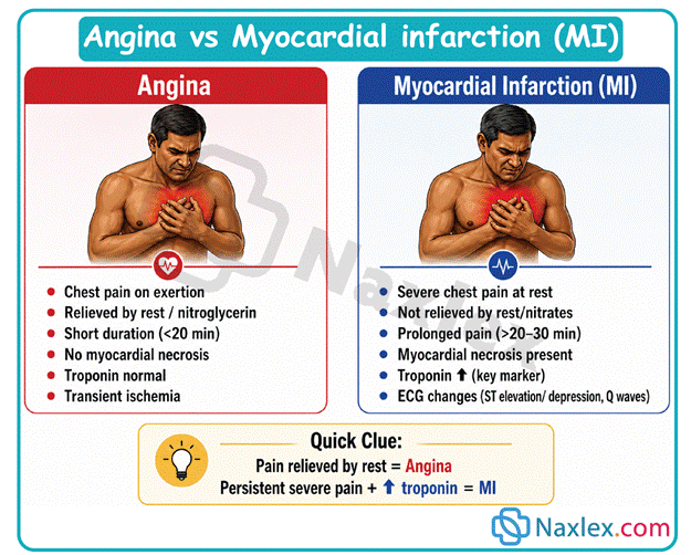
Assessment
Risk Factors
- Male gender or postmenopausal women
- Hypertension
- Tobacco use
- Hyperlipidemia
- Metabolic disorders (diabetes mellitus, hyperthyroidism)
- Methamphetamine or cocaine use
- Stress (occupational, physical exercise, sexual activity)
- An increased risk of coronary artery disease exists for older adult clients who are physically inactive, have one or more chronic diseases (hypertension, heart failure, and diabetes mellitus), or have lifestyle (smoking and diet) habits that contribute to atherosclerosis. Atherosclerotic changes related to aging predispose the heart to poor blood perfusion and oxygen delivery.
- The incidence of cardiac disease increases with age, especially in the presence of hypertension, diabetes mellitus, hypercholesterolemia, elevated homocysteine, and highly sensitive C-reactive protein (HS-CRP).
Subjective Data
- Anxiety, feeling of impending doom
- Chest pain (substernal or precordial)
- Pain can radiate down the shoulder or arm, or may present in the form of jaw pain.
- Pain may be described as a crushing or aching pressure.
- Nausea
- Dizziness
Objective Data
- Physical Assessment Findings
- Pallor, and cool, clammy skin
- Tachycardia and/or heart palpitations
- Diaphoresis
- Vomiting
- Decreased level of consciousness
Diagnostic Tests
- Laboratory Tests
- Cardiac enzymes released with cardiac muscle injury:
- Myoglobin: Earliest marker of injury to cardiac or skeletal muscle. Levels no longer evident after 24 hr.
- Creatine kinase-MB: Peaks around 24 hr after onset of chest pain. Levels no longer evident after 3 days.
- Troponin I or T: Any positive value indicates damage to cardiac tissue and should be reported.
- Troponin I: Levels no longer evident after 7 days.
- Troponin T: Levels no longer evident after 14 to 21 days.
- Cardiac enzymes released with cardiac muscle injury:
- Diagnostic Procedures
- Electrocardiogram (ECG): Recording of electrical activity of the heart over time
- Nursing Actions:
- Assess for changes on serial ECGs.
- Client who has angina: ST depression and/or T-wave inversion indicates presence of ischemia.
- Client who has MI: T-wave inversion indicates ischemia; ST-segment elevation indicates injury; abnormal Q-wave indicates necrosis.
- Nursing Actions:
- Electrocardiogram (ECG): Recording of electrical activity of the heart over time
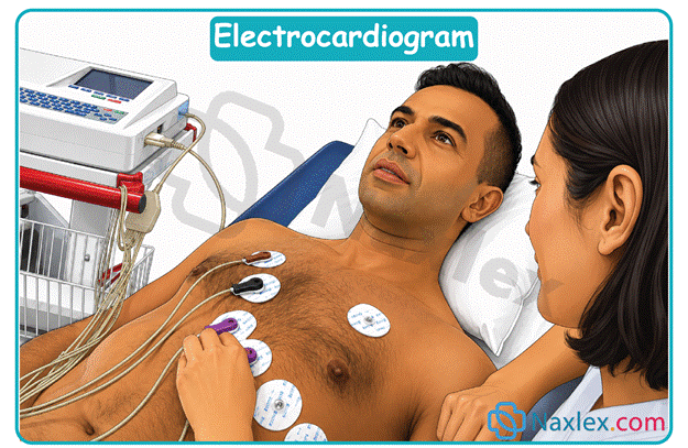
-
- Stress test: Also known as exercise electrocardiography. Client tolerance of activity is tested using a treadmill, bicycle, or medication to evaluate response to increased heart rate.
- Thallium scan: Assesses for ischemia or necrosis. Radioisotopes cannot reach areas with decreased or absent perfusion, and the areas appear as “cold spots.”
- Nursing Actions: Instruct the client to avoid smoking and consuming caffeinated beverages 4 hr prior to the procedure. These can affect the test.
- Cardiac catheterization:
- A coronary angiogram, also called a cardiac catheterization, is an invasive diagnostic procedure used to evaluate the presence and degree of coronary artery blockage.
- Angiography involves the insertion of a catheter into a femoral (sometimes a brachial) vessel and threading it into the right or left side of the heart. Coronary artery narrowing and occlusions are identified by the injection of contrast media under fluoroscopy.
- Nursing Actions:
- Ensure the client understands the procedure prior to signing informed consent.
- Ensure that the client remains NPO 8 hr prior to procedure.
- Assess that the client and family understand the procedure.
- Assess for iodine/shellfish allergy (contrast media).
Management
Nursing Care
- Monitor:
- Vital signs every 15 min until stable, then every hour
- Serial ECG, continuous cardiac monitoring
- Location, precipitating factors, severity, quality, and duration of pain
- Hourly urine output – greater than 30 mL/hr indicates renal perfusion
- Laboratory data (cardiac enzymes, electrolytes, ABGs)
- Administer oxygen
- Obtain and maintain IV access
- Promote energy conservation
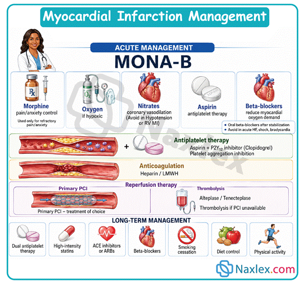
Medications
- Vasodilators
- Nitroglycerin (Nitrostat) prevents coronary artery vasospasm and reduces preload and afterload, decreasing myocardial oxygen demand.
- Nursing Considerations:
- Used to treat angina and help control blood pressure.
- Used cautiously with other antihypertensive medications.
- Vasodilators can cause orthostatic hypotension.
- Client Education:
- Client education regarding response to chest pain:
- Stop activity and rest.
- Place nitroglycerin tablet under tongue to dissolve (quick absorption).
- If pain is unrelieved in 5 min, the client should call 911 or be driven to an emergency department.
- The client can take up to two more doses of nitroglycerin at 5-min intervals.
- Remind the client that a headache is a common side effect of this medication.
- Encourage the client to sit and lie down slowly.
- Client education regarding response to chest pain:
- Analgesics
- Morphine sulfate is an opioid analgesic used to treat moderate to severe pain. Analgesics act on the mu and kappa receptors that help alleviate pain.
- Activation of these receptors produces analgesia (pain relief), respiratory depression, euphoria, sedation, and a decrease in gastrointestinal (GI) motility.
- Use cautiously with clients who have asthma or emphysema due to the risk of respiratory depression.
- Nursing Considerations:
- For the client having chest pain, assess pain every 5 to 15 min.
- Watch for manifestations of respiratory depression, especially in older adults. If respirations are 12/min or less, stop medication, and notify the provider immediately.
- Monitor vital signs for signs of hypotension and decreased respirations.
- Assess for nausea and vomiting.
- Client Education:
- If nausea and vomiting persist, advise the client to notify a nurse.
- Teach the client to use the PCA pump, if applicable.
- The client is the only person who should push the medication administration button. Reassure the client that the safety lockout mechanism on the PCA pump prevents overdosing of the medication.
- Beta-blockers
- Metoprolol tartrate (Lopressor) has antidysrhythmic and antihypertensive properties that decrease the imbalance between myocardial oxygen supply and demand by reducing afterload and slowing heart rate.
- In an acute MI, beta-blockers decrease infarct size and improve short- and long-term survival rates.
- Nursing Considerations:
- Beta-blockers can cause bradycardia and hypotension. Hold the medication if the apical pulse rate is less than 60/min, and notify the provider.
- Avoid giving to clients who have asthma. Cardioselective beta blockers (affect only beta1 receptors) are preferred to minimize effects on the respiratory system.
- Use with caution in clients who have heart failure.
- Client Education:
- Encourage the client to sit and lie down slowly.
- Remind the client to notify the provider immediately if shortness of breath, edema, weight gain, or cough occur.
- Thrombolytic agents
- Streptokinase (Streptase) and alteplase (Activase) are used to break up blood clots.
- Thrombolytic agents have similar side effects and contraindications as anticoagulants.
- For best results, give within 6 hr of infarction.
- Nursing Considerations:
- Assess for contraindications (active bleeding, peptic ulcer disease, history of CVA, recent trauma).
- Monitor for effects of bleeding (mental status changes, hematuria).
- Monitor bleeding times – PT, aPTT, INR, fibrinogen levels, and CBC.
- Monitor for same side effects as anticoagulants (thrombocytopenia, anemia, hemorrhage).
- Administer streptokinase slowly to prevent hypotension.
- Client Education:
- Remind the client of the risk for bruising and bleeding while on this medication.
- Antiplatelet agents
- Aspirin (Ecotrin) and clopidogrel (Plavix) prevent platelets from forming together, which can produce arterial clotting.
- Aspirin prevents vasoconstriction. Due to this and antiplatelet effects, it should be administered with nitroglycerin at the onset of chest pain.
- Nursing Considerations:
- Antiplatelet agents can cause GI upset.
- Use cautiously with clients who have a history of GI ulcers.
- Tinnitus, ringing in the ears, can be a sign of aspirin toxicity.
- Client Education:
- Remind the client of the risk for bruising and bleeding while on this medication.
- Encourage the client to use aspirin tablets with enteric coating and to take with food.
- Tell the client to report ringing in the ears.
- Anticoagulants
- Heparin and enoxaparin (Lovenox) are used to prevent clots from becoming larger or other clots from forming.
- Nursing Considerations:
- Assess for contraindications (active bleeding, peptic ulcer disease, history of CVA, recent trauma).
- Monitor bleeding times – PT, aPTT, INR, and CBC.
- Monitor for adverse effects of anticoagulants (thrombocytopenia, anemia, hemorrhage).
- Client Education:
- Remind the client of the risk for bruising and bleeding while on this medication.
- Glycoprotein IIB/IIIA inhibitors
- Eptifibatide (Integrilin) is used to prevent binding of fibrogen, in turn blocking platelet aggregation. In combination with aspirin therapy, IIB/IIA inhibitors are standard therapy.
- Nursing Considerations:
- This medication can cause active bleeding.
- Client Education:
- Instruct the client to report evidence of bleeding during medication therapy.
Teamwork and Collaboration
- Pain management services can be consulted if pain persists and/or is uncontrolled.
- Cardiac rehabilitation care can be consulted if the client has prolonged weakness and needs assistance with increasing level of activity.
- Nutritional services can be consulted for diet modification to promote low-sodium and low-saturated fat food choices.
Surgical Interventions
- Percutaneous transluminal coronary angioplasty (PTCA)
- Bypass graft (also known as CABG)
Care after Discharge
- Cardiac rehabilitation should be consulted for a specific exercise program related to the heart.
- Nutritional services, such as a dietitian, can be consulted for diet modification or weight management.
- Client Education:
- Instruct the client to monitor and report signs of infection, such as fever, incisional drainage, and redness.
- Teach the client to avoid straining, strenuous exercise, or emotional stress when possible.
- Client education regarding response to chest pain – follow instructions on use of sublingual nitroglycerin.
- If client is a smoker, encourage smoking cessation.
- Encourage the client to remain active and to exercise regularly.
Complications
- Acute MI: A complication of angina not relieved by rest or nitroglycerin.
- Nursing Actions:
- Administer oxygen to the client.
- Notify the provider immediately.
- Nursing Actions:
- Heart failure/cardiogenic shock
- Injury to the left ventricle can lead to decreased cardiac output and heart failure.
- Progressive heart failure can lead to cardiogenic shock.
- This is a serious complication of pump failure, commonly following an MI of 40% blockage.
- Manifestations include tachycardia; hypotension; inadequate urinary output; altered level of consciousness; respiratory distress (crackles and tachypnea); cool, clammy skin; decreased peripheral pulses; and chest pain.
- Nursing Actions:
- Administration of oxygen; possible intubation and ventilation may be required.
- IV administration of morphine, diuretics, and/or nitroglycerin to decrease preload; IV administration of vasopressors and/or positive inotropes to increase cardiac output and to maintain organ perfusion.
- Maintain continuous hemodynamic monitoring.
- Ischemic mitral regurgitation: Evidenced by development of a new cardiac murmur.
- Nursing Actions:
- Administer oxygen to the client.
- Notify the provider immediately.
- Nursing Actions:
- Ventricular aneurysms/rupture: May be due to necrosis from MI; can present as sudden chest pain, dysrhythmias, and severe hypotension.
- Nursing Actions:
- Administer oxygen to the client.
- Notify the provider immediately.
- Nursing Actions:
- Dysrhythmias
- An inferior wall MI may lead to an injury to the AV node, resulting in bradycardia and second-degree AV heart block.
- An anterior wall MI may lead to an injury to the ventricle, resulting in premature ventricular contractions, bundle branch, or complete heart block.
- Nursing Actions:
- Monitor ECG and vital signs.
- Administer oxygen.
- Administer antidysrhythmic medications as indicated.
- Prepare for cardiac pacemaker if needed.
Summary
- Acute coronary syndrome describes the progression from angina to myocardial infarction which is caused by an imbalance between myocardial oxygen demand and supply.
- Early recognition is life-saving because the majority of myocardial infarction related deaths occur within the first hour of symptom onset.
- Angina is usually relieved by rest or nitroglycerin within fifteen minutes while myocardial infarction pain lasts over thirty minutes and requires opioid intervention.
- Angina is classified as stable exertional angina, unstable preinfarction angina, or variant coronary artery spasm angina.
- Diagnosis relies on subjective reports of crushing chest pressure, objective physical findings like diaphoresis, and the presence of elevated cardiac enzymes.
- Electrocardiograms provide essential diagnostic data with ST depression indicating ischemia, ST elevation showing injury, and Q waves marking permanent necrosis.
- Management utilizes a specific medication protocol including vasodilators, morphine analgesics, beta blockers, thrombolytics, antiplatelets, and anticoagulants.
- Cardiac catheterization and coronary angiography are standard invasive interventions used to identify the location and degree of arterial blockages.
- Nursing care requires vigilant monitoring for pump failure, cardiogenic shock, ischemic mitral regurgitation, and lethal dysrhythmias.
- Long term patient outcomes are improved by smoking cessation, regular exercise, dietary modifications, and strict adherence to medication regimens.