





 Video Library


Please set your exam date
Mechanisms of Labor, Leopold’s Maneuvers
Study Questions
Practice Exercise 1
A nurse is caring for a client in active labor whose fetus is confirmed to be in a transverse lie. Which of the following delivery methods should the nurse anticipate as the primary plan of care for this client?
Explanation
Transverse lie represents a critical obstetric situation where the long axis of the fetus lies perpendicular to the maternal longitudinal axis, making vaginal delivery impossible because the fetal body cannot descend through the pelvic inlet. The shoulder typically acts as the presenting part, which exponentially increases the risk of umbilical cord prolapse and uterine rupture if labor progresses without intervention. Therefore, an immediate cesarean delivery remains the definitive management standard to optimize clinical outcomes and eliminate catastrophic maternal-fetal injury.
Rationale for correct answer:
4. A transverse lie creates a mechanical impossibility for safe vaginal passage because the structural diameter of the fetal trunk cannot clear the pelvic boundaries. Attempting an unassisted labor course with this positioning compromises uterine wall integrity and leads to obstructed labor patterns. Immediate surgical intervention is universally indicated because it bypasses the impassable pelvic inlet and directly prevents uterine rupture during active contractions. Consequently, preparing the client for an operative birth stands as the primary plan of care to ensure safe maternal-fetal outcomes.
Rationale for incorrect answers:
1. Manual internal rotation during active labor carries an unacceptably high risk of severe cervical lacerations and immediate uterine trauma. Trying to forcefully manipulate a fetus from a transverse to a longitudinal orientation while contractions are established frequently triggers placental abruption due to shearing forces. Modern obstetric standards favor non-invasive external version before labor or surgical delivery during labor to prevent these iatrogenic injuries.
2. Forceps application requires a longitudinal cephalic presentation where the fetal head has successfully engaged deep within the maternal pelvis. Attempting to apply forceps blades to a fetus situated transversely is technically unfeasible and causes catastrophic fetal skull fractures upon instrument closure. Because the head remains high above the pelvic brim in a transverse lie, operative vaginal instrumentation is absolutely contraindicated to prevent severe maternal tissue lacerations.
3. Vacuum extraction is strictly dependent on the direct application of the suction cup onto the occiput of a deeply engaged cephalic fetus. In a transverse lie, the fetal head is located in the maternal flank rather than the birth canal, making suction cup attachment completely impossible. Utilizing a vacuum extractor under these malpresentation conditions violates core safety guidelines and causes severe neonatal scalp trauma.
Test-taking strategy:
- Analyze the scenario/question: The prompt describes a client in active labor with a fetus in a transverse lie and asks for the primary delivery method the nurse must anticipate. The question requires identifying the safest definitive intervention for a high-risk mechanical malpresentation during active labor.
- Apply Knowledge of Fetal Presentation: Fetal lie describes the relationship between the long axis of the fetus and the long axis of the mother. A transverse lie means the fetus is positioned horizontally across the uterus, preventing the head or buttocks from entering the pelvis to dilate the cervix. Because active labor has already commenced, external manipulative versions are contraindicated due to the risk of membrane rupture and cord accidents, necessitating an alternate delivery route.
- Rule in Choice 4: Cesarean delivery is the only viable method because a transverse fetus cannot physically pass through the pelvic birth canal.
- Rule out Choice 1: Manual internal rotation is an obsolete, highly dangerous maneuver that causes immediate uterine rupture and severe maternal hemorrhage.
- Rule out Choice 2: Forceps-assisted delivery requires a fully engaged cephalic presentation and cannot be physically or safely applied to a transverse trunk.
- Rule out Choice 3: Vacuum extraction requires a vertex presentation at a low pelvic station, making suction application impossible when the fetus lies horizontally.
Take home points
- Transverse lie occurs when the fetal long axis is perpendicular to the maternal spine, rendering safe vaginal delivery completely impossible.
- Active labor in a transverse lie drastically increases the immediate risk of umbilical cord prolapse and maternal uterine rupture.
- Forceps and vacuum extractors are strictly contraindicated unless the fetus is in a longitudinal, engaged cephalic presentation.
- Cesarean delivery remains the mandatory primary method of care for any live fetus confirmed in a transverse lie during labor.
A nurse is assessing a client who has just been admitted to the labor and delivery unit in the active phase of labor. Which of the following anatomical findings would indicate that the fetus is in a position or attitude that is highly favorable for a successful, uncomplicated vaginal delivery? Select all that apply
Explanation
Fetal positioning determines mechanical compatibility with the maternal pelvis during parturition. A longitudinal lie paired with an attitude of flexion optimizes the presenting skull diameters for descent. These relationships minimize maternal tissue trauma and decrease the likelihood of dystocia during active labor.
Rationale for correct answers:
1. A longitudinal lie occurs when the fetal long axis runs parallel to the maternal long axis. This alignment is highly favorable because it directs the fetus directly into the pelvic inlet during contractions. It prevents mechanical obstructions associated with transverse positions and allows normal cardinal movements to take place. Consequently, this relationship forms the foundational requirement for an uncomplicated vaginal birth process.
2. The suboccipitobregmatic diameter is the smallest anteroposterior parameter of the fetal head, measuring approximately 9.5 centimeters. When this diameter presents, it allows the skull to navigate the pelvic floor with minimal resistance. This optimal orientation occurs exclusively when the fetal head achieves complete flexion before entering the birth canal. Choosing this pathway minimizes the risk of prolonged descent and limits maternal soft-tissue trauma.
4. Full flexion represents the ideal fetal attitude where the chin is tucked tightly against the chest. This posture presents the narrowest possible vertex profile to the maternal bony pelvis during labor. It effectively prevents malpresentations like brow or face variations that significantly complicate vertex descent patterns. The nurse recognizes this anatomical configuration as a primary indicator of a highly favorable cephalic presentation.
Rationale for incorrect answers:
3. When the fetal occiput is directed toward the maternal sacrum, it designates an occiput posterior position. This orientation is unfavorable because it forces a larger cranial diameter to rotate through the narrow pelvic outlet during labor. It frequently induces severe maternal back labor and significantly prolongs the second stage of labor. Therefore, this finding represents a malposition that often requires manual or operative intervention.
5. Palpation of the acromion process indicates that the fetus is positioned in a shoulder presentation within the uterus. This finding confirms a transverse lie where the fetal long axis is perpendicular to the maternal spine, making vaginal delivery mechanically impossible without intervention. Attempting to progress through labor with this presentation exposes the client to an extreme risk of uterine rupture. The nurse must immediately anticipate surgical intervention rather than an uncomplicated vaginal delivery.
Test-taking strategy:
- Analyze the scenario/question: The prompt asks for anatomical findings during a labor assessment that indicate a highly favorable fetal position or attitude for a successful, uncomplicated vaginal delivery. This is a select-all-that-apply question requiring the discrimination between optimal mechanical alignments and various pelvic malpresentations or malpositions.
- Apply Knowledge of Fetal Positioning: Fetal orientation is defined by lie, presentation, attitude, and position relative to the maternal pelvis. The optimal combination requires a parallel alignment of spines, universal joint flexion to present the smallest cranial diameters, and an anterior orientation of the landmark. Deviations from these parameters increase pelvic resistance, prolong delivery stages, and necessitate operative or surgical interventions.
- Rule in Choice 1: A parallel relationship between the long axes confirms a longitudinal lie, which is mandatory for any normal head-first or breech vaginal delivery.
- Rule in Choice 2: The suboccipitobregmatic diameter represents the smallest possible presentation profile of the fetal cranium, ensuring optimal transit through the birth canal.
- Rule out Choice 3: Directing the occiput toward the maternal sacrum creates an occiput posterior position, which induces severe back labor and causes prolonged pelvic descent.
- Rule in Choice 4: A fully flexed head with the chin tucked tightly against the breastbone establishes the ideal vertex presentation necessary for a streamlined delivery.
- Rule out Choice 5: Palpating the acromion process identifies a shoulder presentation associated with a transverse lie, which represents an absolute mechanical contraindication to vaginal birth.
Take home points
- A longitudinal lie and vertex presentation are the most favorable configurations for an uncomplicated vaginal delivery.
- Complete flexion of the fetal head presents the suboccipitobregmatic diameter, which is the smallest profile of the fetal skull.
- Occiput posterior positions direct the fetal landmark toward the maternal sacrum, resulting in prolonged labor and intense back pain.
- An acromion presentation indicates a transverse lie, which creates a mechanical impossibility for vaginal birth and requires a cesarean section.
A nurse is performing a sterile vaginal examination on a laboring client and notes that the fetal presenting part is the sacrum, with the fetal hips fully flexed and both knees completely extended upward toward the face. Which of the following terms should the nurse use to document this specific fetal presentation?
Explanation
Malpresentation occurs when the fetus displays a non-cephalic presenting part, significantly increasing risks for umbilical cord prolapse and birth asphyxia. Frank breech involves a longitudinal lie where the buttocks present due to complete thigh flexion and knee extension. This malpresentation often requires cesarean delivery to mitigate severe mechanical birth trauma.
Rationale for correct answer:
1. The fetal presenting part is the sacrum with hips flexed and knees extended, which defines a frank breech presentation. This specific orientation places the lower extremities directly against the fetal torso, positioning the buttocks as the lowest descending pole. Recognizing this anatomy is crucial because it alters labor progression and increases the risk of fetal entrapment during vaginal delivery.
Rationale for incorrect answers:
2. In a complete breech presentation, the fetus sits cross-legged because both the hips and knees are fully flexed. The presenting part features both the buttocks and feet rather than just the buttocks with extended legs. This structural difference alters the mechanism of labor and changes the risk profile for cord compression compared to the frank variety.
3. A footling breech presentation occurs when one or both of the fetal lower extremities are extended downward below the buttocks. One or both feet present into the cervical os, creating an incomplete wedge that significantly increases the danger of cervical cord prolapse during membranes rupture. The scenario specifically describes knees extended upward toward the face, which eliminates this lower limb dependency.
4. A sinciput cephalic presentation represents a head-first orientation where the fetal head is only partially flexed or military. The presenting part is the large fontanelle rather than the sacrum, meaning the fetal vertex enters the pelvis first. Because the prompt explicitly establishes the sacrum as the presenting landmark, any cephalic classification is pathophysiologically incorrect.
Test-taking strategy:
- Analyze the scenario/question: The prompt asks for the precise documentation term for a laboring client whose fetus presents with the sacrum, flexed hips, and completely extended knees. The question requires differentiating specific types of longitudinal fetal malpresentations based on lower extremity positioning.
- Apply Knowledge of Fetal Presentation: Fetal presentation denotes the foremost anatomical structure entering the maternal pelvic inlet first during labor. Breech presentations are classified entirely by the degree of flexion or extension at the fetal hip and knee joints, which determines the mechanical configuration of the descending fetus. Understanding these spatial relationships allows the clinician to accurately anticipate mechanical obstructions and delivery complications.
- Rule in Choice 1: The combination of flexed hips and extended knees upward to the face precisely satisfies the diagnostic criteria for a frank breech.
- Rule out Choice 2: Complete breech requires full flexion of both hips and knees, creating a cross-legged posture that is absent in this clinical scenario.
- Rule out Choice 3: Footling breech requires extension of the lower limbs downward below the level of the buttocks, contradicting the upward extension described.
- Rule out Choice 4: Sinciput cephalic describes a head-first presentation, which is completely impossible given that the sacrum is explicitly noted as the presenting part.
Take home points
- Frank breech presentation features fully flexed fetal hips and completely extended knees with the sacrum acting as the presenting part.
- Fetal malpresentation significantly elevates the clinical risk for umbilical cord prolapse and birth trauma during labor descent.
- Complete breech is differentiated by the universal flexion of both the hip joints and the knee joints in a cross-legged sitting posture.
- Accurate documentation of fetal presentation guides obstetric management and helps determine the safety of vaginal versus surgical delivery pathways.
Practice Exercise 2
A nurse is monitoring a client in the second stage of labor and notes that the fetal head has emerged, but it immediately retracts tightly back against the maternal perineum. Which of the following clinical conditions should the nurse suspect based on this presentation?
Explanation
Shoulder dystocia constitutes a critical obstetric emergency occurring during the second stage of labor when the anterior fetal shoulder becomes mechanically impacted behind the maternal pubic symphysis. The pathognomonic sign is the turtle sign, where the delivered fetal head retracts tightly against the perineum due to persistent shoulder impaction. Immediate recognition is vital because this obstruction can cause rapid fetal acidemia and irreversible brachial plexus injury if not resolved swiftly. Management demands emergency maneuvers rather than fundal pressure to prevent maternal-fetal trauma and ensure successful neonatal resuscitation.
Rationale for correct answer:
2. The immediate retraction of the delivered fetal head back against the maternal perineum defines the classic turtle sign of shoulder dystocia. This retraction occurs because the fetal anterior shoulder becomes wedged behind the maternal pubic bone, preventing downward descent of the torso. The nurse must recognize this finding instantly because delayed intervention leads to severe umbilical cord compression and subsequent neonatal hypoxia. Immediate implementation of corrective maneuvers is required to disimpact the shoulder and prevent permanent brachial plexus damage.
Rationale for incorrect answers:
1. Restitution is a normal cardinal movement of labor where the fetal head rotates 45 degrees after birth to realign with the shoulders. This movement occurs freely without the head snapping backward or pressing tightly against the maternal perineal tissues. Because restitution indicates a lack of mechanical obstruction within the birth canal, it does not cause the tightly bound retraction pattern seen here. Misidentifying an impaction as restitution dangerously delays necessary emergency interventions.
3. External rotation represents the natural progression following restitution, where the shoulders rotate internally into an anteroposterior diameter, causing the head to turn further externally. This process occurs smoothly as the next contraction pushes the body downward, rather than pulling the head backward into the pelvic outlet. A normal progression is characterized by continuous downward advancement of the fetus rather than a sudden halt in descent.
4. Arrest of internal rotation occurs earlier in the second stage of labor before the fetal head ever emerges from the vaginal introitus. If internal rotation fails, the fetus typically becomes wedged in a transverse position, resulting in an obstructed labor pattern high within the pelvis. Because the prompt explicitly notes that the fetal head has already emerged, an arrest of this specific intra-pelvic cardinal movement is anatomically impossible.
Test-taking strategy:
- Analyze the scenario/question: The prompt describes a client in the second stage of labor whose fetal head emerges but immediately retracts tightly against the maternal perineum. The nurse must identify the specific clinical condition indicated by this distinct presentation, which requires recognizing a classic mechanical emergency.
- Apply Knowledge of Labor Complications: The second stage of labor involves the descent and expulsion of the fetus through a series of coordinated cardinal movements. When the head delivers but immediately snaps backward against the perineum, it signifies that the progressive downward momentum has been abruptly halted by an unyielding bony obstruction. This specific visual phenomenon is known universally as the turtle sign and dictates immediate emergency protocols to relieve shoulder impaction and avoid severe hypoxic or neurological injury.
- Rule in Choice 2: The retraction of the head tightly against the perineum is the hallmark diagnostic indicator of shoulder dystocia.
- Rule out Choice 1: Restitution is a normal, non-obstructed movement where the head untwists to align naturally with the fetal spine.
- Rule out Choice 3: Normal external rotation results in the visible turning of the head accompanying the smooth delivery of the body.
- Rule out Choice 4: Arrest of internal rotation occurs entirely within the birth canal before the head can physically exit the vulva.
Take home points
- The retraction of the emerged fetal head against the maternal perineum is called the turtle sign and indicates shoulder dystocia.
- Shoulder dystocia occurs when the anterior fetal shoulder becomes mechanically wedged behind the maternal pubic symphysis.
- Failure to rapidly resolve shoulder dystocia can lead to severe neonatal complications including brachial plexus injury and birth asphyxia.
- Fundal pressure is strictly contraindicated in shoulder dystocia because it further wedges the shoulder and increases the risk of uterine rupture.
A nurse performing a vaginal examination on a client in active labor determines that the lowest portion of the fetal presenting part is located 2 centimeters below the level of the maternal ischial spines. Which of the following stations should the nurse document in the client's medical record?
Explanation
Fetal station measures the descent of the presenting part through the birth canal relative to the maternal ischial spines. The spines serve as the definitive zero landmark, representing the narrowest diameter of the pelvis. Any location superior to these landmarks is designated with a negative number, whereas descent inferior to them is marked with a positive number. Accurate station assessment is critical to monitor labor progression and identify risks for obstructed labor or pelvic disproportion.
Rationale for correct answer:
3. When the lowest portion of the fetal presenting part is located 2 centimeters below the level of the ischial spines, it is documented as station +2. Positive numbers signify that the fetus has successfully navigated past the narrowest portion of the midpelvis. This progressive descent indicates that the pelvic inlet is adequate to accommodate the presenting diameters. Accurate recording of this advancement is crucial for confirming normal labor progression toward delivery.
Rationale for incorrect answers:
1. A designation of station -2 indicates that the leading edge of the fetal presenting part is located 2 centimeters above the maternal ischial spines. This negative value reflects a fetus that is floating or unengaged within the pelvic inlet. Documenting a negative station when the fetus has actually descended below the spines represents a significant clinical error. Misjudging this position can lead to inappropriate interventions for suspected failure to descend.
2. Documenting station 0 signifies that the lowest presenting part of the fetus is aligned directly with the maternal ischial spines. At this landmark plane, the fetal presentation has achieved engagement, meaning the widest biparietal diameter has entered the pelvis. Because the prompt specifies that the fetus has traveled 2 centimeters past this zero reference point, classifying it at the spines is anatomically incorrect.
4. A measurement of station +4 indicates that the fetal presenting part has descended 4 centimeters below the ischial spines, placing it at the pelvic outlet. At this deep station, the fetus is typically crowning and visible at the vaginal introitus, signaling that birth is imminent. Since the assessment reveals the fetus is only 2 centimeters below the spines, this selection overestimates fetal descent and delivery readiness.
Test-taking strategy:
- Analyze the scenario/question: The prompt asks for the correct documentation of the fetal station when the lowest portion of the presenting part is felt 2 centimeters below the level of the maternal ischial spines. The question requires applying standard obstetric measurement rules to translate physical findings into numerical values.
- Apply Knowledge of Fetal Station: Fetal station quantified in centimeters evaluates the spatial relationship of the presenting part to an imaginary line drawn between the ischial spines. The ischial spines represent the narrowest plane of the midpelvis and are mathematically designated as station zero. Measurements above this landmark are assigned negative numbers, while measurements below are assigned positive numbers matching the distance in centimeters.
- Rule in Choice 3: The location of 2 centimeters below the reference plane directly translates to a positive 2, which is documented as station +2.
- Rule out Choice 1: Station -2 describes a fetus positioned 2 centimeters above the spines, which is the exact opposite of the described downward descent.
- Rule out Choice 2: Station 0 means the fetus is resting perfectly level with the spines, failing to account for the additional 2 centimeters of movement.
- Rule out Choice 4: Station +4 describes a fetus that has advanced much further down to the perineum, which overstates the current anatomical position.
Take home points
- The maternal ischial spines serve as the zero reference landmark for determining fetal station during labor.
- Positions above the ischial spines are documented as negative numbers, whereas positions below the spines are documented as positive numbers.
- Engagement is achieved when the presenting part reaches station 0, indicating the widest part of the fetal head has entered the pelvic inlet.
- Progressive advancement from negative to positive stations serves as a primary indicator of successful labor descent and pelvic adequacy.
A nurse is teaching a student nurse about the structural changes that occur during the intrapartum period. Which of the following statements best explains the primary mechanical purpose of fetal head flexion during the cardinal movements of labor?
Explanation
Fetal head flexion is a critical cardinal movement triggered by resistance from the maternal pelvic floor, cervix, or pelvic walls during descent. This passive mechanical adjustment shifts the fetal head from a military or extended attitude into a fully flexed attitude, which optimizes the cranial profile. By tucking the chin tightly against the chest, the fetus substitutes larger cranial diameters for the narrowest possible configuration, preventing obstructed labor patterns. Consequently, this movement facilitates smooth passage through the bony pelvis and decreases the likelihood of soft tissue trauma or operative delivery interventions.
Rationale for correct answer:
3. Flexion serves the primary mechanical purpose of presenting the suboccipitobregmatic diameter, which is the absolute smallest anteroposterior diameter of the fetal skull, to the maternal pelvis. Measuring approximately 9.5 centimeters, this reduced dimension allows the head to navigate the narrow pelvic boundaries far more easily than an extended head would. Without adequate flexion, wider cranial dimensions attempt to enter the pelvic inlet, which frequently causes a complete arrest of descent. Thus, this positional adjustment maximizes mechanical efficiency to ensure a successful and uncomplicated vaginal birth process.
Rationale for incorrect answers:
1. Turning the fetal occiput toward the maternal sacrum describes the development of an occiput posterior position, which is an unfavorable malposition. Flexion does not seek to cause this posterior orientation; rather, it occurs along the longitudinal axis to decrease the presenting cranial circumference regardless of rotation. An occiput posterior position actually increases pelvic resistance, which stands in direct opposition to the primary space-reducing benefit achieved by head flexion.
2. Realignment of the fetal head with the long axis of the shoulders describes the process of restitution, which occurs externally after the head is completely delivered. Flexion is an intra-pelvic maneuver that occurs early during the descent phase of labor, long before the head exits the vaginal introitus. Confusing an early descent mechanism with an external realigning movement reflects a basic misunderstanding of the chronological sequence of cardinal movements.
4. Guiding the acromion process beneath the maternal pubic symphysis refers to the mechanism of shoulder expulsion, where the anterior shoulder slips under the pubic bone. Fetal head flexion acts exclusively on the cranial structures and is entirely completed before the shoulders ever engage deep within the pelvic cavity. The acromion process relates directly to a transverse lie or shoulder presentation, whereas flexion is a mechanism specific to a longitudinal cephalic presentation.
Test-taking strategy:
- Analyze the scenario/question: The prompt asks for the statement that best explains the primary mechanical purpose of fetal head flexion during the cardinal movements of labor. The question requires identifying how this specific alteration in fetal attitude facilitates mechanical transit through the birth canal.
- Apply Knowledge of Cardinal Movements: The cardinal movements of labor represent the positional changes that the fetus undergoes as it travels through the birth canal. Flexion occurs passively as the descending head encounters resistance from maternal tissues, forcing the fetal chin to tuck tightly against the sternum. This structural adaptation replaces the larger occipitofrontal diameter with the minimal suboccipitobregmatic diameter, allowing the cranium to match the narrowest dimensions of the maternal pelvic floor.
- Rule in Choice 3: Tucking the chin changes the presenting skull alignment to the suboccipitobregmatic diameter, presenting the smallest possible profile to the pelvis.
- Rule out Choice 1: Rotating the occiput toward the sacrum describes an occiput posterior malposition, which prolongs labor and is unrelated to axial flexion.
- Rule out Choice 2: Realigning the head with the shoulders defines restitution, an external movement that happens after the head has completely emerged.
- Rule out Choice 4: Managing the acromion process under the pubic bone describes shoulder delivery, which occurs long after cranial flexion has taken place.
Take home points
- Fetal head flexion is a passive cardinal movement caused by the mechanical resistance of the maternal cervix, pelvic walls, and pelvic floor.
- Flexion replaces the larger occipitofrontal diameter with the suboccipitobregmatic diameter, which is the smallest profile of the fetal head.
- Presenting the smallest cranial diameter minimizes mechanical resistance and drastically reduces the incidence of prolonged or obstructed labor.
- Flexion alters the fetal attitude from a neutral or military posture into a universal flexion posture, which is ideal for cephalic descent.
A nurse is analyzing the physiological mechanisms that contribute to the descent of the fetus through the birth canal during labor. Which of the following factors should the nurse identify as primary forces that directly drive fetal descent? Select all that apply
Explanation
Fetal descent relies on a combination of mechanical forces that progressively push the fetus through the pelvic birth canal during labor. The continuous progress of descent is driven by structural forces that build intrauterine pressure during contractions. These mechanisms ensure that the fetus advances downward, overcoming the pelvic tissue resistance to facilitate a successful vaginal delivery.
Rationale for correct answers:
1. During a contraction, the amniotic fluid becomes compressed within the intact amniotic sac, creating a uniform hydraulic pressure system. This hydrostatic force transmits energy evenly across the entire surface of the fetus, pushing it downward into the dilating cervix. Even after the membranes rupture, this fluid pressure mechanism continues to act as a primary driver of fetal engagement. Understanding this fluid dynamic allows the nurse to comprehend how contractions effectively advance the fetus without causing localized mechanical trauma.
2. As the uterus contracts, the muscle fibers of the upper uterine segment shorten permanently, exerting a direct downward pressure against the fetal rump. This direct force pushes the fetal body along the longitudinal axis of the birth canal, causing the presenting part to advance through the maternal pelvis. This upper-segment mechanical force represents the primary driving engine of fetal descent during the first and second stages of labor. It ensures that the uterine contractions are effectively converted into progressive, downward propulsion of the fetus.
Rationale for incorrect answers:
3. Continuous voluntary bearing-down efforts are strictly restricted to the second stage of labor and are not primary forces throughout the entire descent process. Initiating voluntary pushing efforts prematurely during the first stage of labor causes maternal exhaustion and severe cervical edema without advancing the fetus. The nurse knows that primary descent is driven entirely by involuntary uterine contractions rather than continuous maternal pushing efforts.
4. The structural shape and resistance of the maternal bony pelvis act as an opposing impediment to descent rather than a propulsive force. While pelvic architecture forces the fetus to undergo mandatory cardinal movements, it functions as a restrictive boundary that the fetus must navigate. Increased pelvic resistance actually slows down labor progression and can lead to obstructed labor if cephalopelvic disproportion is present.
5. Hyperextension of the fetal neck is a specific positional adjustment that occurs during the mechanism of extension as the head emerges. This anatomical movement is a passive response to pelvic floor resistance rather than a primary force that drives downward descent. It represents a localized directional pivot under the pubic arch rather than a generalized propelling power that moves the fetus through the entire birth canal.
Test-taking strategy:
- Analyze the scenario/question: The prompt asks for the factors that the nurse should identify as the primary forces that directly drive fetal descent through the birth canal during labor. This is a select-all-that-apply question requiring the identification of active propelling forces versus passive mechanisms or resistant structures.
- Apply Knowledge of Fetal Descent: Fetal descent is propelled by specific forces: the pressure of the amniotic fluid, the direct pressure of the contracting fundus on the fetus, the contraction of the maternal diaphragm and abdominal muscles (during the second stage), and the extension and straightening of the fetal body. The question asks for the primary forces driving descent; the pelvis provides resistance and shape, which shapes the descent path but does not drive it, and neck extension is a passive cardinal movement. Continuous voluntary pushing is incorrect because it is only appropriate during the second stage and is not continuous throughout labor.
- Rule in Choice 1: The hydraulic pressure of the amniotic fluid during a contraction acts as a primary hydrostatic force pushing the fetus downward.
- Rule in Choice 2: The direct downward pressure exerted by the shortening fibers of the uterine fundus against the fetal breech directly propels the fetus downward.
- Rule out Choice 3: Continuous voluntary bearing-down efforts are inappropriate during the first stage of labor and do not serve as a continuous primary force.
- Rule out Choice 4: The structural shape and resistance of the maternal bony pelvis provide mechanical resistance and direction, not an active driving force.
- Rule out Choice 5: Hyperextension of the fetal neck is a passive positional change during the extension phase, not a force driving generalized labor descent.
Take home points
- Fetal descent is primarily driven by the involuntary mechanical forces of uterine contractions throughout the stages of labor.
- Hydrostatic pressure from the amniotic fluid distributes contraction forces uniformly to push the fetus into the lower uterine segment.
- Direct fundal pressure against the fetal breech column converts uterine contractions into linear descent along the birth canal.
- Maternal pelvic structures and fetal neck extension provide resistance and positional adaptation rather than active propulsive forces.
A nurse is caring for a nulliparous client who has reached the second stage of labor. Which of the following clinical events are classified as sequential cardinal movements of labor that the fetus must perform to navigate the maternal pelvic outlet? Select all that apply
Explanation
The mechanical passage of the fetus through the birth canal requires a series of adaptive positional changes known collectively as the cardinal movements of labor. These passive adjustments are driven by uterine contractions and maternal pelvic resistance, enabling the fetus to present its smallest anatomical diameters. Proper navigation of these steps ensures progressive descent and prevents obstructed labor patterns during childbirth. Failing to progress through these sequential movements often indicates a mechanical mismatch requiring emergency operative delivery interventions.
Rationale for correct answers:
1. Internal rotation is a critical cardinal movement where the fetal head rotates from a transverse position to an anteroposterior orientation. This rotation allows the longest diameter of the fetal head to align perfectly with the longest diameter of the maternal pelvic outlet. It occurs as the presenting part encounters the resistance of the muscular pelvic floor during active descent. Without this essential realignment, the fetus cannot successfully pass beneath the pubic arch, leading to an arrest of labor.
3. Extension occurs when the sharply flexed fetal head reaches the maternal perineum and pivots beneath the pubic symphysis. The resistance of the pelvic floor forces the fetal head to push upward and outward, resulting in the delivery of the brow, face, and chin. This movement represents a complete reversal of the initial cranial attitude from flexion to extension. The nurse monitors this mechanism as the crowning head emerges completely through the vaginal introitus.
4. Restitution describes the immediate external rotation of the delivered fetal head by 45 degrees to realign itself with its shoulders. When the head is inside the pelvis, the shoulders remain transverse; once the head is freed externally, it untwists to resume a natural anatomic alignment with the torso. This movement is followed immediately by external rotation of the head as the shoulders turn internally to prepare for expulsion. The nurse documents this step as confirmation that the shoulders are navigating the bony pelvis successfully.
Rationale for incorrect answers:
2. Cervical effacement refers to the progressive thinning and shortening of the cervix that takes place during the first stage of labor. While it is a foundational physiological change necessary to allow the fetus to enter the birth canal, it is a maternal structural change rather than a fetal positional movement. It must be fully completed before the client can transition into the second stage of labor. Consequently, it is never classified as an active or passive fetal cardinal movement.
5. Placental separation represents the third stage of labor, during which the placenta detaches from the uterine wall following the complete birth of the neonate. This process is driven by post-delivery uterine remodeling and sudden volume reduction rather than any fetal mechanical navigation. Because the fetus has already exited the birth canal completely before this event occurs, it is chronologically and conceptually separate from the positional mechanisms of labor.
Test-taking strategy:
- Analyze the scenario/question: The prompt asks for the clinical events that are classified as sequential cardinal movements of labor that the fetus must perform to navigate the birth canal. This is a select-all-that-apply question requiring the differentiation of passive fetal positional adaptations from maternal physiological changes and third-stage labor events.
- Apply Knowledge of Cardinal Movements: The cardinal movements of labor describe the specific path and positional adaptations a fetus takes as it moves through the birth canal. The standard sequence includes engagement, descent, flexion, internal rotation, extension, restitution, external rotation, and expulsion. To select the correct answers, the nurse must exclude any options that describe maternal structural changes or events that happen after the delivery of the neonate.
- Rule in Choice 1: Internal rotation is the intra-pelvic movement where the fetal head turns to align with the anteroposterior diameter of the pelvic outlet.
- Rule out Choice 2: Cervical effacement is a maternal physiological process involving the thinning of the cervix during the first stage of labor, not a fetal movement.
- Rule in Choice 3: Extension is the positional change that occurs as the fetal head pivots under the pubic arch and extends upward to deliver the face.
- Rule in Choice 4: Restitution is the external movement where the delivered head untwists by 45 degrees to realign naturally with the fetal shoulders inside the pelvis.
- Rule out Choice 5: Placental separation is an event belonging to the third stage of labor that occurs after the fetus has already been completely delivered.
Take home points
- Cardinal movements represent the sequential, passive positional adjustments the fetus makes to successfully navigate the maternal pelvis.
- Internal rotation aligns the longest diameter of the fetal head with the largest diameter of the maternal pelvic outlet.
- Extension is the mechanical pivoting of the fetal head under the pubic arch, allowing the face and chin to emerge over the perineum.
- Restitution is an external cardinal movement where the born head rotates slightly to realign anatomically with the shoulders still inside the birth canal.
Practice Exercise 3
A nurse is performing Leopold's maneuvers on a client at 39 weeks of gestation who has been admitted in early labor. While performing the first maneuver, the nurse palpates a smooth, hard, round, and highly ballottable mass in the uterine fundus. Which of the following interpretations should the nurse derive from this clinical finding?
Explanation
Leopold's maneuvers determine fetal orientation via systematic abdominal palpation. The first maneuver identifies the contents of the uterine fundus to ascertain the fetal lie. Palpating a rigid, mobile structure indicates the fetal head occupies the upper segment, confirming a breech presentation.
Rationale for correct answer:
1. Palpating a ballottable mass at the fundus indicates the head is located superiorly. This confirms the lower extremities or buttocks occupy the maternal pelvic inlet. This configuration establishes a longitudinal lie because the fetal spine parallels the maternal spine. Consequently, the nurse must interpret this finding as a breech presentation with high fetal impaction risk.
Rationale for incorrect answers:
2. A transverse lie occurs when the fetal long axis is perpendicular to the maternal spine, leaving the fundus empty. In this orientation, the nurse would find a shoulder presentation or acromion process lower in the uterus. The cranial vault would be located laterally in the flank rather than superiorly. Therefore, finding a ballottable mass at the top rules out this horizontal orientation.
3. In a cephalic presentation, the fetal head enters the pelvic inlet first, meaning it would not reside in the upper uterus. The first maneuver would instead reveal the soft, irregular contours of the fetal breech within the fundus. Finding a hard, round mass at the top indicates the vertex is not presenting. Thus, assuming a head-first descent contradicts the presence of the cranium at the uterine apex.
4. An oblique lie represents a transient, diagonal orientation where the fetal axis rests at an angle. The head would typically be located in the maternal iliac fossa rather than centered at the fundus. This variation results in an unstable presentation that usually shifts as active uterine contractions begin. Finding a centered, ballottable mass at the fundal boundary rules out this diagonal malposition.
Test-taking strategy:
- Analyze the scenario/question: The prompt describes a nurse performing the first Leopold's maneuver and finding a smooth, hard, round, ballottable mass in the uterine fundus. The question requires interpreting this specific anatomical finding to determine the correct fetal lie and presentation.
- Apply Knowledge of Leopold's Maneuvers: Leopold's maneuvers are four specific palpations used to map out fetal position systematically. The first maneuver focuses entirely on the fundus to differentiate between the round, hard, freely movable head and the soft, irregular, stationary breech. Correctly identifying which fetal pole occupies the upper segment allows the clinician to deduce what is presenting at the maternal pelvic inlet.
- Rule in Choice 1: A hard, round, ballottable mass in the fundus identifies the head, meaning the buttocks are down, confirming a longitudinal lie with a breech presentation.
- Rule out Choice 2: A transverse lie would leave the fundus empty because the fetus is positioned horizontally, presenting the shoulder instead of the head or breech.
- Rule out Choice 3: A cephalic presentation would mean the head is down in the pelvis, which would cause the softer breech to be felt in the fundus.
- Rule out Choice 4: An oblique lie places the head diagonally in the maternal flank or hip area, preventing a well-defined mass from being felt at the fundal midpoint.
Take home points
- The first Leopold's maneuver evaluates the uterine fundus to determine fetal lie and identify which fetal pole occupies the upper uterus.
- A hard, round, smooth, and ballottable mass at the fundus indicates the fetal head is up, diagnostic of a breech presentation.
- In a cephalic presentation, the fundus contains the fetal buttocks, which palpate as soft, irregular, and non-ballottable structures.
- Transverse and oblique lies are characterized by an empty or poorly occupied fundus because the fetal long axis does not parallel the maternal spine.
A nurse is preparing to perform the third Leopold maneuver on a laboring client to evaluate the degree of fetal engagement. Which of the following technical actions should the nurse implement to execute this maneuver correctly?
Explanation
Leopold maneuvers provide a systematic method for determining fetal position, presentation, and engagement through abdominal palpation. The third maneuver, also known as the Pawlik grip, specifically evaluates the presenting part at the pelvic inlet to determine if fetal engagement has occurred. This assessment is vital to verify that the widest transverse diameter of the fetal skull has successfully navigated past the pelvic brim. Performing this step correctly allows the clinician to gauge mobility and anticipate potential complications like obstructed labor patterns before advanced descent.
Rationale for correct answer:
2. The third maneuver requires the clinician to grasp the lower uterine segment directly above the symphysis pubis using the thumb and fingers of a single hand. If the presenting part is unengaged, it will feel like a mobile mass that can be easily nudged upward, away from the pelvic brim. If engagement has occurred, the presenting structure is firmly fixed within the pelvic inlet and cannot be moved or ballotted upon palpation. Executing this step precisely confirms whether the fetus is safely anchored for descent or still floating high within the maternal pelvis.
Rationale for incorrect answers:
1. Facing the client's feet and sliding both hands down the lower abdomen describes the technique for the fourth Leopold maneuver, or the pelvic grip. The fourth maneuver aims to determine fetal attitude and identify the cephalic prominence by assessing which side meets early bony resistance. The nurse performs the third maneuver while still facing the client's face, using only a single hand to evaluate mobility at the pelvic inlet. Confounding these two maneuvers leads to an incorrect assessment of engagement parameters and misinterprets the true fetal presentation.
3. Placing both palms flat on the superior aspect of the fundus represents the first Leopold maneuver, which determines fetal lie and fundal characteristics. The first maneuver identifies whether the soft, irregular breech or the hard, round cranial vault occupies the upper segment of the uterus. This step is completed at the very beginning of the assessment series and focuses entirely on the uterine apex. It cannot evaluate lower pelvic entry or determine if the fetus has successfully entered the lower birth canal.
4. Applying steady lateral pressure with one hand while palpating the opposite side describes the second Leopold maneuver, or the umbilical grip. The second maneuver identifies the location of the smooth fetal back versus the nodular small parts, which helps determine fetal position and lateral orientation. This bilateral palpation maps the long axis of the fetus along the sides of the maternal abdominal wall. It does not provide any diagnostic data regarding the entry or fixation of the presenting part at the pubic symphysis.
Test-taking strategy:
- Analyze the scenario/question: The prompt asks for the correct technical action a nurse must implement to execute the third Leopold maneuver correctly on a laboring client. The question requires differentiating the specific hand placements and clinician orientations used across the four distinct steps of abdominal palpation.
- Apply Knowledge of Leopold Maneuvers: Leopold maneuvers must be performed in a precise, numerical sequence to systematically map out uterine contents. The first maneuver explores the fundus, the second assesses the lateral abdominal walls, the third utilizes a single hand above the pubis to gauge engagement, and the fourth requires facing the feet to evaluate the cephalic prominence. Understanding the technical boundaries of each grip allows the nurse to isolate the single-handed technique as the definitive method for checking pelvic entry.
- Rule in Choice 2: Grasping the lower uterine segment with the thumb and fingers of one hand is the signature technique for Pawlik's grip to test for engagement.
- Rule out Choice 1: Facing the client's feet and utilizing both hands describes the fourth maneuver, which assesses fetal attitude rather than engagement mobility.
- Rule out Choice 3: Palpating the superior aspect of the fundus with both palms describes the first maneuver, which identifies the contents of the upper uterine pole.
- Rule out Choice 4: Exerting lateral pressure with one hand while the other feels for the back describes the second maneuver, which maps out fetal position.
Take home points
- The third Leopold maneuver utilizes a single hand placed directly above the symphysis pubis to evaluate the presentation and engagement of the fetus.
- Mobility or ballotability of the presenting part during the third maneuver indicates that fetal engagement has not yet occurred.
- If the presenting part is firmly fixed and cannot be displaced upward, it confirms the fetus has successfully engaged at station zero.
- The first, second, and third maneuvers require facing the client's face, whereas the fourth maneuver requires the nurse to turn and face the client's feet.
A nurse is performing abdominal palpations via Leopold's maneuvers on a client at term. During the execution of the fourth maneuver, the nurse notes that the fingers meet an early anatomical resistance at the cephalic prominence on the same side of the abdomen as the fetal small parts. Which of the following fetal attitudes does this finding indicate?
Explanation
The fourth Leopold maneuver evaluates fetal attitude by identifying the cephalic prominence relative to lateral landmarks. A fully flexed head presents the smallest cranial diameter for a vertex presentation, ensuring optimal mechanical passage through the pelvic inlet.
Rationale for correct answer:
2. When the fetal head is completely flexed, the sinciput or forehead forms the prominent bony landmark that arrests the downward movement of the clinician's fingers. This anatomical prominence is positioned on the opposite side of the smooth fetal back, placing it directly on the same side as the fetal small parts. Finding the resistance here confirms that the narrowest suboccipitobregmatic diameter is preparing to enter the birth canal. This optimal alignment indicates a classic vertex presentation that minimizes the risk of mechanical pelvic dystocia.
Rationale for incorrect answers:
1. A fully extended head positions the occiput as the primary cephalic prominence rather than the sinciput forehead structure. In this hyperextended attitude, the back of the skull bends backward sharply to touch the fetal spine, shifting the pelvic resistance toward the fetal back instead of the limbs. This malpresentation results in a face presentation where the fragile facial bones emerge as the leading denominator. This configuration significantly increases delivery resistance and frequently mandates an operative intervention due to obstructed labor patterns.
3. A partially extended head occurs when the fetus fails to achieve complete flexion or extension, causing the brow to present first. The cephalic prominence becomes less distinctly localized because the head occupies a transitional posture between the vertex and face profiles. This position presents the large occipitofrontal diameter, which significantly exceeds the normal dimensions of the maternal pelvic floor. This mechanical mismatch causes a brow presentation that typically arrests during descent and requires manual or surgical correction.
4. A neutral head orientation describes a military presentation where the fetal head is neither flexed nor extended toward the torso. The fingers descending along the lower uterine segment encounter equal resistance on both sides simultaneously because the head sits perfectly straight. This posture presents the occipitofrontal diameter to the pelvic inlet, which delays normal internal rotation movements. This lack of axial flexion prevents a single distinct prominence from dominating the abdominal palpation findings.
Test-taking strategy:
- Analyze the scenario/question: The prompt describes a nurse finding a cephalic prominence on the same side as the fetal small parts during the fourth Leopold maneuver. The question requires interpreting this specific palpatory finding to determine the correct fetal attitude and presentation.
- Apply Knowledge of Leopold Maneuvers: The fourth maneuver requires the clinician to face the client's feet and slide both hands down the lower maternal abdomen toward the pelvis. The cephalic prominence is the portion of the fetal head that prevents the hand from descending smoothly into the pelvic inlet. By evaluating whether this bony resistance aligns with the rigid fetal back or the movable small parts, the nurse can accurately deduce the degree of cranial flexion or extension.
- Rule in Choice 2: In a fully flexed vertex presentation, the forehead protrudes on the side of the small parts, meeting the examining fingers early.
- Rule out Choice 1: A fully extended head creates a face presentation where the occiput protrudes on the same side as the fetal back.
- Rule out Choice 3: A partially extended head results in a brow presentation where the prominence is poorly defined on the abdominal grid.
- Rule out Choice 4: A neutral head orientation creates a military presentation where both sides of the skull present equal anatomical resistance.
Take home points
- The fourth Leopold maneuver is performed by facing the client's feet to locate the cephalic prominence and evaluate fetal attitude.
- In a fully flexed head, the cephalic prominence is the sinciput, which is palpated on the same side as the fetal small parts.
- If the cephalic prominence is felt on the same side as the fetal back, it indicates a hyperextended head and a face presentation.
- A neutral or partially extended attitude increases the presenting skull diameter, which often leads to prolonged or obstructed labor patterns.
A nurse has completed the four steps of Leopold's maneuvers on a client admitted to the intrapartum unit. Which of the following clinical purposes represent primary objectives achieved by utilizing this systemic abdominal palpation technique? Select all that apply
Explanation
Leopold maneuvers provide a non-invasive, systematic method for mapping out uterine contents via maternal abdominal palpation. Performing these sequential grips allows the clinician to identify structural relationships before advanced labor descent. This physical assessment provides essential data required to formulate a safe intrapartum plan and optimize overall maternal-fetal monitoring.
Rationale for correct answers:
1. Differentiating between the hard, round cranial vault and the soft, irregular breech allows the nurse to confirm the specific fetal presentation. Identifying what part enters the pelvic inlet is vital because malpresentations like breech or face significantly alter labor management. Recognizing these anatomical variations early ensures the team can prepare for potential complications like obstructed labor. This definitive identification represents a cornerstone objective of completing the systemic palpation protocol.
2. Mapping out the fetal back versus the small parts allows the nurse to determine the exact fetal position within the uterus. Fetal heart tones transmit most clearly through the rigid scapular region of the fetal back rather than the limbs. Pinpointing this area guides the precise placement of external electronic monitoring transducers on the maternal abdomen. This objective ensures high-quality data collection and prevents continuous signal loss during active maternal-fetal tracking.
4. Assessing the longitudinal orientation of the fetal pole relative to the maternal spine determines the exact fetal lie. A parallel alignment confirms a longitudinal lie, whereas a perpendicular alignment identifies a transverse lie that makes vaginal delivery impossible. Knowing the lie is a prerequisite for interpreting subsequent presentation parameters and predicting labor safety. This foundational axis determination represents a primary clinical objective achieved during the initial abdominal sweeps.
Rationale for incorrect answers:
3. Evaluation of cervical dilation and effacement requires a direct, sterile vaginal examination rather than external abdominal palpation. Leopold maneuvers cannot assess the internal consistency, opening, or shortening of the maternal cervical os during labor. Relying on external palpation to estimate these pelvic parameters introduces significant diagnostic error and delays tracking labor transition phases. Thus, these specific tissue changes remain completely outside the scope of abdominal maneuvers.
5. External manual palpation can identify gross deviations like polyhydramnios but cannot provide an exact measurement of amniotic fluid volume. Precise quantification requires an ultrasound assessment to calculate the amniotic fluid index or measure the single deepest pocket. Abdominal maneuvers lack the technical refinement to isolate fluid volume from uterine and fetal tissue density. Therefore, estimating exact fluid parameters is clinically impossible using this manual method alone.
Test-taking strategy:
- Analyze the scenario/question: The prompt asks for the primary clinical objectives achieved by utilizing the four steps of Leopold maneuvers on a laboring client. This is a select-all-that-apply question requiring the discrimination of data obtained via external abdominal palpation from data requiring internal exams or ultrasound imaging.
- Apply Knowledge of Leopold Maneuvers: Leopold maneuvers are designed strictly as an external, manual assessment technique to determine fetal lie, presentation, position, and engagement. The first maneuver checks the fundus for lie, the second locates the back for position and fetal heart tone monitoring, the third gauges pelvic engagement, and the fourth evaluates cranial attitude. The nurse must exclude any parameters that require invasive internal visualization, digital pelvic exploration, or advanced diagnostic ultrasound technology.
- Rule in Choice 1: The series of maneuvers systematically isolates the presenting part at the pelvic brim, satisfying the core objective of determining fetal presentation.
- Rule in Choice 2: Identifying the location of the smooth, firm fetal back directly provides the ideal anatomical landmark for placing the fetal heart rate transducer.
- Rule out Choice 3: Assessing cervical dilation and effacement can only be accomplished via an internal, digital sterile vaginal exam, making this option incorrect.
- Rule in Choice 4: The first maneuver directly establishes whether the fetal axis runs parallel or perpendicular to the maternal spine, confirming the fetal lie.
- Rule out Choice 5: Determining the exact volume of amniotic fluid requires an ultrasound calculation of the amniotic fluid index rather than simple manual palpation.
Take home points
- Leopold maneuvers are a non-invasive, four-step abdominal palpation method used to determine fetal lie, presentation, position, and engagement.
- Identifying the location of the fetal back using the second maneuver provides the optimal site for monitoring fetal heart tones.
- Internal parameters such as cervical dilation, effacement, and membrane status cannot be assessed using external Leopold maneuvers.
- Exact amniotic fluid volume quantification requires diagnostic ultrasound imaging and cannot be measured via manual abdominal inspection.
A nurse is preparing to perform Leopold's maneuvers on a client who is at 38 weeks of gestation. Which of the following interventions should the nurse perform to promote client safety, ensure physical comfort, and maintain structural accuracy during the assessment? Select all that apply
Explanation
Patient preparation optimization minimizes physiological stressors and enhances palpatory diagnostic sensitivity during abdominal examinations. Implementing precise positioning modifications prevents supine hypotensive syndrome resulting from great vessel occlusion by the gravid uterus. These supportive actions maintain maternal hemodynamic stability, reduce abdominal wall tension, and optimize conditions for accurate fetal mapping during the subsequent four-step assessment series.
Rationale for correct answers:
1. Instructing the client to completely empty their bladder prior to the procedure directly enhances maternal comfort and ensures structural palpation accuracy. An engorged bladder physically displaces the lower uterine segment upward and laterally, which severely distorts the anatomical baseline of the pelvic inlet. It also causes significant pain during deep pelvic grips, which induces voluntary abdominal muscle guarding that obscures fetal parts. Therefore, ensuring an empty bladder is essential to accurately evaluate engagement and presentation parameters.
3. Placing a small rolled towel under the right hip induces a left lateral uterine tilt that prevents aortocaval compression by the heavy, term uterus. Lying flat without this wedge allows the uterine mass to compress the inferior vena cava, drastically reducing venous return and triggering sudden maternal hypotension. This simple mechanical intervention maintains robust placental perfusion and ensures maternal hemodynamic safety throughout the abdominal manipulation series. The nurse incorporates this step as a standard safety practice during any late-pregnancy supine assessment.
4. Explaining each manual step beforehand reduces maternal anxiety and secures essential cooperation, which actively prevents defensive abdominal wall tensing. Voluntary or involuntary contractions of the rectus abdominis muscles create an impenetrable physical barrier that prevents the nurse from deeply palpating the underlying fetal poles. Clear communication allows the client to relax their musculature, ensuring the nurse can clearly distinguish the fetal back from small parts. This supportive interaction directly optimizes the diagnostic yield of the four systemic maneuvers.
5. Ensuring the nurse's hands are thoroughly warmed before touching the abdomen prevents triggering an involuntary abdominal muscle contraction known as guarding. Cold hands stimulate superficial cutaneous thermal receptors, inducing immediate localized muscle spasms that mask the underlying uterine contours and fetal landmarks. Warming the hands ensures a soothing touch that preserves the relaxed state of the maternal abdominal field. This basic procedural refinement is critical for maintaining tactile sensitivity and achieving an accurate fetal orientation mapping.
Rationale for incorrect answers:
2. Assisting the client into a rigid, flat supine position for the entire duration of the exam is strictly contraindicated due to the risk of supine hypotension syndrome. Prolonged, unmitigated vena cava compression causes rapid maternal dizziness, diaphoresis, and a dangerous drop in cardiac output that compromises fetal oxygenation status. The nurse must always avoid a completely flat posture in the third trimester, instead using a lateral wedge or a semi-Fowler position with knees slightly flexed to maximize safety.
Test-taking strategy:
- Analyze the scenario/question: The prompt asks for interventions a nurse should perform to promote safety, comfort, and structural accuracy before and during Leopold's maneuvers at 38 weeks of gestation. This is a select-all-that-apply question that requires identifying procedural steps that optimize maternal physiology and abdominal accessibility while avoiding dangerous positioning.
- Apply Knowledge of Assessment Preparation: Preparing a client for late-pregnancy abdominal palpation requires balancing maternal comfort with cardiovascular safety. A term uterus exerts substantial pressure on the retroperitoneal vessels, meaning a flat supine position must be modified using a hip wedge to maintain cardiac return. Furthermore, any factor that increases abdominal wall tension—such as an unevacuated bladder, maternal anxiety, or cold hands—will cause guarding and render deep manual palpation impossible.
- Rule in Choice 1: Emptying the bladder eliminates a mechanical barrier, preventing uterine displacement and increasing tactile accuracy during pelvic grips.
- Rule out Choice 2: Placing the client completely flat on their back induces severe aortocaval compression, making this a highly dangerous selection.
- Rule in Choice 3: Utilizing a rolled towel under the right hip tilts the uterus off the vena cava, preserving maternal-fetal blood flow.
- Rule in Choice 4: Explaining the procedure fosters relaxation and prevents abdominal tensing, which is necessary for clear physical assessment.
- Rule in Choice 5: Warming the hands avoids a sudden cold reflex spasm, preserving abdominal wall relaxation for effective palpation.
Take home points
- Emptying the bladder before Leopold's maneuvers prevents uterine displacement and minimizes maternal discomfort during deep pelvic palpation.
- A right hip wedge must be utilized during late-pregnancy supine exams to prevent maternal aortocaval compression and supine hypotension.
- Maternal relaxation achieved through clear communication and warm hands prevents abdominal muscle guarding, ensuring accurate fetal mapping.
- A flat, unmodified supine position is strictly contraindicated in the third trimester due to the risk of compromised placental perfusion.
Comprehensive Questions
A nurse is performing an assessment on a client in active labor and determines that the fetal presenting part is located 2 centimeters below the level of the maternal ischial spines. Which of the following designations represents the correct clinical interpretation of this fetal station finding?
Explanation
Fetal station quantifies the descent of the presenting part through the birth canal relative to the maternal ischial spines. The spines serve as the definitive zero landmark, representing the narrowest diameter of the pelvis. Any location superior to these landmarks is designated with a negative number, whereas descent inferior to them is marked with a positive number. Accurate station assessment is critical to monitor labor progression and identify risks for obstructed labor or pelvic disproportion.
Rationale for correct answer:
3. When the lowest portion of the fetal presenting part is located 2 centimeters below the level of the ischial spines, it is documented as station +2. Positive numbers signify that the fetus has successfully navigated past the narrowest portion of the midpelvis. This progressive descent indicates that the pelvic inlet is adequate to accommodate the presenting diameters. Accurate recording of this advancement is crucial for confirming normal labor progression toward delivery.
Rationale for incorrect answers:
1. A designation of station -2 indicates that the leading edge of the fetal presenting part is located 2 centimeters above the maternal ischial spines. This negative value reflects a fetus that is floating or unengaged within the pelvic inlet. Documenting a negative station when the fetus has actually descended below the spines represents a significant clinical error. Misjudging this position can lead to inappropriate interventions for suspected failure to descend.
2. Documenting station 0 signifies that the lowest presenting part of the fetus is aligned directly with the maternal ischial spines. At this landmark plane, the fetal presentation has achieved engagement, meaning the widest biparietal diameter has entered the pelvis. Because the prompt specifies that the fetus has traveled 2 centimeters past this zero reference point, classifying it at the spines is anatomically incorrect.
4. A measurement of station +4 indicates that the fetal presenting part has descended 4 centimeters below the ischial spines, placing it at the pelvic outlet. At this deep station, the fetus is typically crowning and visible at the vaginal introitus, signaling that birth is imminent. Since the assessment reveals the fetus is only 2 centimeters below the spines, this selection overestimates fetal descent and delivery readiness.
Test-taking strategy:
- Analyze the scenario/question: The prompt asks for the correct documentation of the fetal station when the lowest portion of the presenting part is felt 2 centimeters below the level of the maternal ischial spines. The question requires applying standard obstetric measurement rules to translate physical findings into numerical values.
- Apply Knowledge of Fetal Station: Fetal station quantified in centimeters evaluates the spatial relationship of the presenting part to an imaginary line drawn between the ischial spines. The ischial spines represent the narrowest plane of the midpelvis and are mathematically designated as station zero. Measurements above this landmark are assigned negative numbers, while measurements below are assigned positive numbers matching the distance in centimeters.
- Rule in Choice 3: The location of 2 centimeters below the reference plane directly translates to a positive 2, which is documented as station +2.
- Rule out Choice 1: Station -2 describes a fetus positioned 2 centimeters above the spines, which is the exact opposite of the described downward descent.
- Rule out Choice 2: Station 0 means the fetus is resting perfectly level with the spines, failing to account for the additional 2 centimeters of movement.
- Rule out Choice 4: Station +4 describes a fetus that has advanced much further down to the perineum, which overstates the current anatomical position.
Take home points
- The maternal ischial spines serve as the zero reference landmark for determining fetal station during labor.
- Positions above the ischial spines are documented as negative numbers, whereas positions below the spines are documented as positive numbers.
- Engagement is achieved when the presenting part reaches station 0, indicating the widest part of the fetal head has entered the pelvic inlet.
- Progressive advancement from negative to positive stations serves as a primary indicator of successful labor descent and pelvic adequacy.
A nurse is caring for a client in labor and is evaluating the mechanical forces that drive the intrapartum process. Which of the following factors should the nurse identify as the primary force that drives fetal descent through the birth canal during the first stage of labor?
Explanation
Fetal descent during the first stage of labor depends entirely on involuntary mechanical forces that propel the fetus downward. The primary driver during this stage is the high intrauterine pressure generated by the upper uterine segment. These involuntary forces ensure the fetus advances through the birth canal, overcoming opposing maternal boundaries to achieve successful cervical dilation.
Rationale for correct answer:
2. Involuntary uterine contractions constitute the primary propulsive force driving fetal descent and cervical changes during the first stage of labor. As the smooth muscle fibers of the fundus contract and shorten, they exert direct linear pressure against the fetal breech, pushing the presenting part downward. This mechanical force also builds hydrostatic pressure within the amniotic sac to dilate the cervix. Without these rhythmic contractions, descent cannot occur, resulting in a complete arrest of labor.
Rationale for incorrect answers:
1. The maternal pelvic shape serves as a structural guide that dictates the specific path and rotation the fetus must perform rather than an active propulsive force. While pelvic architecture determines mechanical compatibility with the fetal skull, it acts as a passive container rather than a driving engine. If the pelvic diameter is inadequate, it creates an impediment that halts labor progression rather than accelerating fetal advancement.
3. The fetal head size is an anatomical factor that influences the degree of resistance encountered during transit rather than a force that drives downward movement. A larger head diameter increases the mechanical difficulty of passage and requires greater molding to navigate the pelvic boundaries. The cranium itself remains a passive structure that relies entirely on external forces to push it through the birth canal.
4. Maternal soft tissue resistance acts as an opposing friction barrier that the descending fetus must overcome during labor. The muscles of the pelvic floor, the vagina, and the cervix present physical resistance that actually slows down the rate of fetal descent. This resistance forces the fetus to undergo mandatory cardinal movements, functioning as a restrictive constraint rather than a propelling power.
Test-taking strategy:
- Analyze the scenario/question: The prompt asks for the primary force that drives fetal descent through the birth canal during the first stage of labor. The question requires isolating the active propulsive mechanism from passive structural factors or opposing forces.
- Apply Knowledge of Labor Forces: The intrapartum process relies on primary and secondary forces to achieve delivery. Primary powers are the involuntary uterine contractions responsible for effacement, dilation, and descent during the first stage. Secondary powers involve voluntary maternal bearing-down efforts, which only become appropriate and effective during the second stage of labor after full dilation.
- Rule in Choice 2: Uterine contractions represent the primary power of labor that directly propels the fetus downward during the first stage.
- Rule out Choice 1: Maternal pelvic shape determines the path of descent and provides passive resistance rather than active downward force.
- Rule out Choice 3: Fetal head size is a structural variable that affects resistance and pelvic fit, not an active propulsive force.
- Rule out Choice 4: Maternal soft tissue resistance is an opposing force that retards descent rather than driving the fetus forward.
Take home points
- Involuntary uterine contractions are the primary powers of labor that drive fetal descent and cervical dilation during the first stage.
- Voluntary maternal bearing-down efforts act as secondary powers and are restricted entirely to the second stage of labor.
- Pelvic shape and fetal head size are passive structural variables that determine mechanical compatibility rather than propulsive energy.
- Soft tissue resistance from the cervix and pelvic floor acts as an opposing force that the fetus must overcome via contraction pressure.
A nurse is performing a sterile vaginal examination on a client in labor and notes that the fetal head is fully flexed with the chin tucked tightly against the chest. Which of the following mechanisms of labor is directly associated with this specific fetal finding?
Explanation
During parturition, cardinal movements represent passive structural adaptations required for fetal transit. Maternal pelvic resistance forces continuous postural adjustments, presenting the smallest fetal cranial diameter to prevent obstructed labor and ensure optimal vaginal delivery without severe tissue trauma.
Rationale for correct answer:
2. Flexion occurs when the descending cranium meets resistance from the maternal pelvic floor, forcing the chin to tuck tightly against the sternum. This structural adaptation presents the narrowest suboccipitobregmatic diameter, significantly reducing mechanical friction to facilitate continuous fetal descent.
Rationale for incorrect answers:
1. Descent describes the continuous downward progression of the fetus through the pelvic birth canal driven by uterine contractions. While essential for delivery, it represents generalized linear movement rather than the specific postural adaptation of a tucked fetal chin.
3. Internal rotation involves the turning of the fetal cranium to align the sagittal suture with the anteroposterior diameter of the pelvis. This twisting mechanism navigates the structural constraints of the ischial spines rather than altering the cranial attitude.
4. Extension occurs when the deeply engaged cranium pivots directly under the pubic symphysis to emerge from the vaginal introitus. This terminal movement lifts the chin entirely away from the chest, directly reversing the necessary flexion posture established during early labor.
Test-taking strategy:
- Analyze the scenario/question: The prompt requires identifying the labor mechanism associated with a fetal head fully flexed with the chin tucked tightly against the chest. This requires differentiating specific intrapartum positional adjustments.
- Apply knowledge of Cardinal Movements: The mechanisms of labor describe the necessary positional changes the fetus undergoes to navigate the maternal pelvis. As the descending head encounters resistance, it tucks the chin toward the chest to reduce its presenting profile. This structural alteration minimizes mechanical pelvic dystocia to promote a streamlined vaginal birth.
- Rule out Choice 1: Descent is the overall downward progression through the birth canal, not the specific action of tucking the chin.
- Rule in Choice 2: Flexion is the exact anatomical mechanism defined by the chin tucking tightly against the chest to present the smallest cranial profile.
- Rule out Choice 3: Internal rotation is the twisting movement aligning the cranium anteroposteriorly, completely unrelated to axial chin positioning.
- Rule out Choice 4: Extension is the upward pivoting motion under the pubic bone that reverses flexion, presenting the face over the maternal perineum.
Take home points
- Fetal head flexion occurs passively when the descending cranium encounters structural resistance from the maternal pelvic floor.
- This mechanism forces the chin against the chest to present the suboccipitobregmatic diameter to the maternal pelvis.
- Presenting this minimal cranial profile reduces mechanical friction and prevents obstructed labor during the intrapartum period.
- Extension represents the direct physiological opposite of flexion and occurs later as the head crowns at the perineum.
A nurse is conducting a prenatal education session for a group of clients and discusses the various configurations of the passenger within the birth canal. Which of the following configurations represents the optimal fetal position for a successful, uncomplicated vaginal delivery?
Explanation
Fetal position describes the structural relationship between the maternal bony pelvis and the fetal presenting part denominator. Cephalic presentations rely heavily on spontaneous internal rotation to ensure the occiput navigates smoothly beneath the maternal pubic arch. Malpositions cause severe mechanical pelvic dystocia, prolonging active labor and elevating emergency surgical delivery risks.
Rationale for correct answer:
3. Occiput anterior positioning represents the ideal orientation because the fetal face turns completely toward the maternal sacrum. This allows the smooth occipital bone to glide efficiently beneath the rigid maternal pubic symphysis. It presents the minimal 9.5 cm suboccipitobregmatic diameter to the maternal pelvic inlet. This alignment optimizes downward descent, minimizes soft tissue trauma, and ensures an uncomplicated vaginal birth.
Rationale for incorrect answers:
1. Left occiput posterior placement forces the fetal occiput directly against the rigid maternal sacrum, creating a malposition. This poor alignment presents a much larger cranial diameter than an anterior position. It leads to severe, persistent maternal back labor and significant progression delays. This mechanical mismatch frequently requires manual rotation or operational interventions due to obstructed labor.
2. Right occiput transverse indicates that the fetal denominator is oriented sideways toward the maternal right iliac fossa. While frequent in early labor, failure to rotate internally out of this plane causes transverse arrest. This halts downward progress because the wide biparietal diameter cannot clear the narrow bispinous diameter. It results in secondary labor dystocia that impedes normal delivery mechanisms.
4. Right occiput posterior positions place the fetal face upward toward the maternal pubic bone, compounding delivery friction. The fetal head is forced into a deflexed attitude, presenting a wider frontoccipital plane to the pelvis. This induces profound maternal exhaustion and elevates the risk of severe perineal lacerations. It remains a major indication for vacuum-assisted extraction or emergency cesarean delivery.
Test-taking strategy:
- Analyze the scenario/question: The question asks the nurse to identify the specific fetal passenger configuration within the maternal birth canal that represents the optimal position for a successful, uncomplicated vaginal delivery.
- Apply Knowledge of Obstetric Mechanics: Understanding fetal positioning codes is crucial for evaluating labor efficiency. The first letter identifies the maternal side, the middle letter identifies the fetal landmark, and the third letter identifies the maternal pelvis orientation. The relationship between the fetal occiput and the maternal pelvis dictates the total diameter presented to the birth canal. Anterior configurations allow the head to adapt naturally to the pelvic curve, minimizing mechanical resistance.
- Rule in Choice 3: This configuration turns the face toward the maternal spine, presenting the smallest suboccipitobregmatic skull diameter to accelerate safe vaginal transit.
- Rule out Choice 1: This configuration creates an occiput posterior malposition, which wedges the hard skull against the sacrum, causing severe back labor.
- Rule out Choice 2: This configuration represents a persistent transverse alignment that stalls descent at the midpelvis unless appropriate internal rotation occurs.
- Rule out Choice 4: This configuration represents a posterior positioning that increases presenting cranial dimensions, driving mechanical labor arrest and maternal fatigue.
Take home points
- The occiput anterior position is the optimal orientation for a normal vaginal delivery because it aligns the fetal head with the largest pelvic diameters.
- Occiput posterior positions place the fetal skull against the maternal sacrum, which induces severe back labor and prolongs the second stage of labor.
- Persistent transverse positions fail to navigate the maternal ischial spines properly, creating a mechanical barrier that can lead to secondary labor arrest.
- Achieving full axial flexion reduces the presenting fetal head diameter to 9.5 cm, minimizing maternal soft tissue trauma and lowering operative delivery rates.
A nurse is reviewing the clinical records of several clients admitted to the labor and delivery unit. Which of the following variations in fetal presentation should the nurse identify as being the most likely to result in a normal, spontaneous vaginal delivery?
Explanation
Fetal presentation indicates the specific anatomical part of the passenger entering the maternal pelvic inlet first. Longitudinal lies with a cephalic presentation optimize pelvic space, whereas deflexed or transverse variations cause significant mechanical pelvic dystocia. Malpresentations increase the risk of cord prolapse, severe tissue trauma, or maternal exhaustion, often requiring surgical interventions to avoid obstructed labor.
Rationale for correct answer:
1. Vertex presentation occurs when the fetal head is completely flexed onto the chest, which is ideal for delivery. This optimal configuration presents the smallest suboccipitobregmatic diameter to the birth canal, measuring approximately 9.5 centimeters. It allows the skull bones to undergo maximum molding while navigating the rigid pelvic boundaries. This presentation minimizes frictional resistance, making it the most likely to achieve a normal spontaneous delivery.
Rationale for incorrect answers:
2. Brow presentation arises when the fetal head is partially extended, presenting the large occipitofrontal plane to the pelvis. This unstable posture increases the presenting diameter to approximately 13.5 centimeters, which exceeds normal pelvic limits. It leads to prolonged labor, severe maternal tensing, and secondary failure to descend. This mechanical mismatch usually converts to another presentation or necessitates a cesarean birth.
3. Face presentation occurs when the fetal head is hyper-extended, causing the face to lead through the pelvis. This configuration presents the submentobregmatic diameter, which can sometimes permit vaginal transit only if the chin rotates anteriorly. However, it results in extensive facial bruising, severe airway edema, and prolonged labor stages. It carries a much higher risk of mechanical arrest compared to a classic vertex delivery.
4. Shoulder presentation happens when the fetus is in a transverse lie, placing the acromion process over the inlet. Vaginal delivery is physically impossible in this horizontal orientation because the torso cannot navigate the pelvic outlet. Attempting labor in this position poses an immediate risk of uterine rupture and fatal umbilical cord prolapse. Therefore, this critical malpresentation always requires an emergency surgical intervention.
Test-taking strategy:
- Analyze the scenario/question: The question asks the nurse to evaluate multiple fetal presentation variations and identify the specific configuration that is most likely to result in a normal, spontaneous vaginal delivery.
- Apply Knowledge of Obstetric Mechanics: Assessing fetal presentation requires evaluating the degree of cranial flexion or fetal lie relative to the maternal pelvis. The passenger must present the absolute smallest diameter to navigate the pelvic outlet without causing maternal tissue trauma or mechanical arrest. Complete flexion maximizes compatibility with pelvic architecture, whereas extension or transverse positioning introduces massive structural barriers that impede progress.
- Rule in Choice 1: This configuration ensures full cranial flexion, presenting the minimal suboccipitobregmatic diameter to maximize the probability of an uncomplicated vaginal birth.
- Rule out Choice 2: This configuration represents a deflexed posture that presents an excessively wide occipitofrontal diameter, causing profound mechanical labor dystocia.
- Rule out Choice 3: This configuration stems from extreme hyperextension, which causes severe facial trauma and frequently stalls descent due to anatomical resistance.
- Rule out Choice 4: This configuration indicates a perpendicular transverse lie that completely blocks pelvic entry, making spontaneous vaginal birth physically impossible.
Take home points
- The vertex presentation is the most common and optimal configuration for an uncomplicated vaginal delivery because it minimizes the presenting diameter.
- Brow and face presentations occur due to varying degrees of cranial extension, which increases mechanical resistance and elevates operative delivery rates.
- A shoulder presentation is associated with a transverse lie, creating an absolute barrier to vaginal birth that mandates a cesarean delivery.
- Achieving full axial flexion allows the fetal skull to mold effectively, reducing maternal soft tissue trauma and preventing prolonged labor patterns.
A nurse is monitoring a client during the second stage of labor immediately following the emergence of the fetal head from the vulva. Which of the following statements best describes the primary biological purpose of the restitution mechanism during this phase of labor?
Explanation
Restitution describes the spontaneous external rotation of the delivered fetal head to realign itself with the internal shoulders. During the preceding internal rotation, the fetal head twists relative to the torso to clear the narrow pelvic ischial spines. Once the head exits the vulva, it automatically untwists to resolve this temporary neck torsion. This passive mechanical adjustment is a prerequisite for subsequent external rotation and successful fetal expulsion.
Rationale for correct answer:
2. Restitution allows the delivered fetal head to untwist by 45 degrees, returning to a natural, anatomical right angle with the shoulders. While the head rotates internally to navigate the pelvic outlet, the shoulders remain in a transverse orientation within the upper birth canal. Once freed from the vulva, the head spontaneously moves back to its original oblique position to match the torso. This essential untwisting aligns the neck musculature perfectly to prevent injury before the shoulders undergo their own pelvic rotation.
Rationale for incorrect answers:
1. Delivering the fetal shoulders smoothly over the perineal body describes the final mechanism of labor known as expulsion. Expulsion occurs after restitution and external rotation are fully complete, allowing the anterior and posterior shoulders to slide out. Restitution is strictly limited to the external realigning of the head and neck rather than the active delivery of the limbs. Therefore, linking this specific movement to immediate shoulder exit misinterprets the sequential intrapartum phases.
3. Rotating the fetal head completely to the occiput anterior position describes the mechanism of internal rotation, which occurs before delivery of the head. Internal rotation takes place deep within the maternal pelvis during the late first stage or early second stage of labor. Restitution only happens externally after the head has already emerged from the vulva in an anterior position. Consequently, this selection describes an internal movement that is chronologically opposite to the post-emergence phase.
4. Facilitating rapid and continuous fetal descent through the outlet describes the generalized force of descent driven by uterine contractions. While descent happens simultaneously across all internal mechanisms, restitution itself is a passive positional alignment rather than a propulsive driver. Restitution actually pauses rapid downward transit momentarily while the head shifts externally into a resting profile. Assuming this alignment directly accelerates descent confuses positional molding with active labor propulsion.
Test-taking strategy:
- Analyze the scenario/question: The question asks the nurse to identify the statement that best describes the primary biological purpose of the restitution mechanism during the second stage of labor immediately after the head emerges.
- Apply Knowledge of Obstetric Mechanics: The cardinal movements of labor follow a strict chronological sequence: engagement, descent, flexion, internal rotation, extension, restitution, external rotation, and expulsion. Restitution is an external movement that occurs because the head rotated internally inside the pelvis while the shoulders stayed transverse, creating neck torsion. Once the head is delivered through extension, it immediately untwists to align back with the internal shoulders.
- Rule in Choice 2: This statement correctly identifies that restitution is the untwisting of the head to restore a natural right angle alignment with the shoulders.
- Rule out Choice 1: This statement describes expulsion, which is the final cardinal movement involving the delivery of the torso, not the alignment of the head.
- Rule out Choice 3: This statement describes internal rotation, an intra-pelvic movement that happens prior to the head crowning and emerging from the vulva.
- Rule out Choice 4: This statement describes descent, which is the baseline propulsive movement throughout labor rather than a localized positional alignment.
Take home points
- Restitution is the passive external untwisting of the fetal head by 45 degrees immediately following its emergence from the vulva.
- This movement corrects the temporary neck torsion created when the head rotated internally while the shoulders remained transverse.
- Restitution allows the fetal head to return to a natural anatomical alignment with the bisacromial diameter of the shoulders.
- This step must occur before external rotation, which brings the shoulders into an anteroposterior alignment for final expulsion.
A nurse is preparing a clinical care plan for a client whose fetus is confirmed to be in a breech presentation. Which of the following options should the nurse identify as a primary risk directly associated with this type of presentation?
Explanation
Fetal breech presentation occurs when the caudal pole enters the maternal pelvic inlet first, disrupting normal intrapartum mechanics. Longitudinal breech configurations fail to form an effective, fluid-tight seal against the lower uterine segment, leaving open spaces near the cervical os. This structural deficiency significantly increases the risk of occult or overt umbilical cord prolapse when the amniotic membranes rupture. This critical obstetric emergency compromises vital placental perfusion, necessitating immediate digital elevation of the presenting part and emergency surgical delivery to prevent severe neonatal asphyxia.
Rationale for correct answer:
2. Umbilical cord prolapse risk increases drastically because the irregular buttocks or feet do not fill the lower maternal pelvis completely. Unlike a hard, smooth cranial vault that blocks the cervix, the breech presentation leaves substantial spaces around the pelvic brim. When the membranes rupture, the sudden hydrostatic rush of amniotic fluid can sweep the umbilical cord down ahead of the fetus. This compression cuts off the fetal oxygen supply, transforming the labor into an immediate life-threatening obstetric emergency.
Rationale for incorrect answers:
1. Reduced risk of requiring a Cesarean delivery is completely false because a breech presentation heavily increases the likelihood of a surgical birth. Vaginal delivery of a breech fetus carries profound risks, including head entrapment, hyperextension injuries, and brachial plexus damage. Because of these severe neonatal safety hazards, standard obstetric protocols almost universally dictate an elective or emergency Cesarean delivery. Classifying this malpresentation as an indicator for less surgical risk contradicts established evidence-based intrapartum management.
3. Easier and more rapid passage through the pelvic inlet does not occur because the breech configuration creates an inefficient, prolonged labor pattern. The soft podalic pole is a poor dilating wedge compared to the rigid, heavy fetal cranium, which leads to slow cervical progression. Furthermore, the largest and least compressible part of the fetus—the head—is delivered last, risking sudden mechanical entrapment behind an incompletely dilated cervix. This inversion of delivery diameters complicates transit and significantly increases the incidence of labor dystocia.
4. Decreased maternal pain experience during contractions is incorrect because breech presentations are frequently associated with increased, poorly coordinated maternal distress. The irregular presenting parts exert uneven, asymmetric pressure on the lower uterine segment and cervical nerve plexuses, which can stall smooth dilation. This mechanical inefficiency leads to prolonged, exhausting labor stages that typically require higher rates of oxytocin augmentation. These augmented, hypertonic contractions substantially worsen the maternal sensory pain experience during active labor.
Test-taking strategy:
- Analyze the scenario/question: The question asks the nurse to evaluate the clinical care plan for a client with a confirmed fetal breech presentation and identify a primary risk directly associated with this type of presentation.
- Apply Knowledge of Obstetric Mechanics: A breech malpresentation occurs when the fetal buttocks or lower extremities enter the pelvis before the head. This structure prevents the fetus from tightly sealing the cervix, which increases the space available around the lower uterine segment. Evaluating these mechanical differences allows the nurse to prioritize life-threatening complications, such as umbilical cord descent below the presenting part upon amniotic membrane rupture.
- Rule in Choice 2: This option correctly identifies that the irregular shape of the breech presentation allows the umbilical cord to slip past the presenting part, causing an immediate prolapse.
- Rule out Choice 1: This option is incorrect because breech presentations carry a very high rate of surgical intervention, making a Cesarean delivery the standard of care.
- Rule out Choice 3: This option is incorrect because the head is delivered last rather than first, which can lead to life-threatening head entrapment and prolonged pelvic passage.
- Rule out Choice 4: This option is incorrect because uneven cervical pressure and prolonged labor stages generally cause increased, uncoordinated maternal pain.
Take home points
- Breech presentations carry a high risk of umbilical cord prolapse because the irregular podalic pole fails to form a tight seal against the maternal cervix.
- Spontaneous rupture of membranes in a breech labor requires an immediate assessment of fetal heart tones to rule out umbilical cord compression.
- Vaginal delivery of a breech fetus significantly increases the risk of head entrapment, asphyxia, and severe birth trauma, which usually necessitates a Cesarean birth.
- The fetal head represents the largest, least compressible diameter, meaning its position at the fundus during early labor delays normal cervical dilation.
A nurse is conducting a sterile vaginal examination on a client in active labor and successfully palpates the fetal occiput located in the right anterior quadrant of the maternal pelvis. Which of the following three-letter abbreviations should the nurse select to accurately document this fetal position?
Explanation
Fetal position documentation utilizes a standardized three-letter abbreviation system to describe the precise spatial relationship between the passenger and the maternal pelvis. The first letter denotes whether the fetal reference point is oriented toward the right or left side of the maternal pelvis, while the second letter isolates the specific fetal presenting denominator landmark. The final letter indicates the location of this landmark relative to the anterior, posterior, or transverse segments of the maternal pelvic girdle. Accurate charting of these spatial axes ensures precise communication among the intrapartum team to monitor spontaneous rotation or anticipate mechanical pelvic dystocia.
Rationale for correct answer:
4. The abbreviation ROA indicates that the fetal position is right occiput anterior, which perfectly matches the digital palpation findings. The first letter, R, establishes that the landmark is positioned on the maternal right side, while the second letter, O, confirms that the occiput is the leading bony denominator in this cephalic presentation. The third letter, A, identifies that the occipital bone resides within the anterior quadrant of the pelvic circle, facing toward the maternal pubic bone. This configuration represents a highly favorable orientation that optimizes mechanical passage and promotes a streamlined vaginal delivery.
Rationale for incorrect answers:
1. The abbreviation LOA represents a left occiput anterior position, where the fetal landmark is located within the left anterior segment of the maternal pelvis. While this orientation is also highly optimal for a smooth delivery, it places the occipital bone on the exact opposite lateral side described in the clinical record. Charting a left-sided orientation when physical palpation explicitly identifies a right-sided landmark introduces an anatomical error. This misdocumentation undermines the tracking of necessary cardinal movements.
2. The abbreviation ROT describes a right occiput transverse position, indicating that the fetal denominator is directed completely toward the mother's right hip. In this transverse plane, the sagittal suture runs horizontally across the pelvis rather than angling toward the anterior pubic arch. Although the fetus occupies the right side of the pelvis, it lacks the necessary anterior rotation described in the assessment. Relying on this designation misrepresents a transitional or potentially stalled alignment as an anterior presentation.
3. The abbreviation ROP signifies a right occiput posterior position, commonly referred to as a "sunny-side up" fetal orientation. This configuration turns the fetal occiput backward toward the maternal sacroiliac joints, forcing the hard skull flat against the rigid sacrum. This posterior alignment causes intense back labor and prolongs the deceleration phase due to an increased presenting cranial profile. Because the prompt specifies that the occiput is located anteriorly, selecting a posterior designation contradicts the palpation data.
Test-taking strategy:
- Analyze the scenario/question: The prompt asks the nurse to select the correct three-letter abbreviation to document a fetal position where the occiput is palpated in the right anterior quadrant of the maternal pelvis.
- Apply Knowledge of Obstetric Notation: Deciphering fetal position abbreviations requires breaking down each letter sequentially from left to right. The first letter is determined by the maternal left (L) or right (R) side, the middle letter represents the fetal denominator (O for occiput in vertex presentations), and the last letter reflects the pelvic quadrant (A for anterior, P for posterior, T for transverse). By isolating "right" as R, "occiput" as O, and "anterior" as A, the nurse can systematically construct the correct abbreviation.
- Rule in Choice 4: Right (R), Occiput (O), Anterior (A) directly combines to form ROA, providing the accurate clinical designation.
- Rule out Choice 1: Left (L), Occiput (O), Anterior (A) positions the fetus on the opposite side of the maternal pelvis.
- Rule out Choice 2: Right (R), Occiput (O), Transverse (T) describes a sideways orientation facing the hip rather than the front.
- Rule out Choice 3: Right (R), Occiput (O), Posterior (P) describes a backward-facing orientation toward the spine instead of the pubis.
Take home points
- Fetal position abbreviations are always recorded from the perspective of the maternal pelvis, not the clinician's hand.
- The three-letter system represents the maternal side (first letter), the fetal denominator (middle letter), and the pelvic quadrant (third letter).
- The ROA and LOA positions are the most common and optimal orientations for reducing mechanical resistance during delivery.
- Misdocumenting fetal position can lead to a failure to anticipate prolonged labor patterns associated with posterior or transverse malpositions.
A nurse is analyzing an ultrasound report for a client admitted with irregular uterine contractions and notes that the fetal spine is oriented at a 90-degree angle to the maternal spine. Which of the following terms should the nurse use to document this specific fetal lie?
Explanation
Fetal lie characterizes the precise relationship between the long axis of the fetus and the long axis of the mother. When the fetal spine is oriented at a 90-degree angle to the maternal spine, it forms a perpendicular intersection across the abdominal cavity. This horizontal configuration completely disrupts normal labor mechanics and prevents pelvic engagement of a cephalic or podalic pole. Managing this malalignment requires early identification to prevent life-threatening maternal-fetal trauma during active labor.
Rationale for correct answer:
3. A transverse lie occurs when the long axis of the fetus runs completely perpendicular to the long axis of the maternal spine. In this horizontal alignment, the fetus spans across the maternal abdomen, placing the shoulder or acromion process as the presenting part over the pelvic inlet. Because the fetal torso cannot physically navigate the birth canal, a normal vaginal delivery is impossible. Recognizing this perpendicular orientation allows the healthcare team to plan an emergency or scheduled Cesarean delivery.
Rationale for incorrect answers:
1. A longitudinal lie describes a parallel configuration where the fetal spine runs vertically alongside the maternal spine. This optimal alignment allows either the head or the buttocks to enter the pelvic inlet, facilitating a vertex or breech presentation. Because the prompt explicitly describes a perpendicular, 90-degree angular relationship, documenting this finding as a longitudinal axis is structurally incorrect. Misclassifying this presentation would lead to an inappropriate trial of labor for a blocked pelvis.
2. An oblique lie represents a diagonal orientation where the fetal spine crosses the maternal spine at an angle of approximately 45 degrees. This transitional posture is unstable and typically converts into either a longitudinal or a transverse position during active contractions. Since the ultrasound report explicitly confirms a strict 90-degree perpendicular intersection, selecting an oblique designation underestimates the mechanical severity of the crosswise positioning and misrepresents the fetal plane.
4. Vertical lie is an obsolete, non-standard obstetric term that is not recognized within standard clinical documentation systems. The correct medical jargon used to describe a vertical, parallel alignment between the maternal and fetal spines is a longitudinal lie. Including non-standard descriptions in the electronic medical record introduces communication confusion among the intrapartum team. Therefore, this option must be excluded in favor of standardized terminology.
Test-taking strategy:
- Analyze the scenario/question: The prompt asks the nurse to select the correct clinical term to document a fetal lie where the fetal spine is oriented at a 90-degree angle to the maternal spine.
- Apply Knowledge of Fetal Lie: Fetal lie is evaluated by comparing the structural spinal axes of the mother and the fetus. A parallel alignment (0 degrees) indicates a longitudinal lie, a diagonal alignment (~45 degrees) indicates an oblique lie, and a perpendicular alignment (90 degrees) indicates a transverse lie. Isolating the 90-degree parameter allows the nurse to immediately identify the transverse layout, where vaginal passage is mechanically impossible.
- Rule in Choice 3: A perpendicular 90-degree spinal intersection is the definitive anatomical definition of a transverse lie.
- Rule out Choice 1: A longitudinal lie requires a parallel, 0-degree alignment rather than a right-angle crossover.
- Rule out Choice 2: An oblique lie requires a diagonal, 45-degree cross-axis rather than a perfect perpendicular orientation.
- Rule out Choice 4: A vertical lie is an incorrect, unapproved term that fails to use proper obstetric medical jargon.
Take home points
- A transverse lie is defined by a perpendicular, 90-degree relationship between the fetal long axis and the maternal spine.
- Vaginal delivery is physically impossible in a persistent transverse lie, making a Cesarean delivery mandatory to prevent uterine rupture.
- External abdominal inspection of a transverse lie often reveals a wider maternal abdominal contour and a low fundal height.
- The shoulder or acromion process acts as the primary presenting part when a fetus remains fixed in a horizontal transverse plane.
A nurse is monitoring the delivery of a newborn and observes that immediately after the fetal head is born, it spontaneously rotates to realign itself with the fetal shoulders that remain inside the maternal pelvis. Which of the following terms should the nurse use to identify this specific cardinal movement?
Explanation
The cardinal movements represent passive, sequential adaptations of the passenger navigating the maternal birth canal. As the fetal head exits the vulva, it must realign with its underlying skeletal frame to relieve the mechanical forces imposed during descent. Misinterpreting these dynamic adjustments compromises the tracking of labor progression and delays appropriate manual assistance during fetal delivery.
Rationale for correct answer:
2. Restitution describes the spontaneous, 45-degree external untwisting of the newborn head immediately following its birth. During internal rotation within the midpelvis, the fetal head twists relative to the torso to clear the narrow ischial spines, creating temporary neck torsion while the shoulders remain transverse. Once the head is completely freed from the vulva via extension, it automatically returns to an oblique angle to realign with the shoulders. This crucial mechanical adjustment is a prerequisite for subsequent external rotation and successful fetal expulsion.
Rationale for incorrect answers:
1. External rotation occurs after restitution when the fetal shoulders internally rotate from a transverse to an anteroposterior diameter within the maternal pelvis. This internal shoulder movement causes the delivered head to rotate an additional 45 degrees externally on the perineum, turning the face directly toward one of the mother's thighs. While closely linked, external rotation represents a separate, subsequent phase driven by shoulder movement rather than the initial untwisting of the fetal neck.
3. Expulsion represents the final cardinal movement of labor, characterized by the active delivery of the fetal torso and lower extremities. It occurs after the anterior shoulder glides beneath the pubic arch and the posterior shoulder clears the perineal body, allowing the rest of the body to exit rapidly. Restitution is strictly limited to the post-emergence positional realignment of the head and neck rather than the active exit of the fetal limbs.
4. Internal rotation describes the twisting of the fetal head within the maternal pelvic cavity to align the sagittal suture with the anteroposterior pelvic diameter. This intra-pelvic adjustment typically takes place during the late first stage or early second stage of labor before the head crowns. Restitution occurs externally after the head has already emerged from the vulva, making internal rotation chronologically opposite to this post-emergence phase.
Test-taking strategy:
- Analyze the scenario/question: The question asks the nurse to identify the specific cardinal movement where the delivered fetal head spontaneously rotates to realign itself with the internal shoulders.
- Apply Knowledge of Cardinal Movements: The mechanisms of labor follow a strict chronological sequence: engagement, descent, flexion, internal rotation, extension, restitution, external rotation, and expulsion. Restitution is an external movement that occurs because the head rotated internally inside the pelvis while the shoulders stayed transverse, creating neck torsion. Once the head is delivered through extension, it immediately untwists to align back with the internal shoulders.
- Rule in Choice 2: Restitution is the exact anatomical mechanism defined by the spontaneous untwisting of the head to restore a natural right angle alignment with the shoulders.
- Rule out Choice 1: External rotation occurs after restitution, reflecting the internal movement of the shoulders rather than the release of neck torsion.
- Rule out Choice 3: Expulsion is the final mechanism involving the delivery of the torso, completely separate from post-emergence head alignment.
- Rule out Choice 4: Internal rotation is an intra-pelvic movement that happens prior to the head crowning and emerging from the vulva.
Take home points
- Restitution is the passive external untwisting of the fetal head by 45 degrees immediately following its emergence from the vulva.
- This movement corrects the temporary neck torsion created when the head rotated internally while the shoulders remained transverse.
- Restitution allows the fetal head to return to a natural anatomical alignment with the bisacromial diameter of the shoulders.
- This step must occur before external rotation, which brings the shoulders into an anteroposterior alignment for final expulsion.
A nurse is performing a physical assessment on a client in labor and determines that the fetus is in a vertex presentation. Which of the following clinical findings should the nurse identify as typical characteristics associated with this specific presentation? Select all that apply
Explanation
Vertex presentation optimizes mechanical compatibility between the passenger and the maternal birth canal by minimizing the presenting cranial profile. Achieving full axial flexion allows the rigid fetal skull bones to mold effectively against the maternal pelvis, reducing frictional resistance during descent. This optimal relationship minimizes maternal soft tissue trauma, shortens labor duration, and lowers the clinical incidence of emergency surgical delivery.
Rationale for correct answers:
1. The fetal head is fully flexed with the chin tucked tightly to the chest represents the baseline attitude required for a vertex presentation. This complete flexion is a passive response to the resistance encountered from the maternal pelvic floor during early descent. Tucking the chin eliminates wider cranial planes, allowing the smoothly rounded occiput to lead the fetus through the pelvic inlet. This posture is essential for maintaining normal labor progression.
2. The suboccipitobregmatic diameter functions as the presenting part when the fetal head is fully flexed onto the sternum. This minimal transverse axis measures approximately 9.5 centimeters, making it the smallest possible dimension of the mature fetal skull. Presenting this narrow profile enables the head to navigate the tight boundaries of the midpelvis and ischial spines successfully. This reduction in diameter prevents mechanical pelvic dystocia.
4. It represents the most common and favorable presentation for a vaginal delivery, occurring in over 95% of all term labors. This high prevalence reflects the natural geometric fit of the egg-shaped fetus within the inverted pear-shaped uterine cavity. Because it presents the smallest skull diameter and allows maximum cranial molding, it carries the highest probability of an uncomplicated spontaneous birth.
Rationale for incorrect answers:
3. A partially deflexed head with the forehead presenting first describes a brow presentation rather than a vertex presentation. In a brow presentation, the head is in a neutral or slightly extended attitude, presenting the wide occipitofrontal diameter of approximately 13.5 centimeters. This malpresentation introduces a massive structural barrier that frequently stalls descent, making it an entirely distinct obstetric variation.
5. It significantly increases the structural risk for an immediate umbilical cord prolapse is incorrect because vertex presentations actually provide protection against this emergency. The smooth, spherical, and heavily engaged vertex forms a tight, symmetric plug against the lower uterine segment and maternal cervix. This firm physical seal leaves no open space for the cord to slip through upon membrane rupture, minimizing prolapse risks.
Test-taking strategy:
- Analyze the scenario/question: The question asks the nurse to select all the typical characteristics associated with a vertex presentation in a laboring client. This requires distinguishing optimal cephalic parameters from deflexed malpresentations or pathological complications.
- Apply Knowledge of Obstetric Mechanics: A vertex presentation is a cephalic orientation defined by complete flexion of the fetal head. This posture ensures that the smallest cranial diameter enters the pelvic inlet first, optimizing the fit within the birth canal and creating a physical block that prevents the umbilical cord from descending below the fetus.
- Rule in Choice 1: Complete flexion with the chin tucked tightly against the sternum is the definitive anatomical marker of a vertex presentation.
- Rule in Choice 2: Full flexion presents the minimal suboccipitobregmatic diameter of 9.5 centimeters to the maternal birth canal.
- Rule out Choice 3: A partially deflexed posture with the forehead leading defines a brow presentation, which is a problematic cephalic malpresentation.
- Rule in Choice 4: The vertex presentation is the statistical norm and the most structurally favorable position for an uncomplicated vaginal delivery.
- Rule out Choice 5: The vertex presentation forms a tight pelvic seal that actively lowers, rather than increases, the risk of an umbilical cord prolapse.
Take home points
- The vertex presentation requires complete cranial flexion, which positions the fetal chin tightly against the chest.
- This configuration presents the suboccipitobregmatic diameter, which is the smallest skull measurement at approximately 9.5 centimeters.
- Vertex presentation is the most common and favorable orientation, maximizing the likelihood of a successful vaginal delivery.
- The snug fit of the vertex against the maternal cervix creates an effective physical barrier that prevents an umbilical cord prolapse.
A nurse is conducting an admission assessment on a client whose fetus is confirmed to be in a breech presentation. Which of the following potential complications should the nurse identify as risks directly associated with a breech delivery? Select all that apply
Explanation
Fetal breech presentation occurs when the caudal pole enters the maternal pelvic inlet first, disrupting normal intrapartum mechanics. Longitudinal breech configurations fail to form an effective, fluid-tight seal against the lower uterine segment, leaving open spaces near the cervical os. This structural deficiency significantly increases the risk of occult or overt umbilical cord prolapse when the amniotic membranes rupture. This critical obstetric emergency compromises vital placental perfusion, necessitating immediate digital elevation of the presenting part and emergency surgical delivery to prevent severe neonatal asphyxia.
Rationale for correct answers:
1. Umbilical cord prolapse risk increases drastically because the irregular buttocks or feet do not fill the lower maternal pelvis completely. Unlike a hard, smooth cranial vault that blocks the cervix, the breech presentation leaves substantial spaces around the pelvic brim. When the membranes rupture, the sudden hydrostatic rush of amniotic fluid can sweep the umbilical cord down ahead of the fetus. This compression cuts off the fetal oxygen supply, transforming the labor into an immediate life-threatening obstetric emergency.
2. Acute neonatal head entrapment during delivery represents a major hazard of breech birth because the largest fetal diameter emerges last. The soft podalic pole can slip through a partially dilated cervix, but the rigid, hard fetal head can become wedged behind the remaining cervical rim. This mechanical arrest compresses the umbilical cord within the birth canal, cutting off blood supply while the head is still trapped. This severe complication can lead to hypoxic-ischemic encephalopathy or fatal intracranial hemorrhage.
5. Potential necessity for an operative or emergency Cesarean delivery is a primary risk due to the mechanical hazards of vaginal breech transit. Because of the dangers of head entrapment, cord compression, and brachial plexus injury, standard obstetric guidelines almost universally mandate surgical intervention. If a trial of labor fails to progress or signs of fetal distress occur, the team must rapidly perform an emergency surgical birth.
Rationale for incorrect answers:
3. Easier and more efficient mechanical passage through the pelvic canal does not occur because the breech configuration creates an inefficient, prolonged labor pattern. The soft podalic pole is a poor dilating wedge compared to the rigid, heavy fetal cranium, which leads to slow cervical progression. Furthermore, the largest and least compressible part of the fetus—the head—is delivered last, risking sudden mechanical entrapment behind an incompletely dilated cervix. This inversion of delivery diameters complicates transit and significantly increases the incidence of labor dystocia.
4. Higher likelihood of achieving a successful, unassisted vaginal birth is completely false because a breech presentation heavily increases the likelihood of a surgical birth. Vaginal delivery of a breech fetus carries profound risks, including head entrapment, hyperextension injuries, and brachial plexus damage. Because of these severe neonatal safety hazards, standard obstetric protocols almost universally dictate an elective or emergency Cesarean delivery. Classifying this malpresentation as an indicator for less surgical risk contradicts established evidence-based intrapartum management.
Test-taking strategy:
- Analyze the scenario/question: The question asks the nurse to evaluate the clinical care plan for a client with a confirmed fetal breech presentation and identify primary risks directly associated with this type of presentation.
- Apply Knowledge of Obstetric Mechanics: A breech malpresentation occurs when the fetal buttocks or lower extremities enter the pelvis before the head. This structure prevents the fetus from tightly sealing the cervix, which increases the space available around the lower uterine segment. Evaluating these mechanical differences allows the nurse to prioritize life-threatening complications, such as umbilical cord descent below the presenting part upon amniotic membrane rupture or delivery of the torso through an incompletely dilated cervix.
- Rule in Choice 1: This option correctly identifies that the irregular shape of the breech presentation allows the umbilical cord to slip past the presenting part, causing an immediate prolapse.
- Rule in Choice 2: This option is correct because the unyielding fetal skull is delivered last, which can lead to life-threatening head entrapment and prolonged pelvic passage.
- Rule out Choice 3: This option is incorrect because the head is delivered last rather than first, which complicates transit and causes structural mismatches.
- Rule out Choice 4: This option is incorrect because a breech presentation carries an extremely low probability of a safe, unassisted vaginal birth, almost always requiring surgical delivery.
- Rule in Choice 5: This option is correct because the inherent mechanical risks of breech transit routinely demand operative intervention via emergency Cesarean birth.
Take home points
- Breech presentations carry a high risk of umbilical cord prolapse because the irregular podalic pole fails to form a tight seal against the maternal cervix.
- Spontaneous rupture of membranes in a breech labor requires an immediate assessment of fetal heart tones to rule out umbilical cord compression.
- Vaginal delivery of a breech fetus significantly increases the risk of head entrapment, asphyxia, and severe birth trauma, which usually necessitates a Cesarean birth.
- The fetal head represents the largest, least compressible diameter, meaning its position at the fundus during early labor delays normal cervical dilation.
A nurse is reviewing intrapartum assessment documentation guidelines with a group of orienting student nurses. Which of the following physiological elements should the nurse identify as core components determined during a Leopold maneuver? Select all that apply
Explanation
Leopold maneuvers comprise a systematic series of four specific abdominal palpations performed to determine the orientation of the fetus within the uterine cavity. This manual external assessment maps the anatomical contours of the passenger relative to the maternal torso and pelvic girdle. Gathering these baseline spatial characteristics allows the intrapartum team to anticipate mechanical compatibility, locate the optimal site for electronic fetal monitoring, and identify potential malpresentations that could cause obstructed labor.
Rationale for correct answers:
1. Fetal presentation is determined primarily during the first maneuver, which involves palpating the uterine fundus to identify the leading pole, and the third maneuver, which assesses the lower uterine segment. The nurse differentiates between a soft, irregular podalic pole and a hard, round, mobile cephalic mass at the pelvic inlet. Confirming which structural part leads into the pelvis is essential for establishing the baseline safety parameters for vaginal transit.
2. Fetal position is mapped using the second maneuver, which locates the smooth, resistant fetal back versus the small, irregular, mobile extremities, combined with the third and fourth maneuvers. Pinpointing whether the occiput faces anteriorly, posteriorly, or transversely relative to the maternal pelvic quadrants provides critical predictive data regarding labor efficiency. This mapping helps clinicians anticipate prolonged labor patterns associated with posterior malpositions.
4. Fetal attitude is evaluated during the fourth maneuver, where the nurse presses deeply with fingertips above the pubic symphysis to locate the cephalic prominence. If the prominence is felt on the same side as the small parts, the head is flexed in a vertex presentation; if it is on the same side as the back, the head is extended. Determining this degree of cranial flexion is crucial because extension drastically increases the presenting diameter.
5. Fetal lie is established during the first two maneuvers by assessing whether the fetal long axis runs parallel or perpendicular to the maternal spine. If the fundus contains a distinct pole and the sides reveal a long, smooth back opposite mobile limbs, a longitudinal lie is confirmed. If the fundus feels empty and the fetal mass is oriented horizontally across the abdomen, a transverse lie is identified, indicating an absolute barrier to spontaneous birth.
Rationale for incorrect answers:
3. Fetal heart rate cannot be measured, quantified, or directly determined via manual abdominal palpation alone. While the second Leopold maneuver identifies the fetal back—which is the optimal anatomical site to position a transducer—the actual heart rate must be measured using a Doppler device or a fetoscope. Palpation evaluates skeletal and tissue anatomy rather than the metabolic or electrical activity of the fetal myocardium.
Test-taking strategy:
- Analyze the scenario/question: The question asks the nurse to select all the core physiological elements that are directly determined by performing a Leopold maneuver during an intrapartum assessment.
- Apply Knowledge of Leopold Maneuvers: Leopold maneuvers are strictly a physical palpation technique used to evaluate the spatial, mechanical, and structural properties of the fetus. The four steps gather physical data about the passenger (lie, presentation, position, and attitude). They do not measure internal electronic signatures or acoustic physiological rates, which require specialized auscultation equipment.
- Rule in Choice 1: The first and third maneuvers identify whether the head or buttocks occupy the lower uterine segment, confirming presentation.
- Rule in Choice 2: The second maneuver locates the back and limbs, which allows the nurse to deduce the fetal orientation within the pelvic quadrants.
- Rule out Choice 3: Fetal heart rate requires auscultation or electronic monitoring equipment and cannot be measured via external tactile palpation.
- Rule in Choice 4: The fourth maneuver detects the cephalic prominence to evaluate whether the head is flexed or extended, establishing attitude.
- Rule in Choice 5: The first and second maneuvers determine whether the fetal spine runs parallel or perpendicular to the maternal spine, establishing lie.
Take home points
- Leopold maneuvers are a non-invasive, four-step abdominal palpation method used to determine fetal lie, presentation, position, and attitude.
- The first maneuver assesses the fundus, the second identifies the back and limbs, the third evaluates the lower pole, and the fourth determines the cephalic prominence.
- Identifying the location of the smooth fetal back during the second maneuver guides the precise placement of external fetal heart rate monitors.
- Leopold maneuvers provide immediate diagnostic data regarding structural positioning but cannot measure functional metrics like the fetal heart rate.
A nurse is preparing an educational module detailing the mechanical processes of the intrapartum period. Which of the following biological adjustments should the nurse classify as recognized cardinal movements of labor that the fetus performs during delivery? Select all that apply
Explanation
The cardinal movements of labor represent the sequential, passive positional adjustments the fetus undergoes to successfully navigate the rigid maternal pelvic architecture. These positional shifts are driven by the mechanical forces of involuntary uterine contractions and voluntary maternal bearing-down efforts. Accurate clinical tracking of these structural adaptations is vital for evaluating normal intrapartum progression and identifying mechanical obstacles early to prevent obstructed labor.
Rationale for correct answers:
2. Engagement is the initial cardinal movement that occurs when the largest transverse diameter of the fetal presenting part passes through the maternal pelvic inlet. In a cephalic presentation, this is typically the biparietal diameter, which officially positions the fetus at a 0 station relative to the ischial spines. This baseline stabilization confirms that the pelvic inlet is structurally adequate to allow the initiation of the downward descent.
4. Flexion occurs passively when the descending fetal head encounters resistance from the maternal tissues, cervix, or pelvic floor walls. This mechanical pressure forces the fetal chin to tuck tightly against the sternum, altering the presentation profile. This crucial adjustment swaps wider cranial planes for the minimal suboccipitobregmatic diameter of 9.5 centimeters, drastically reducing friction to allow continued passage through the midpelvis.
5. Expulsion represents the final cardinal movement of labor, characterized by the active exit of the remainder of the fetal body. Immediately following external rotation, the anterior shoulder glides under the maternal pubic symphysis, followed closely by the posterior shoulder clearing the perineal body. Once the shoulder girdle is completely freed, the rest of the torso and limbs slide out rapidly to complete the fetal delivery.
Rationale for incorrect answers:
1. Dilation describes the progressive widening and opening of the maternal external cervical os from closed to 10 centimeters. While it is a foundational metric used to track the progression of the first stage of labor, it is an anatomical alteration of the maternal cervix rather than a movement executed by the fetus. Classifying maternal tissue widening as a fetal positional shift misattributes maternal physiological changes to the passenger axis.
3. Effacement refers to the shortening, thinning, and drawing up of the maternal cervix as labor advances, measured clinically from 0% to 100%. Like dilation, this process is an involuntary maternal tissue response driven by fundal contractions pulling the lower uterine segment upward. It does not represent a passive behavioral or structural adjustment performed by the fetus during its descent, making it a maternal metric rather than a cardinal movement.
Test-taking strategy:
- Analyze the scenario/question: The question asks the nurse to select all recognized cardinal movements of labor that the fetus performs during delivery from a list of intrapartum processes.
- Apply Knowledge of Cardinal Movements: The mechanisms of labor describe specific positional adaptations executed by the fetus (passenger) as it descends. The standard sequence includes engagement, descent, flexion, internal rotation, extension, restitution, external rotation, and expulsion. Differentiating these fetal movements from maternal tissue modifications (such as changes to the cervix) allows for systematic elimination.
- Rule out Choice 1: Dilation is the widening of the maternal cervix, representing a maternal change rather than a fetal positional movement.
- Rule in Choice 2: Engagement is the entry of the biparietal diameter into the pelvic inlet, marking the first true cardinal movement.
- Rule out Choice 3: Effacement is the thinning of the maternal cervix, which is a maternal physiological change, not a fetal adjustment.
- Rule in Choice 4: Flexion is the passive tucking of the fetal chin against the chest to minimize the presenting cranial diameter.
- Rule in Choice 5: Expulsion is the final cardinal movement involving the delivery of the fetal shoulders and body from the birth canal.
Take home points
- The cardinal movements of labor are passive, sequential adjustments made by the fetus as it navigates the maternal birth canal.
- The standard sequence of these fetal mechanisms includes engagement, descent, flexion, internal rotation, extension, restitution, external rotation, and expulsion.
- Flexion is essential because it presents the smallest cranial diameter (suboccipitobregmatic) to overcome pelvic floor resistance.
- Cervical dilation and effacement are maternal physiological changes driven by uterine contractions, not cardinal movements performed by the fetus.
A nurse is evaluating the clinical charts of several prenatal clients to identify potential structural delivery complications. Which of the following options should the nurse classify as recognized variations belonging to a cephalic presentation? Select all that apply.
Explanation
Cephalic presentation encompasses any fetal orientation where the head enters the maternal pelvic inlet first. The specific variation within this classification is determined by the fetal attitude, which is the degree of flexion or extension of the head relative to the body. Identifying these cranial variations allows the intrapartum team to anticipate mechanical compatibility, plan for prolonged labor patterns, or implement necessary obstetric interventions to prevent obstructed labor.
Rationale for correct answers:
2. Vertex presentation is the most common and favorable variation of a cephalic presentation, occurring when the head is fully flexed. Tucking the chin tightly against the chest allows the occipital bone to lead through the birth canal, presenting the smallest cranial profile. This configuration maximizes the efficiency of uterine contractions, promotes effective molding of the skull, and ensures an uncomplicated vaginal birth.
3. Face presentation is a cephalic variation that occurs when the fetal head is hyper-extended rather than flexed. In this position, the occiput dips back toward the spine, forcing the face to lead through the pelvic inlet first. While vaginal transit is sometimes possible if the chin rotates anteriorly, it introduces significant resistance, delays descent, and causes extensive facial bruising and airway edema.
5. Brow presentation is a cephalic variation resulting from a partially extended or neutral fetal head attitude. This incomplete flexion causes the forehead or brow area to become the leading presenting part over the cervix. This orientation presents an exceptionally wide cranial diameter to the pelvis, which typically stalls downward progress and frequently necessitates a cesarean delivery.
Rationale for incorrect answers:
1. Frank breech presentation belongs to the podalic or breech classification rather than a cephalic presentation. In this orientation, the fetal buttocks enter the pelvic inlet first with the hips flexed and knees extended flat against the torso. Because the head is positioned at the uterine fundus instead of leading into the birth canal, it introduces entirely different risks, such as head entrapment.
4. Footling breech presentation is another variation belonging exclusively to the breech presentation category. This alignment is characterized by one or both fetal feet presenting first through the cervix into the lower vaginal canal. This loose structural configuration provides a highly inefficient dilating wedge and dramatically elevates the immediate risk for an emergency umbilical cord prolapse.
Test-taking strategy:
- Analyze the scenario/question: The question asks the nurse to evaluate prenatal charts and select all recognized variations that belong to a cephalic (head-first) presentation.
- Apply Knowledge of Fetal Presentation: Fetal presentations are broadly categorized by the leading anatomical structure: cephalic (head), breech (pelvis/feet), or shoulder (transverse). Cephalic presentations are further sub-classified based on the degree of neck flexion or extension (attitude). Isolating options where the head leads allows for the systematic exclusion of pelvic presentations.
- Rule out Choice 1: This option describes a breech presentation where the buttocks lead, which is the opposite of a cephalic presentation.
- Rule in Choice 2: This option is a cephalic presentation characterized by full neck flexion, making it the most optimal birthing alignment.
- Rule in Choice 3: This option is a cephalic presentation characterized by extreme neck hyperextension, forcing the face to present first.
- Rule out Choice 4: This option describes a breech presentation where the lower extremities lead, eliminating it from the cephalic category.
- Rule in Choice 5: This option is a cephalic presentation characterized by moderate neck extension, causing the forehead to lead into the pelvis.
Take home points
- Cephalic presentations are classified based on the fetal attitude, ranging from full flexion to complete hyperextension.
- The three primary variations of a cephalic presentation include the vertex, brow, and face presentations.
- Vertex presentation presents the smallest cranial diameter, whereas brow and face presentations increase pelvic resistance.
- Frank breech and footling breech presentations are podalic variations where the lower body enters the pelvic inlet first.
Exams on Mechanisms of Labor, Leopold’s Maneuvers
Custom Exams
Login to Create a Quiz
Click here to loginLessons
 Naxlex
Just Now
Naxlex
Just Now
Notes Highlighting is available once you sign in. Login Here.
Objectives
- Analyze the mechanical and anatomical relationships between the fetus and the maternal pelvis during the intrapartum period, focusing on how alterations in fetal orientation impact labor progression.
- Differentiate the clinical characteristics, maternal risks, and neonatal complications associated with variations in fetal lie, presentation, attitude, and position.
- Correlate the sequential cardinal movements of labor with the structural adaptations required for successful transition through the pelvic birth canal.
- Synthesize the diagnostic findings obtained from abdominal palpation via Leopold's maneuvers to determine fetal alignment, engagement, and descent accurately.
- Apply the National Council Licensure Examination (NCLEX) clinical judgment framework to prioritize nursing interventions for laboring clients experiencing mechanical dystocia, malpresentation, or pelvic engagement delays.
Introduction
- Labor is a complex physiological process requiring a coordinated mechanical interaction between the passenger (fetus), passage (maternal pelvis and soft tissues), powers (uterine contractions and maternal pushing efforts), psyche (maternal psychological state), and position (maternal physical positioning).
- Successful vaginal delivery depends heavily on the fetus adjusting its anatomical orientation to navigate the rigid constraints of the bony maternal pelvis.
- Assessing the mechanical relationship between the fetus and the maternal birth canal provides vital clinical data used to predict the ease or difficulty of labor progression, identify potential complications early, and guide supportive nursing interventions.
- The primary components evaluated to gauge this relationship include fetal lie, presentation, attitude, and position, which are determined clinically using abdominal palpation (Leopold's maneuvers), vaginal examinations, or bedside ultrasound assessments.
- Understanding these foundational concepts allows the nurse to interpret clinical findings accurately, optimize maternal positioning, anticipate collaborative needs, and ensure safe outcomes for both the laboring client and the newborn.
Fetal Landmarks, Lie, Presentation, and Attitude
3.1 Fetal Lie: Longitudinal, Transverse, and Oblique
- Definition: Fetal lie denotes the spatial relationship between the long axis (spine) of the fetus and the long axis (spine) of the maternal client.
- Longitudinal Lie:
- The long axis of the fetus is completely parallel to the long axis of the maternal spine, accounting for >99% of term pregnancies.
- This orientation allows for either a cephalic (head-first) or breech (pelvis-first) presentation, creating a highly favorable alignment for the initiation of labor.
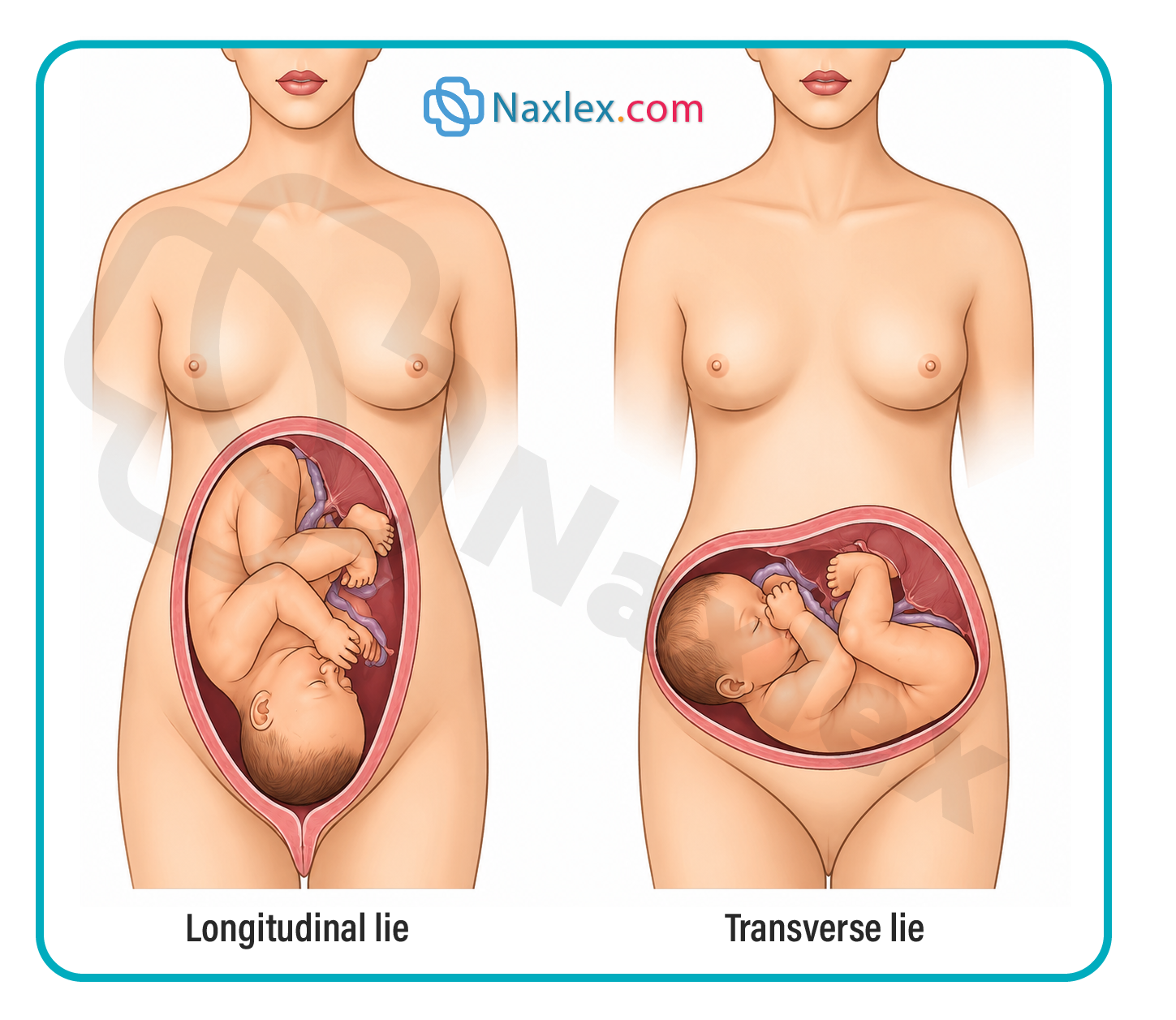
- Transverse Lie:
- The long axis of the fetus is completely perpendicular (at a 90° angle) to the long axis of the maternal spine.
- The fetal long axis crosses the maternal spine horizontally, meaning a shoulder or arm typically acts as the presenting part at the pelvic inlet.
- Vaginal delivery is mechanically impossible with a persistent transverse lie because the fetus cannot descend through the pelvic canal in this orientation; failure to intervene carries an absolute risk of uterine rupture and severe fetal compromise.
- Oblique Lie:
- The long axis of the fetus is positioned at an angle between a longitudinal lie and a transverse lie, creating a diagonal alignment across the maternal abdomen.
- An oblique lie is inherently unstable and typically converts spontaneously into either a longitudinal or a transverse lie during the active phase of labor as uterine contractions force the fetus downward.
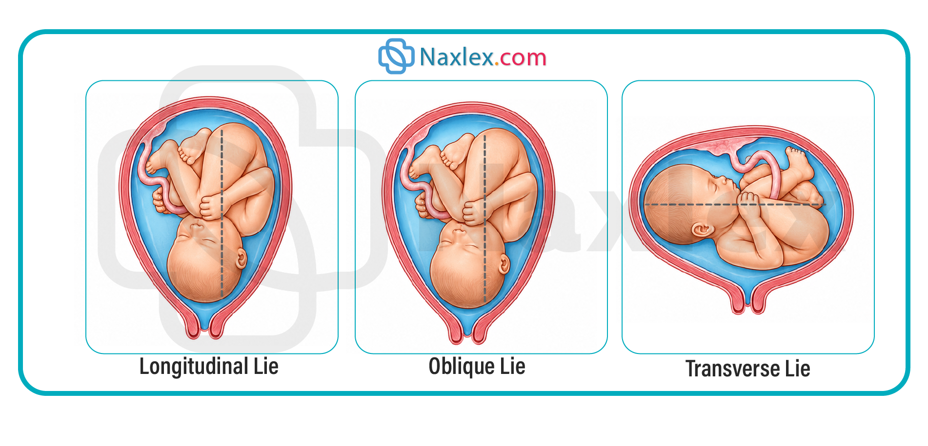
Nursing Insights
- The nurse must recognize that a persistent transverse lie at term represents a structural barrier to delivery that always necessitates a scheduled or emergent Cesarean birth to prevent catastrophic maternal-fetal injury.
- When a client presents in labor with a transverse lie, the nurse's priority actions are to avoid any artificial rupture of membranes (AROM) due to an extreme risk of umbilical cord prolapse, maintain the client on strict bed rest, and immediately notify the primary health care provider to prepare for surgical intervention.
3.2 Fetal Presentation: Cephalic, Breech, and Shoulder
- Definition: Fetal presentation identifies the specific anatomical part of the fetus that enters the maternal pelvic inlet first and leads through the birth canal during a vaginal delivery.
- Cephalic Presentation:
- The fetal head enters the pelvic inlet first, representing approximately 96% of all term deliveries.
- Classification depends on the degree of flexion or extension of the fetal head, with vertex (full flexion) being the most common and optimal variation.
- Breech Presentation:
- The fetal buttocks, feet, or both enter the pelvic inlet first, occurring in approximately 3% to 4% of term singleton pregnancies.
- Frank Breech: The fetal hips are fully flexed, and both knees are completely extended, positioning the feet flat against the face and leaving the buttocks as the sole presenting part.
- Complete Breech: The fetal hips and knees are both fully flexed, causing the thighs to sit tightly against the abdomen and the legs crossed, presenting the buttocks and feet together.
- Footling (Incomplete) Breech: One or both fetal feet extend downward below the buttocks, presenting into the cervix or vagina first and creating a direct path for cord descent.
- Shoulder Presentation:
- The acromion process or scapula presents at the pelvic inlet, directly associated with a transverse fetal lie.
- Clinical signs include an asymmetrical maternal abdominal shape, a low fundal height measurement relative to gestational age, and the inability to palpate a fetal pole at the pelvic inlet during a vaginal exam.
Nursing Insights
- Breech presentations carry a significantly elevated risk for umbilical cord prolapse because the irregular shape of the lower fetal body does not seal the cervix tightly, allowing room for the cord to slip downward ahead of the fetus.
- The nurse must understand that a footling breech presentation carries the absolute highest risk for umbilical cord prolapse due to the lack of space filling the lower uterine segment, requiring immediate notification of the primary health care provider if membranes rupture.
- In breech deliveries, there is a severe risk of head entrapment, where the cervix dilates enough to pass the soft breech and trunk but clamps down around the larger, unyielding fetal head, resulting in acute neonatal hypoxia.
3.3 Fetal Attitude and Head Diameters
- Definition: Fetal attitude describes the spatial relationship of the fetal body parts to one another, reflecting the overall degree of flexion or extension maintained by the fetus.
- Normal Attitude (Vertex):
- Characterized by full flexion of the fetal head, where the chin is tucked tightly against the chest, the arms are folded over the thorax, and the knees are drawn up toward the abdomen.
- This optimal posture presents the suboccipitobregmatic diameter, which measures approximately 9.5 cm.
- The suboccipitobregmatic diameter represents the smallest possible anteroposterior diameter of the fetal skull, allowing it to navigate the narrow dimensions of the maternal pelvis smoothly.
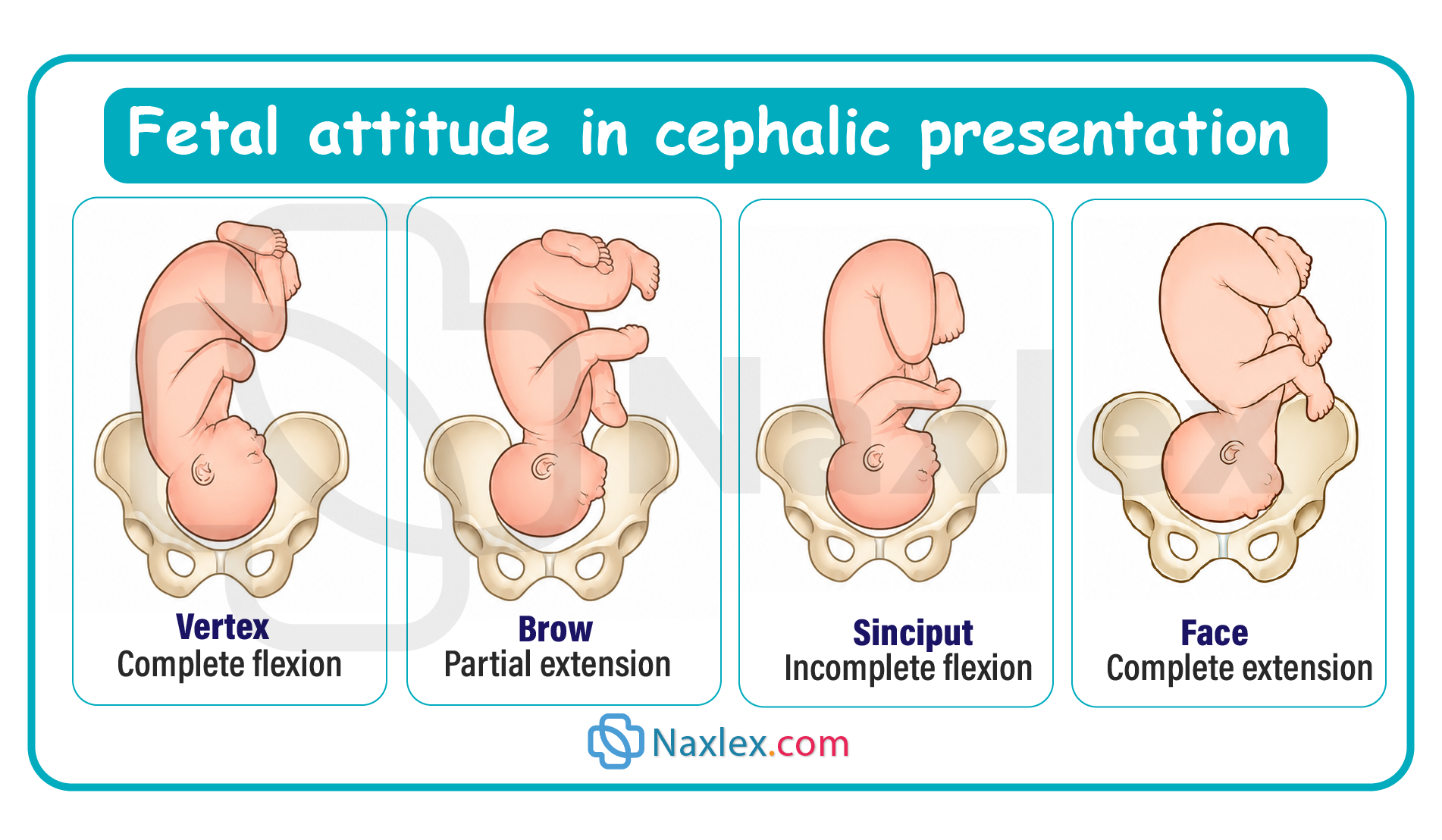
- Abnormal Attitudes:
- Military Presentation (Sinciput): The fetal head is completely neutral, neither flexed nor extended, presenting the occipitofrontal diameter (approximately 11.75 cm) and often resulting in prolonged labor.
- Brow Presentation: The fetal head is partially extended, presenting the occipitomental diameter (approximately 13.5 cm), which is the largest anteroposterior diameter of the fetal head and usually requires surgical intervention due to cephalopelvic disproportion (CPD).
- Face Presentation: The fetal head is fully hyperextended, causing the occiput to touch the fetal back and presenting the submentobregmatic diameter (approximately 9.5 cm), which can allow for a vaginal delivery if the mentum (chin) rotates anteriorly.
Nursing Insights
- The nurse must know that a vertex presentation with full flexion is critical because it utilizes the minimal 9.5 cm suboccipitobregmatic diameter, whereas deflexion or partial extension increases the presenting skull diameter, creating a high risk for cephalopelvic disproportion (CPD) and labor arrest.
- During a face presentation, if the mentum (chin) rotates posteriorly into the sacral hollow (mentum posterior), the fetal neck cannot hyperextend any further to clear the pubic symphysis, making a successful vaginal birth anatomically impossible and necessitating an immediate Cesarean section.
3.4 Fetal Position and Pelvic Quadrants
- Definition: Fetal position describes the relationship of a chosen designated landmark on the fetal presenting part to the specific front, back, or side quadrants of the maternal pelvis.
- Designated Landmarks:
- O (Occiput): Used for vertex cephalic presentations.
- M (Mentum): Used for face cephalic presentations.
- S (Sacrum): Used for breech presentations.
- A (Acromion process): Used for shoulder presentations.
- Three-Letter Abbreviation System:
- First Letter: Indicates the side of the maternal pelvis where the fetal landmark resides: Right (R) or Left (L).
- Second Letter: Indicates the specific landmark on the presenting part: Occiput (O), Mentum (M), Sacrum (S), or Acromion (A).
- Third Letter: Indicates the location of the landmark relative to the maternal pelvis: Anterior (A), Posterior (P), or Transverse (T).
- Clinical Comparisons:
- Left Occiput Anterior (LOA): The fetal occiput points toward the front-left quadrant of the mother's pelvis; this is the most common, favorable, and ideal orientation for a rapid vaginal delivery.
- Occiput Posterior (OP): The fetal occiput faces the maternal sacrum (back of the pelvis), frequently referred to as a "sunny-side-up" position.
- Occiput Transverse (ROT/LOT): The fetal occiput is directed toward the right or left lateral wall of the maternal pelvis, acting as an intermediate stage during internal rotation.
Nursing Insights
- An occiput posterior (OP) position places intense mechanical pressure on the maternal sacral nerves, causing severe, continuous lower back pain during and between uterine contractions, a clinical phenomenon widely known as "back labor."
- The nurse can actively promote the rotation of a fetus from an occiput posterior (OP) position to an optimal occiput anterior (OA) position by avoiding a supine maternal posture and instead implementing frequent position changes, such as hands-and-knees positioning, pelvic rocking, or a side-lying lateral Sims position on the side opposite the fetal occiput.
Mechanisms Of Labor (Cardinal Movements)
Mechanisms of labor (cardinal movements) are the sequential positional and attitudinal changes that the fetus undergoes as it passes through the maternal pelvis during vaginal delivery.
These movements allow the fetus to adapt to the shape of the birth canal and facilitate a safe birth.
They include:
- Engagement – fetal head enters the pelvic inlet
- Descent – downward movement through the pelvis
- Flexion – chin moves toward chest to present a smaller diameter
- Internal rotation – head rotates to align with the maternal pelvis
- Extension – head extends as it passes under the pubic symphysis
- External rotation (restitution) – head realigns with shoulders after delivery
- Expulsion – delivery of the shoulders and the rest of the body
5.1 Engagement and Descent
- Engagement Definition: Engagement occurs when the widest transverse diameter of the fetal presenting part—specifically the biparietal diameter in a cephalic presentation—has completely passed through the maternal pelvic inlet.
- In clinical practice, engagement is established when the lowest portion of the fetal presenting part reaches the level of the maternal ischial spines, which represents a fetal station of 0.
- In primigravid clients, engagement typically occurs several weeks before the onset of active labor due to structural adaptations and abdominal muscle tone, whereas in multigravid clients, it frequently occurs during the active phase or after the onset of labor.
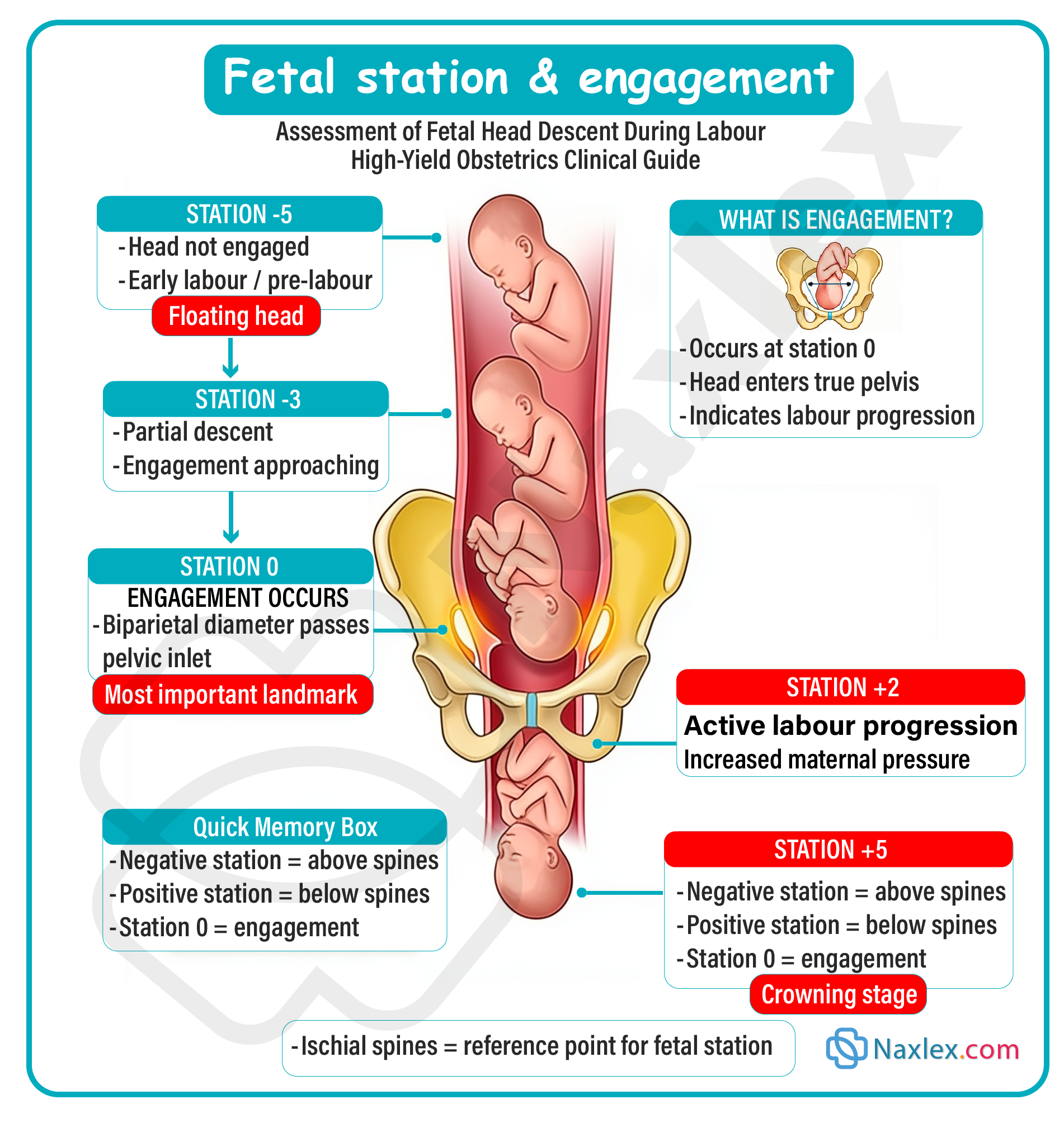
- Descent Definition: Descent is the continuous downward progress of the fetal presenting part through the maternal pelvic architecture during labor, acting as the baseline vehicle for all other cardinal movements.
- Forces Driving Descent: Fetal descent is directly propelled by a combination of multiple dynamic physical forces:
- The mechanical downward pressure exerted by the amniotic fluid during uterine contractions.
- The direct fundal pressure of uterine contractions pushing down against the fetal rump or breech pole.
- The active, voluntary bearing-down efforts of the maternal abdominal muscles during the second stage of labor.
- The structural configuration and dimensions of the maternal pelvic pathway, alongside the physiological relaxation of maternal soft tissues.
- Clinical Assessment of Station: Fetal station measures the degree of descent by calculating the distance in centimeters between the leading edge of the presenting part and an imaginary line drawn between the rigid maternal ischial spines.
- Negative Station (-5 to -1 cm): The fetal presenting part remains floating above the level of the ischial spines, indicating that the head has not yet achieved engagement within the pelvic inlet.
- Zero Station (0 cm): The leading edge of the presenting part is perfectly aligned with the maternal ischial spines, indicating that complete engagement has been reached.
- Positive Station (+1 to +5 cm): The presenting part has descended below the level of the ischial spines toward the pelvic floor, with +4 cm to +5 cm indicating that the fetus is actively crowning at the vaginal introitus.
Nursing Insights
- The nurse must understand that assessing engagement via a 0 or positive station is a vital milestone prior to any artificial rupture of membranes, because a negative station means the head is unengaged, leaving a large physical gap through which the umbilical cord can wash down, resulting in an immediate, life-threatening cord prolapse.
- If a primigravid client experiences a prolonged early stage of labor with a fetus remaining at a high, non-engaged negative station despite strong, frequent uterine contractions, the nurse must suspect cephalopelvic disproportion and closely monitor for signs of secondary labor arrest.
5.2 Flexion and Internal Rotation
- Flexion Definition: Flexion occurs as the descending fetal head encounters structural resistance from the rigid maternal bony pelvis, the muscular pelvic floor, or the fibromuscular cervix, forcing the fetal neck to flex until the chin is tucked tightly against the sternum.
- Mechanical Significance of Flexion: This passive adjustment shifts the presenting aspect of the fetal skull from a wider anteroposterior diameter to the minimal suboccipitobregmatic diameter, which measures approximately 9.5 cm.
- By minimizing the physical profile of the head, flexion ensures that the smallest possible cranial surface area tries to traverse the strict boundaries of the mid-pelvis.
- Internal Rotation Definition: Internal rotation consists of the progressive turning of the fetal head from the transverse or oblique position in which it entered the pelvic inlet to an anteroposterior position, aligning with the maternal pelvis.
- Anatomical Pathway: The pelvic inlet is widest along its transverse axis, but the pelvic outlet is widest along its anteroposterior axis.
- To adapt to this structural twist, the fetal head must rotate—most commonly 45° to the right or left—until the occiput is positioned directly beneath the maternal pubic symphysis (Occiput Anterior).
- This critical alignment occurs during active descent through the mid-pelvis as the involuntary contractions push the fetal skull down against the sloping, V-shaped levator ani muscles of the pelvic floor.
Nursing Insights
- The nurse must recognize that persistent failure of internal rotation often leads to a persistent occiput posterior position, where the fetal skull becomes jammed against the maternal sacrum, arresting labor progress and requiring the nurse to use upright, asymmetrical positions like lateral lunge positioning to prompt rotation.
- During a sterile vaginal examination, the nurse must use the open sutures and fontanelles of the fetal skull to track the progress of flexion and internal rotation; finding a large diamond-shaped anterior fontanelle toward the front of the pelvis tells the nurse the head is deflexed, warning of a prolonged labor path.
5.3 Extension and Restitution (External Rotation)
- Extension Definition: Extension is the cardinal movement where the fully descended, flexed fetal head pivots under the rigid maternal pubic symphysis and emerges through the vaginal opening.
- Mechanical Mechanism: Once the fetal occiput reaches the lower border of the pubic symphysis, it acts as a mechanical fulcrum or pivot point.
- The resistance of the firm perineal body pushes the head upward and forward, forcing the neck to extend as the face, forehead, nose, mouth, and chin successively sweep over the stretched perineum.
- Restitution Definition: Restitution describes an immediate, spontaneous 45° turn of the emerged fetal head to the right or left, returning it to a natural right angle with the fetal shoulders that are still contained within the birth canal.
- This movement untwists the fetal neck, which had been forced into an artificial alignment during the internal rotation phase.
- External Rotation Definition: External rotation occurs as the fetal shoulders inside the pelvis undergo their own internal rotation to align their wide biacromial diameter with the wide anteroposterior diameter of the maternal pelvic outlet.
- As the internal shoulders turn, this mechanical force causes the external fetal head to rotate another 45° externally, tracking the orientation of the torso.
Nursing Insights
- The nurse must remain highly alert immediately following the extension phase for the "turtle sign"—where the delivered fetal head retracts tightly back against the maternal perineum—which indicates that the anterior shoulder has become impacted behind the pubic symphysis, signaling an acute shoulder dystocia emergency.
- During the extension phase, the nurse must provide controlled perineal support using modified Ritgen’s maneuver to prevent explosive extension of the head, thereby minimizing severe maternal third- and fourth-degree perineal lacerations.
5.4 Expulsion
- Expulsion Definition: Expulsion is the final cardinal movement of labor, characterized by the exit of the entire remaining fetal trunk and extremities from the maternal birth canal.
- Sequence of Shoulder Delivery: Following external rotation, the anterior fetal shoulder slips beneath the maternal pubic symphysis first and is delivered downward to protect the brachial plexus nerves.
- The posterior shoulder is then delivered upward over the curved maternal perineum, clearing the structural boundary of the lower pelvic outlet.
- Final Delivery: Once both shoulders are completely free of the pelvic bones, the rest of the flexible fetal body, abdomen, hips, and legs are expelled rapidly and smoothly with minimal resistance, concluding the second stage of labor.
Nursing Insights
- During the expulsion phase, the nurse's critical duty is to avoid applying undue or forceful downward traction on the newly delivered fetal head, as excessive lateral force can stretch and permanently damage the delicate roots of the brachial plexus, leading to Erb-Duchenne paralysis.
- Immediately following the complete expulsion of the newborn, the nurse must prioritize clearing the airway if necessary and initiating immediate skin-to-skin contact with the maternal client to promote endogenous oxytocin release, which aids in uterine contraction and mitigates post-delivery hemorrhage risks.
Leopold’s Maneuvers And Clinical Assessment
Leopold’s maneuvers are a systematic series of four abdominal palpation techniques performed by a healthcare provider to assess the fetus during pregnancy.
They are used to determine:
- Fetal position (where the back and limbs are)
- Fetal presentation (cephalic, breech, etc.)
- Fetal lie (longitudinal, transverse, oblique)
- Fetal descent into the pelvis
The maneuvers involve palpating different areas of the maternal abdomen in a specific sequence to identify the location of the fetal head, back, and presenting part.
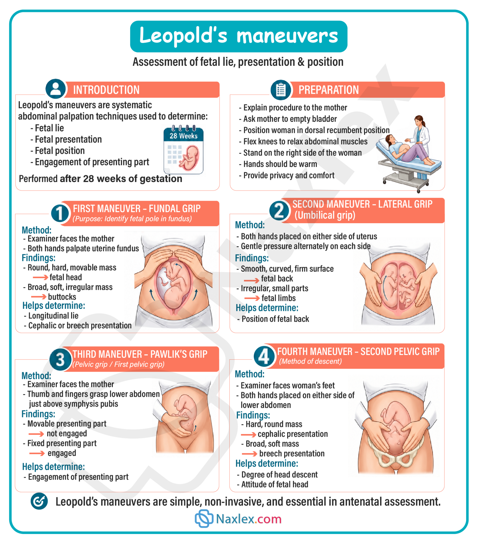
7.1 First Maneuver: Fetal Presentation and Fundal Palpation
- Definition and Clinical Objective: The first maneuver is executed to identify what occupying fetal pole resides within the fundal region of the uterus, which directly establishes the primary fetal presentation.
- Anatomical Technique: The nurse faces the head of the laboring client and places both palms flat across the superior curvature of the uterine fundus, applying a firm but gentle downward digital pressure to evaluate the shape, consistency, and mobility of the mass.
- Clinical Findings Interpretation:
- Breech Pole Identification: If the fetal buttocks occupy the fundus, the nurse will palpate a large, soft, irregular, nodular, and non-breech-ballottable mass that moves synchronously with the fetal trunk.
- Cephalic Pole Identification: If the fetal head occupies the fundus, the nurse will palpate a smooth, round, hard, unyielding, and highly ballottable mass that moves independently of the fetal torso due to the flexibility of the neck.
Nursing Insights
- The nurse must ensure the client empties their bladder completely before performing Leopold's maneuvers, as an engorged urinary bladder alters uterine height, causes significant maternal discomfort, and obscures the palpation of anatomical fetal structures.
- If the nurse palpates a hard, round, ballotable head in the fundus, this diagnostic finding alerts the clinical team that the fetus is in a breech presentation, prompting immediate preparation for ultrasound confirmation and a potential elective Cesarean delivery to minimize the risk of cord prolapse or head entrapment.
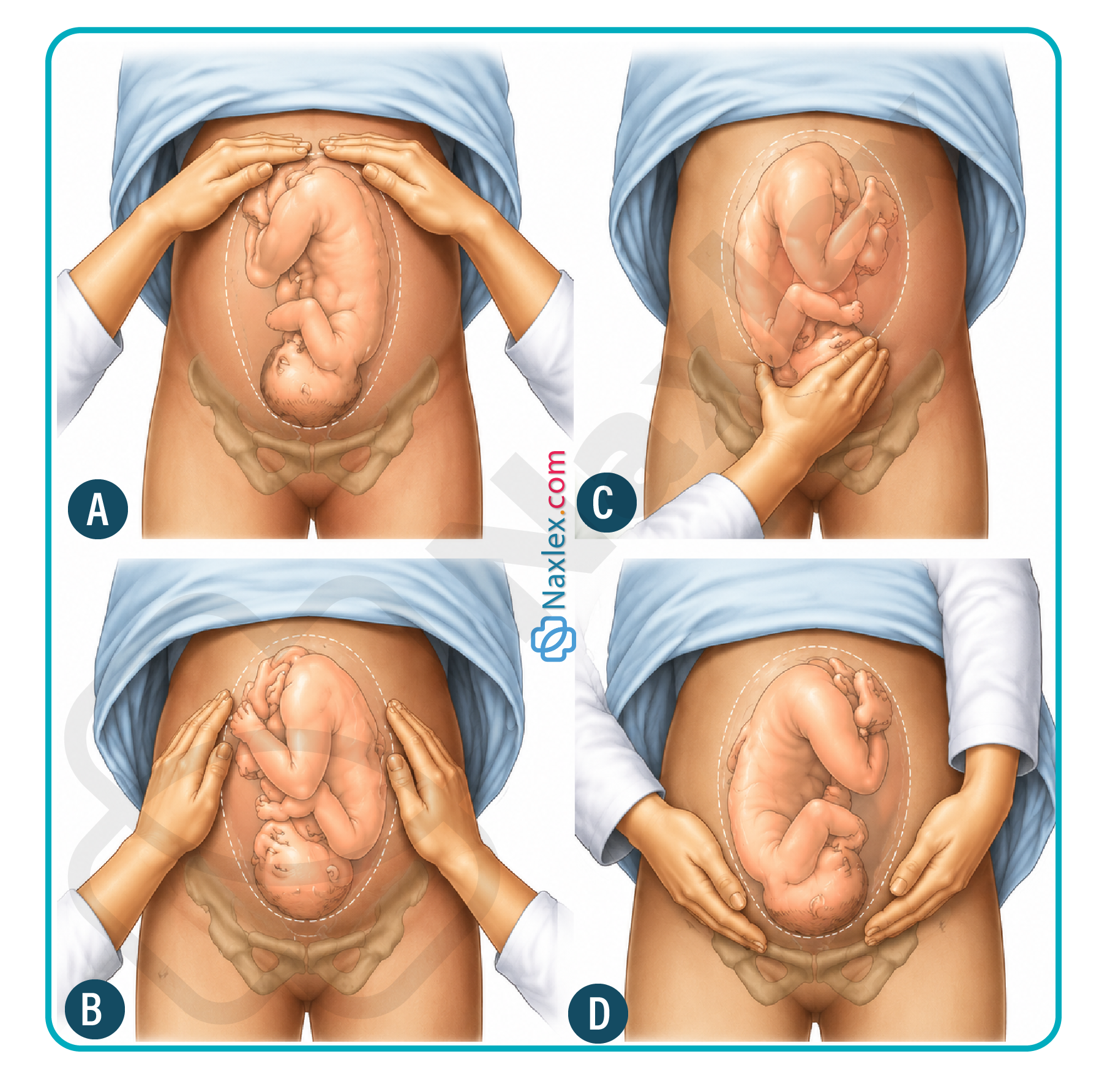
7.2 Second Maneuver: Fetal Lie and Back Localization
- Definition and Clinical Objective: The second maneuver is designed to identify the orientation of the fetal long axis relative to the maternal spine and localize the smooth surface of the fetal back.
- Anatomical Technique: Maintaining the forward-facing position toward the client's head, the nurse moves both hands downward along the lateral walls of the uterus, applying steady pressure with one palm to stabilize the uterus while the opposite hand systematically moves from the top to the bottom of the flank.
- Clinical Findings Interpretation:
- Fetal Back Localization: The fetal back presents as a continuous, firm, smooth, convex, and rigid resistance along one side of the maternal abdomen.
- Fetal Small Parts Localization: The fetal extremities (arms, knees, and feet) present as numerous small, irregular, mobile, bumpy, and sometimes active nodules along the opposite uterine side.
Nursing Insights
- The nurse must accurately localize the smooth, rigid surface of the fetal back during the second maneuver because this anatomical site provides the clearest transmission of fetal heart sounds; the nurse must place the transducer of the external electronic fetal monitor or Doppler device directly over this specific quadrant to ensure optimal tracing.
- If the nurse notes a smooth, firm resistance running horizontally across the lower abdomen rather than vertically along the maternal flanks, this provides key physical evidence of a transverse lie, warning the nurse to avoid vaginal interventions that could trigger a cord prolapse.
7.3 Third Maneuver: Fetal Engagement and Pelvic Inlet Palpation
- Definition and Clinical Objective: The third maneuver, also termed Pawlik's maneuver, aims to identify what fetal pole occupies the lower uterine segment, confirm the presentation at the pelvic inlet, and determine if engagement has occurred.
- Anatomical Technique: The nurse maintains a position facing the client's head and uses a single dominant hand to grasp the lower uterine segment immediately superior to the pubic symphysis, positioning the thumb and fingers wide apart to encircle the presenting part.
- Clinical Findings Interpretation:
- Non-Engaged Fetal Pole: If the presenting part is freely movable, easily displaced upward away from the pelvic brim, and ballottable, engagement has not occurred (negative station).
- Engaged Fetal Pole: If the presenting part feels rigidly fixed, deeply wedged within the pelvic architecture, and cannot be displaced upward by manual pressure, engagement is confirmed (0 or positive station).
Nursing Insights
- The nurse must realize that if the third maneuver demonstrates a freely movable, non-engaged cephalic mass at term, the client remains at high risk for an occult or overt umbilical cord prolapse if spontaneous or artificial rupture of membranes occurs.
- When a client presents with regular contractions and the third maneuver demonstrates a rigid, immovable mass at the pelvic inlet, the nurse can confidently anticipate that the fetus has successfully entered the true pelvis, paving the way for active descent.
7.4 Fourth Maneuver: Fetal Attitude and Cephalic Prominence
- Definition and Clinical Objective: The fourth maneuver is conducted to identify the degree of fetal head flexion or extension (fetal attitude) by locating the cephalic prominence relative to the pelvic inlet.
- Anatomical Technique: The nurse changes position, turning to face away from the client's head and toward the client's feet, placing both palms flat along the lower quadrants of the uterus and sliding the fingers downward along the sides of the pelvis toward the pubic arch.
- Clinical Findings Interpretation:
- Flexed Attitude (Vertex Presentation): The cephalic prominence (the brow/forehead) is located on the same side as the small parts, and the fingers meet resistance early on this side, while the hand on the side of the fetal back slides deeply into the pelvis without obstruction.
- Extended Attitude (Face Presentation): The cephalic prominence (the occiput) is located on the same side as the smooth fetal back, where the neck is hyperextended, creating a rigid anatomical barrier that arrests the downward movement of the fingers on that side.
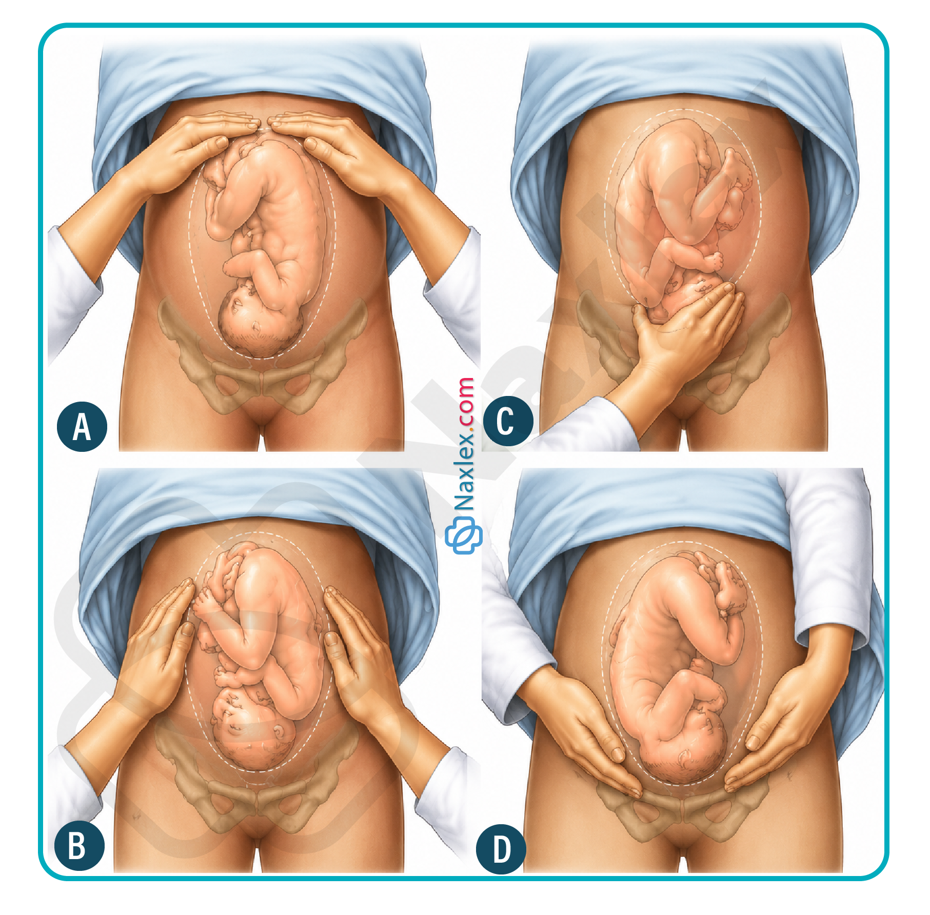
Nursing Insights
- The nurse must understand that if the fingers hit the cephalic prominence on the same side as the fetal back during the fourth maneuver, the fetal head is hyperextended into an abnormal attitude, alerting the care team to expect face presentation complications and potential labor dystocia.
- Mastery of the fourth maneuver enables the nurse to anticipate the ease of the descent phase, as finding the cephalic prominence on the same side as the small parts reassures the nurse that the head is optimally flexed to present its smallest diameter.
Summary
- Fetal Alignment Dynamics: Fetal lie compares the maternal and fetal long axes, where a parallel longitudinal lie (>99%) is mandatory for normal intrapartum progression, whereas a transverse lie (<1%) creates a total mechanical barrier requiring immediate surgical intervention via a Cesarean delivery.
- Anatomical Presentations and Attitudes: Fetal presentation dictates the leading anatomical structure entering the pelvic inlet, which is optimized during a cephalic vertex presentation where an attitude of full flexion presents the minimal 9.5 cm suboccipitobregmatic diameter.
- Positional Geometry: Fetal position utilizes a three-letter abbreviation system to map the fetal landmark to the four quadrants of the maternal pelvis, with Left Occiput Anterior (LOA) being the most common and ideal variant, while persistent Occiput Posterior (OP) positioning causes severe "back labor" and labor arrest.
- Sequential Cardinal Movements: To exit the pelvis, the fetus must perform a precise series of passive mechanical adaptations: engagement, descent, flexion, internal rotation, extension, restitution, external rotation, and final expulsion.
- Clinical Evaluation Milestones: Fetal station measures structural descent in centimeters relative to the zero baseline of the rigid maternal ischial spines, functioning as an essential clinical indicator to gauge pelvic navigation and evaluate the safety of amniotomy.
- Diagnostic Palpation Framework: Leopold's maneuvers provide a systematic four-step abdominal palpation protocol allowing the nurse to determine fetal presentation (first maneuver), identify fetal lie and back orientation for optimal electronic fetal monitoring transducer placement (second maneuver), confirm pelvic inlet engagement (third maneuver), and evaluate cephalic prominence to determine head attitude (fourth maneuver).
Naxlex
Videos
Login to View Video
Click here to loginTake Notes on Mechanisms of Labor, Leopold’s Maneuvers
This filled cannot be empty
Join Naxlex Nursing for nursing questions & guides! Sign Up Now