





 Video Library


Four Stages of Labor Part 2
Study Questions
Practice Exercise 1
A nurse is caring for a client who has just reached complete cervical dilation of 10 cm with 100% effacement. Which of the following statements best describes the beginning of this stage of labor?
Explanation
The second stage of labor is characterized by cervical dilation to 10 cm and full effacement, culminating in fetal descent. Uterine contractions drive expulsion through the birth canal alongside active voluntary maternal bearing-down efforts. Maternal positioning and continuous fetal monitoring remain mandatory parameters to prevent fetal hypoxia during passage.
Rationale for correct answer:
3. Complete cervical dilation marks the exact anatomical onset of the second stage of labor. This phase continues through active fetal expulsion until delivery is achieved. The client transitions from passive dilation to active pushing during this timeframe. Maternal effort directly assists uterine contractions in moving the neonate down the pelvic pathway.
Rationale for incorrect answers:
1. An urge to bear down often accompanies lower fetal station but does not define stage boundaries. The Ferguson reflex triggers this involuntary sensation when the presenting part stretches the pelvic floor. Clients may feel this urge prior to achieving full 10 cm dilation. Pushing before full dilation can cause cervical edema or laceration.
2. A station of +2 indicates significant descent into the ischial spine pathway. While fetal descent progresses rapidly in the second stage, station level alone does not dictate onset. The official threshold relies entirely on cervical measurement criteria rather than station. Beginning pushing solely based on station risks pushing against an incompletely dilated cervix.
4. Rupture of membranes can occur during any stage of labor or prior to onset. Delivery of the placenta defines the third stage of labor rather than the second stage. The second stage terminates strictly with fetal delivery, not placental separation. Confusing these clinical endpoints leads to improper tracking of labor progression.
Test-taking strategy:
- Analyze the scenario/question: The client is at 10 cm dilation and 100% effacement, which defines the start of the second stage of labor. The question asks for the statement that best describes the beginning and boundaries of this specific stage.
- Apply Knowledge of Intrapartum Stages: Obstetric nursing requires precise knowledge of the anatomical definitions for each stage of labor. Stage 1 spans from true labor onset to full dilation, whereas Stage 2 spans from full dilation to fetal delivery. Stage 3 spans from fetal delivery to placental delivery, and Stage 4 covers immediate postpartum recovery.
- Rule out Choice 1: The Ferguson reflex urge to push is a subjective symptom, not the clinical benchmark defining stage onset.
- Rule out Choice 2: Fetal station measures progress within the pelvis, but +2 station does not define the official start of the second stage.
- Rule in Choice 3: Complete dilation (10 cm) to complete newborn expulsion accurately defines the precise boundaries of the second stage of labor.
- Rule out Choice 4: Rupture of membranes is an event that can happen at any point, and placental delivery marks the end of the third stage.
- Select Choice 3 as it correctly provides the exact physiological definition and clinical boundaries for the second stage of labor.
Take home points
- The second stage of labor begins at 10 cm cervical dilation and ends with the birth of the neonate.
- Involuntary urge to push (Ferguson reflex) occurs as the fetal presenting part presses on the pelvic floor muscles.
- Pushing prior to complete 10 cm dilation is contraindicated due to risks of cervical swelling and tissue trauma.
- The third stage of labor begins immediately after the expulsion of the infant and ends with the delivery of the placenta.
A nurse is teaching a nursing student about the cardinal movements of labor. Which of the following sequences correctly reflects the order in which these movements typically occur?
Explanation
The cardinal movements of labor represent the passive positional changes the fetus undergoes while navigating the maternal pelvic canal during birth. Uterine contractions and involuntary maternal forces drive fetal descent through distinct anatomical landmarks. Proper mechanism execution prevents dystocia and minimizes tissue trauma to both the mother and the neonate during the expulsion phase.
Rationale for correct answer:
2. The correct order begins when the biparietal diameter enters the pelvic inlet during engagement, followed by continuous downward movement known as descent. The fetal chin then tucks to present the smallest diameter via flexion, followed by internal rotation, extension, external rotation, and finally full expulsion.
Rationale for incorrect answers:
1. Descent begins before engagement in this listed sequence, which is incorrect because the fetal head must first settle into the pelvic inlet. Engagement must precede significant descent through the midpelvis. Furthermore, flexion typically occurs as the head meets resistance during descent rather than before engagement occurs. Misordering these initial steps disrupts the clinical assessment of fetal station.
3. Placing flexion before descent incorrectly alters the physiological timeline of fetal movement. While engagement occurs first, flexion happens as the descending head encounters resistance from the cervix and pelvic floor. Additionally, extension must occur before external rotation as the head emerges under the pubic arch. Reversing these steps obscures the mechanism of pelvic passage.
4. Flexion cannot occur prior to engagement and descent because pelvic floor resistance is required to force the fetal chin onto the chest. Extension must precede external rotation so the head can negotiate the vulvar outlet before the shoulders rotate. Placing external rotation before extension presents an anatomically impossible sequence for fetal delivery.
Test-taking strategy:
- Analyze the scenario/question: The question asks for the correct sequential order of the cardinal movements of labor as a fetus moves through the birth canal.
- Apply Knowledge of Cardinal Movements of Labor: Successive positional adaptations are required for the fetal head and shoulders to accommodate the varying diameters of the maternal pelvis. Engagement occurs when the biparietal diameter passes the pelvic inlet, followed by descent driven by uterine contractions and amniotic fluid pressure. Flexion reduces the presenting head diameter upon encountering resistance, internal rotation aligns the head with the pelvic outlet, extension allows the head to emerge under the symphysis pubis, external rotation aligns the shoulders, and expulsion completes delivery.
- Rule out Choice 1: Reverses engagement and descent, placing descent prior to the head settling into the pelvic inlet.
- Rule in Choice 2: Accurately lists the seven cardinal movements in their precise physiological sequence from engagement to expulsion.
- Rule out Choice 3: Incorrectly places extension before internal rotation, which contradicts the natural mechanics of pelvic descent.
- Rule out Choice 4: Begins with flexion prior to engagement and places external rotation before extension, creating an incorrect mechanical sequence.
- Select Choice 2 as it accurately represents the standard chronological order of intrapartum fetal progression.
Take home points
- The cardinal movements occur in the order of engagement, descent, flexion, internal rotation, extension, external rotation, and expulsion.
- Engagement occurs when the widest part of the fetal presenting part reaches the level of the maternal ischial spines (station 0).
- Flexion allows the smallest fetal head diameter (suboccipitobregmatic) to present to the maternal pelvis.
- Extension allows the fetal head to pass under the symphysis pubis and emerge through the vaginal opening.
A nurse is monitoring a multiparous client during the second stage of labor without epidural analgesia. Which of the following findings would prompt the nurse to suspect a protraction disorder of descent?
Explanation
A protraction disorder of descent during the second stage of labor is characterized by a pathologically slow progression of fetal descent through the birth canal despite adequate uterine contractions. Normal progress thresholds differ based on parity, requiring multiparous clients to demonstrate faster descent rates than nulliparous clients. Inadequate progress increases the risk of maternal fatigue, uterine rupture, and intrapartum fetal hypoxia.
Rationale for correct answer:
2. Multiparous clients without epidural analgesia are expected to achieve a fetal descent rate of at least 2 cm per hour during active pushing. A rate of 1 cm per hour represents delayed progression, meeting the clinical diagnostic threshold for a protraction disorder of descent. This delay warrants prompt assessment for potential cephalopelvic disproportion or ineffective bearing-down efforts.
Rationale for incorrect answers:
1. A descent rate of 3 cm per hour exceeds the minimum expected standard of 2 cm per hour for a multiparous client. This finding reflects normal, adequate progress through the maternal pelvis. It indicates effective uterine contractions and maternal pushing efforts, requiring no intervention.
3. Advancing from station 0 to +2 within 30 minutes represents a rapid descent rate of 2 cm per half-hour (extrapolating to 4 cm per hour). This rapid progression demonstrates optimal descent without any evidence of labor delay. The fetal head is successfully navigating the pelvic outlet.
4. Moving from station +1 to +3 within 45 minutes equals a progress rate of approximately 2.67 cm per hour, well above the 2 cm per hour threshold. This finding indicates normal advancement during the second stage. It rules out any underlying mechanical dystocia or protraction issues.
Test-taking strategy:
- Analyze the scenario/question: The client is a multiparous woman in the second stage of labor without epidural analgesia. The question asks which finding indicates a protraction disorder of descent, which means identifying a rate of descent slower than the normal physiological threshold for this specific parity.
- Apply Knowledge of Labor Dystocia and Descent Rates: Diagnostic criteria for second-stage labor disorders differentiate between nulliparous and multiparous clients based on rate of station change per hour. A protraction disorder of descent in a multiparous client is defined as a descent rate of less than 2 cm per hour during active pushing (compared to less than 1 cm per hour in nulliparous clients). Recognizing these exact quantitative parameters allows the nurse to identify labor arrest or delay early and prevent maternal and fetal complications.
- Rule out Choice 1: A 3 cm change per hour exceeds the 2 cm per hour baseline, indicating normal second-stage progress.
- Rule in Choice 2: A 1 cm change per hour is below the expected 2 cm per hour rate for a multiparous client, confirming a protraction disorder.
- Rule out Choice 3: Progressing 2 cm in 30 minutes reflects rapid, normal descent through the birth canal.
- Rule out Choice 4: Progressing 2 cm in 45 minutes yields a rate of 2.67 cm per hour, which demonstrates adequate progress.
- Select Choice 2 as it reflects a rate of fetal descent below the expected physiological parameter for a multiparous client, indicating protraction.
Take home points
- A protraction disorder of descent in a multiparous client without an epidural is defined as a descent rate of less than 2 cm per hour during the second stage of labor.
- Multiparous clients generally progress through both the first and second stages of labor at faster physiological rates than nulliparous clients.
- Common causes of protracted descent include fetal malposition, cephalopelvic disproportion, maternal exhaustion, and epidural-induced motor blockade.
- Management of second-stage protraction includes evaluating uterine contraction strength, altering maternal positioning, and assessing the need for operative vaginal delivery or amniotomy.
A client in the second stage of labor is pushing with each contraction using sustained breath-holding for approximately 10 seconds at a time. Which of the following physiologic effects should the nurse anticipate as a result of this pushing technique?
Explanation
Sustained breath-holding during pushing represents closed-glottis pushing, which induces the Valsalva maneuver and increases thoracic pressure. This strain reduces maternal venous return, temporarily compromising cardiac output and uteroplacental perfusion. Consequently, both maternal systemic oxygenation and fetal gas exchange drop, predisposing the fetus to fetal distress and abnormal heart rate patterns during the expulsion stage.
Rationale for correct answer:
2. Sustained breath-holding increases intrathoracic pressure, triggering the Valsalva maneuver and decreasing venous return to the heart. This temporary drop in cardiac output decreases placental perfusion, leading directly to transient hypoxia in both mother and fetus. Open-glottis pushing is preferred to maintain adequate fetal oxygenation.
Rationale for incorrect answers:
1. Sustained closed-glottis pushing decreases venous return to the right atrium, which subsequently diminishes cardiac output. Lower cardiac output reduces blood flow to the placenta, directly impairing fetal oxygenation rather than leaving it unaffected. Maintaining continuous placental blood flow requires avoiding prolonged breath-holding.
3. Sustained breath-holding (apnea during pushing) prevents air exchange, which directly opposes the rapid ventilation seen in maternal hyperventilation. While hyperventilation can occur between contractions, breath-holding during contractions leads to transient hypoxia and potential respiratory acidosis rather than isolated alkalosis. Prolonged pushing strain impairs systemic gas exchange.
4. Bearing down against a closed glottis significantly increases intra-abdominal pressure rather than decreasing it. Although elevated abdominal pressure assists fetal descent, the associated vascular compression compromises uterine artery blood flow. The primary risk of this maneuver remains reduced uteroplacental circulation.
Test-taking strategy:
- Analyze the scenario/question: The client is using sustained breath-holding (closed-glottis pushing for ~10 seconds) during the second stage of labor. The question asks for the physiological effect the nurse should anticipate from this specific pushing method.
- Apply Knowledge of Maternal-Fetal Intrapartum Physiology: Pushing technique directly alters maternal hemodynamics and fetal oxygen delivery. Closed-glottis pushing (Valsalva maneuver) raises intrathoracic and intra-abdominal pressure, compressing the inferior vena cava and decreasing maternal venous return. This drop in preload reduces maternal cardiac output, arterial blood pressure, and uteroplacental blood flow, ultimately leading to transient maternal hypoxemia and fetal hypoxia characterized by late decelerations or bradycardia. Conversely, open-glottis pushing (exhaling while bearing down) preserves maternal cardiac output and optimizes fetal oxygen supply.
- Rule out Choice 1: Closed-glottis pushing decreases maternal cardiac output and adversely impacts fetal oxygen delivery.
- Rule in Choice 2: The Valsalva maneuver impairs uteroplacental perfusion, causing transient hypoxia in both the mother and the fetus.
- Rule out Choice 3: Sustained breath-holding involves prolonged apnea during contractions, which does not cause hyperventilation during the push.
- Rule out Choice 4: Breath-holding increases intra-abdominal pressure rather than decreasing it.
- Select Choice 2 as it accurately describes the negative hemodynamic and oxygenation consequences of sustained closed-glottis pushing.
Take home points
- Sustained breath-holding during pushing triggers the Valsalva maneuver, decreasing maternal venous return and cardiac output.
- Impaired uteroplacental perfusion during closed-glottis pushing can lead to transient fetal hypoxia and non-reassuring heart rate tracings.
- Open-glottis pushing (exhaling while bearing down for 6 to 8 seconds) is recommended to maintain optimal maternal-fetal oxygenation.
- Continuous evaluation of pushing mechanics helps mitigate maternal fatigue, pelvic floor trauma, and intrapartum fetal distress.
A nurse is caring for a client during the second stage of labor. Which of the following findings should the nurse identify as expected maternal physiologic changes during this stage? Select all that apply
Explanation
The second stage of labor involves intense physical exertion, fetal descent through the pelvic floor, and significant maternal hemodynamic shifts. Neuroendocrine surges of catecholamines and endogenous endorphins drive physiological adaptations during active pushing. Expected maternal findings include neuromuscular responses, perineal distension, and marked somatic pressure, while systemic fatigue remains cumulative despite short rest intervals between contractions.
Rationale for correct answers:
1. Heavy muscular exertion and intense nerve compression from the descending fetus cause localized muscle tremor and autonomic diaphoresis. These manifestations reflect physiological responses to peak catecholamine release during active expulsion. The nurse recognizes leg trembling as an expected response during the strenuous pushing phase.
2. As the fetal presenting part descends onto the pelvic floor, the perineal body stretches, flattens, and bulges outward. This anatomical adaptation reflects perineal thinning required to accommodate passage through the vaginal introitus. Continuous monitoring ensures recognition of normal tissue distension versus impending laceration.
4. Direct fetal head pressure against the sacral plexus and rectum creates an intense, involuntary sensation of pressure. This sensory feedback triggers the Ferguson reflex, prompting the client to bear down naturally. Increased rectal pressure serves as a primary clinical marker of advancing fetal station.
Rationale for incorrect answers:
3. Maternal heart rate increases transiently during contractions and pushing efforts due to sympathetic stimulation and elevated cardiac output. A sustained drop in maternal heart rate is abnormal and may indicate severe hemodynamic instability or excessive vagal stimulation. Heart rate elevations match the heavy metabolic demand of second-stage labor.
5. Maternal fatigue accumulates progressively during the second stage due to sustained physical work and energy expenditure. While short rest periods between contractions allow brief relaxation, complete fatigue resolution does not occur until well into the postpartum recovery period. Managing maternal energy requires structured support and encouraging open-glottis pushing.
Test-taking strategy:
- Analyze the scenario/question: The scenario describes a client in the second stage of labor. The question asks the nurse to identify expected maternal physiological changes during this stage from a "Select all that apply" list.
- Apply Knowledge of Maternal Physiology in the Second Stage of Labor: The second stage involves intense physical exertion, neuroendocrine activation, and severe mechanical compression as the fetus passes through the birth canal. Catecholamine surges and muscle fatigue cause trembling and sweating, while direct fetal pressure on the levator ani muscles and rectum causes perineal bulging and severe rectal pressure. Maternal heart rate increases to support a 40% to 50% increase in cardiac output, and muscular exhaustion builds continuously rather than resolving between contractions.
- Rule in Choice 1: Diaphoresis and leg trembling are normal neurovascular responses to intense physical work and fetal nerve compression.
- Rule in Choice 2: Progressive perineal thinning and bulging occur naturally as the fetal presenting part distends the pelvic floor.
- Rule out Choice 3: Maternal heart rate increases during second-stage labor to meet high metabolic demands; a sustained decrease is abnormal.
- Rule in Choice 4: Descending fetal head pressure against the rectum is a cardinal sign of advancing fetal station and triggers the urge to push.
- Rule out Choice 5: Exhaustion accumulates over time during labor, making complete resolution of fatigue between contractions physiologically impossible.
- Select Choices 1, 2, and 4 as they accurately represent normal maternal physiological adaptations during the second stage of labor.
Take home points
- Expected physiological responses during the second stage of labor include diaphoresis, leg trembling, perineal bulging, and severe rectal pressure.
- Fetal head descent onto the pelvic floor triggers the Ferguson reflex, creating an involuntary urge to bear down.
- Maternal heart rate and cardiac output increase significantly during active pushing to meet elevated metabolic demands.
- Fatigue accumulates progressively throughout labor, requiring nursing measures to conserve maternal energy between contractions.
A nurse notes gradual decreases in fetal heart rate that begin after the peak of the client's contractions and slowly return to baseline after the contraction ends. Which of the following actions should the nurse take first?
Explanation
Gradual decreases in fetal heart rate that begin after the peak of a contraction and return to baseline after the contraction finishes define late decelerations, indicating uteroplacental insufficiency. This non-reassuring pattern reflects impaired oxygen transfer to the fetus during peak uterine tension. Immediate intrauterine resuscitation protocols must be initiated to optimize maternal placental blood flow, elevate arterial oxygen content, and mitigate risks of progressive fetal acidemia and systemic hypoxia.
Rationale for correct answer:
3. Late decelerations signify impaired uteroplacental perfusion and demand immediate nursing intervention to restore fetal oxygenation. Repositioning the client to a lateral position relieves inferior vena cava compression, enhancing venous return and placental blood flow. Promptly notifying the provider ensures collaborative medical management for potential fetal distress.
Rationale for incorrect answers:
1. Encouraging continued pushing without intervention exacerbates fetal hypoxia by further compromising uteroplacental blood flow during contractions. Pushing increases intra-abdominal pressure, which worsens fetal acidosis when non-reassuring heart rate patterns are present. Resuscitation measures must precede any further bearing-down efforts.
2. Early decelerations—not late decelerations—are benign patterns caused by fetal head compression during labor. Late decelerations reflect compromised placental exchange rather than a normal mechanical response. Documenting late decelerations as an expected physiological pattern constitutes a failure to recognize severe intrapartum complications.
4. Instructing the client to hold her breath longer induces the Valsalva maneuver, which lowers maternal cardiac output and reduces uterine blood flow. Extended breath-holding worsens maternal hypoxemia and exacerbates fetal hypoxia. Clients experiencing late decelerations require open-glottis breathing and supplemental oxygen administration.
Test-taking strategy:
- Analyze the scenario/question: The client's fetal heart rate tracing demonstrates gradual decelerations that begin after the contraction peak and return to baseline after the contraction ends, which is the classic definition of late decelerations. The question asks for the priority ("first") nursing action.
- Apply Prioritization and Intrauterine Resuscitation Principles: Late decelerations are caused by uteroplacental insufficiency and indicate fetal hypoxia. The priority response follows the steps of intrauterine resuscitation: turn the client onto her side (to maximize uterine blood flow), administer supplemental oxygen, discontinue oxytocin if infusing, increase intravenous fluid rate, and notify the healthcare provider. The nurse must act immediately to improve oxygen delivery before taking secondary actions or continuing labor progress.
- Rule out Choice 1: Continued pushing increases intrathoracic and intra-abdominal pressure, further compromising uteroplacental perfusion during late decelerations.
- Rule out Choice 2: Misidentifies late decelerations as benign early decelerations caused by head compression, delaying essential resuscitation.
- Rule in Choice 3: Repositioning the client directly addresses uteroplacental insufficiency by removing vena cava compression, while immediate provider notification ensures rapid medical backup.
- Rule out Choice 4: Prolonged breath-holding worsens maternal-fetal gas exchange, aggravating late decelerations.
- Select Choice 3 as it represents the immediate, life-saving intrauterine resuscitation intervention required for late decelerations.
Take home points
- Late decelerations are caused by uteroplacental insufficiency and represent a non-reassuring fetal heart rate pattern requiring immediate intervention.
- Priority intrauterine resuscitation steps include lateral positioning, supplemental oxygen, IV fluid bolus, stopping oxytocin, and provider notification.
- Early decelerations mirror the contraction wave and result from head compression, requiring no clinical intervention.
- Prolonged breath-holding during pushing reduces cardiac output and worsens fetal acidemia during late deceleration events.
A nurse is assessing fetal station during the second stage of labor. The fetal presenting part is documented at a station of +3. Which of the following best describes this finding?
Explanation
Fetal station measures the degree of descent of the fetal presenting part through the maternal pelvis relative to the ischial spines. Stations designated with positive numbers indicate that the presenting part has passed below this central anatomical landmark. Progressing to lower positive stations reflects advanced fetal descent, signifying impending delivery as the head approaches the pelvic floor and vulvar introitus during the expulsion stage.
Rationale for correct answer:
3. A station of +3 indicates that the leading edge of the fetal presenting part has descended 3 cm below the maternal ischial spines. Positive numerical values quantify descent toward the vaginal outlet during the second stage of labor. Reaching +3 station signifies that fetal descent is well advanced and birth is imminent.
Rationale for incorrect answers:
1. Designated negative numbers quantify distance above the ischial spines, so 3 cm above the spines corresponds to a station of -3. Negative values indicate that the presenting part remains high in the pelvic inlet. Describing a +3 station as 3 cm above the spines reflects a fundamental misinterpretation of fetal measurement scales.
2. The level of the maternal ischial spines serves as the anatomical zero point, documented precisely as station 0. Station 0 marks the baseline milestone where fetal engagement has occurred. Identifying a +3 station as level with the spines ignores the numerical progression of pelvic station values.
4. Engagement is defined as the arrival of the biparietal diameter at the pelvic inlet, corresponding to station 0. A +3 station indicates deep descent far past the point of initial engagement into the lower pelvic cavity. Stating the head is unengaged contradicts the clinical reality of a positive station value.
Test-taking strategy:
- Analyze the scenario/question: The question asks for the correct interpretation of a documented fetal station of +3 during the second stage of labor.
- Apply Knowledge of Fetal Station Parameters: Fetal station measures the position of the fetal presenting part in centimeters relative to the maternal ischial spines. Station 0 represents the level of the ischial spines (engagement). Negative numbers (-1 to -5) indicate centimeters above the ischial spines, whereas positive numbers (+1 to +5) indicate centimeters below the ischial spines, with +4 to +5 indicating birth at the perineum.
- Rule out Choice 1: Describes a station of -3, where the presenting part remains 3 cm above the ischial spines.
- Rule out Choice 2: Describes station 0, where the presenting part is directly aligned with the ischial spines.
- Rule in Choice 3: Accurately defines a +3 station as 3 cm below the maternal ischial spines, confirming advanced descent.
- Rule out Choice 4: Engagement occurs at station 0; a +3 station means the presenting part has descended well past engagement.
- Select Choice 3 as it correctly interprets the positive numerical value for fetal station relative to the maternal ischial spines.
Take home points
- Station 0 indicates that the fetal presenting part is level with the maternal ischial spines, confirming engagement.
- Negative station numbers (-1 to -5) indicate that the presenting part is located centimeters above the ischial spines.
- Positive station numbers (+1 to +5) indicate that the presenting part has descended centimeters below the ischial spines.
- A station of +3 indicates advanced fetal descent into the pelvic outlet, signaling imminent delivery.
A nurse is caring for a client during the extension phase of the cardinal movements of labor. Which of the following client statements reflects an accurate description of the sensation associated with this movement?
Explanation
During the extension phase of labor, the fetal head passes under the symphysis pubis and emerges through the vulvar outlet, causing significant perineal distension. This stretching triggers intense localized somatic discomfort commonly referred to as the "ring of fire." Effective communication, controlled breathing, and perineal support are essential interventions to manage this maternal pain and prevent perineal lacerations during fetal expulsion.
Rationale for correct answer:
1. Extension occurs when the fetal head negotiates the pubic arch and crowning takes place at the vaginal introitus. The extreme stretching of the vulva and perineal tissues causes a sharp burning sensation and severe somatic pressure. This client statement accurately reflects the physical sensations experienced during fetal extension.
Rationale for incorrect answers:
2. Sudden nausea or vomiting is a common systemic response during the transition phase of the first stage of labor due to rapid cervical dilation. While transient nausea can occur due to pain, it does not specifically characterize the mechanical event of fetal head extension. The dominant sensation during extension remains perineal stretch and severe rectal pressure.
3. Leg cramping during labor typically results from muscle fatigue, electrolyte shifts, or deep pelvic nerve compression during fetal descent. It is not restricted to rest periods between contractions, nor does it define the extension phase. Perineal tissue distension remains the primary physiological driver of sensory feedback during head emergence.
4. Rectal pressure reaches its peak intensity during the extension and expulsion phases due to maximal fetal station descent against the pelvic floor. Claiming a sudden relief from pressure contradicts the mechanical reality of the head distending the lower vaginal canal. Pressure resolves only after full delivery of the neonate occurs.
Test-taking strategy:
- Analyze the scenario/question: The client is in the extension phase of the cardinal movements of labor. The question asks which client statement accurately describes the sensation associated with this specific mechanical movement.
- Apply Knowledge of Mechanics and Sensations of Extension: Cardinal movements describe how the fetus adapts to the birth canal. Extension occurs after internal rotation, as the fetal occiput passes beneath the symphysis pubis and the head emerges by extending upward (occiput, face, and chin). As the fetal head crowns and extends through the vaginal introitus, it maximally stretches the vulvar ring and perineal body. This severe mechanical stretching stimulates somatic pain fibers, causing clients to describe an intense burning, stinging, or stretching sensation ("ring of fire").
- Rule in Choice 1: Accurately describes the intense burning and pressure caused by perineal distension during fetal head extension and crowning.
- Rule out Choice 2: Describes symptoms typical of the transition phase of labor, not the mechanical sensations of the extension phase.
- Rule out Choice 3: Leg cramping is related to localized muscle exertion or nerve pressure and is not the defining sensory experience of head extension.
- Rule out Choice 4: Rectal pressure reaches its maximum peak during extension rather than resolving.
- Select Choice 1 as it correctly identifies the characteristic sensation caused by fetal head extension through the perineum.
Take home points
- Extension occurs as the fetal occiput passes under the symphysis pubis and the head emerges via upward extension.
- Crowning and perineal stretching during extension create a intense burning, stinging sensation ("ring of fire").
- Peak rectal and perineal pressure occurs during the extension phase as the fetal presenting part reaches station +4 to +5.
- Nursing measures during extension include instructing the client to perform controlled short breaths or gentle pushing to allow gradual tissue expansion and prevent lacerations.
Practice Exercise 2
A nurse is coaching a client during the second stage of labor. Which of the following pushing techniques should the nurse encourage to optimize maternal-fetal oxygenation?
Explanation
Spontaneous open-glottis pushing involves bearing down while gradually exhaling air, preserving continuous maternal gas exchange and optimal uteroplacental perfusion. Closed-glottis pushing techniques lower maternal venous return and reduce cardiac output via the Valsalva maneuver. Encouraging short exhalations prevents excessive intrathoracic pressure spikes, protecting against maternal exhaustion, fetal hypoxia, and late decelerations during active fetal expulsion.
Rationale for correct answer:
2. Open-glottis pushing allows the client to release air slowly while bearing down, preventing significant increases in intrathoracic pressure. This technique maintains maternal cardiac output and preserves steady blood flow through the intervillous spaces of the placenta. Optimizing placental blood flow prevents fetal acidosis and promotes efficient oxygenation.
Rationale for incorrect answers:
1. Sustained breath-holding for 10 seconds induces closed-glottis strain, decreasing maternal venous return and lowering arterial blood pressure. This drop in systemic pressure reduces placental blood flow, directly compromising fetal gas exchange during contractions. Repeated breath-holding exacerbates maternal fatigue and fetal hypoxemia.
3. Utilizing a prolonged Valsalva maneuver throughout contractions severely compresses the inferior vena cava and reduces right atrial filling. Decreased cardiac pre-load leads to maternal hypotension and secondary fetal distress. Closed-glottis pushing increases the incidence of non-reassuring fetal heart rate tracings.
4. Initiating immediate breath-holding at full dilation forces closed-glottis pushing before involuntary maternal urges naturally begin. Routine early strain strains the pelvic floor and impairs maternal oxygenation without accelerating labor progression. Breath-holding should be avoided in favor of physiologic pushing.
Test-taking strategy:
- Analyze the scenario/question: The nurse is coaching a client during the second stage of labor. The question asks which pushing technique best optimizes maternal-fetal oxygenation.
- Apply Knowledge of Intrapartum Pushing Physiology: Labor coaching strategies directly impact maternal hemodynamics and oxygen transfer across the placenta. Sustained breath-holding (closed-glottis or Valsalva pushing) causes an abrupt spike in intrathoracic pressure, impairing venous return to the heart and decreasing cardiac output. This transient drop in maternal blood pressure impairs uteroplacental perfusion, leading to fetal hypoxemia and acidemia. Conversely, spontaneous open-glottis pushing—where the client exhales slowly during exertion for 6 to 8 seconds—prevents intrathoracic vascular compression, maintains steady maternal cardiac output, and maximizes oxygen delivery to the fetus.
- Rule out Choice 1: Sustained 10-second breath-holding triggers the Valsalva maneuver, decreasing cardiac output and fetal oxygen supply.
- Rule in Choice 2: Spontaneous open-glottis pushing with exhalation preserves maternal venous return and optimizes uteroplacental gas exchange.
- Rule out Choice 3: A prolonged Valsalva maneuver impairs maternal placental perfusion and predisposes the fetus to late decelerations.
- Rule out Choice 4: Mandatory breath-holding upon reaching complete dilation induces premature strain and decreases oxygenation.
- Select Choice 2 as it accurately describes the evidence-based pushing technique that maximizes maternal and fetal oxygenation.
Take home points
- Open-glottis pushing with exhalation maintains maternal cardiac output and optimizes uteroplacental blood flow.
- Closed-glottis pushing (Valsalva maneuver) increases intrathoracic pressure and decreases maternal venous return.
- Impaired uteroplacental perfusion from sustained breath-holding increases the risk of fetal hypoxia and acidosis.
- Labor coaching should encourage delayed, spontaneous pushing guided by maternal urge rather than forced breath-holding.
A client asks the nurse why warm compresses are being applied to the perineum during pushing. Which of the following responses by the nurse is most accurate?
Explanation
Applying warm compresses to the perineum during the second stage of labor promotes local blood flow, enhances tissue elasticity, and reduces muscle tension. Increasing microvascular circulation allows the perineal body and vulvar tissues to distend smoothly around the advancing fetal head. This evidence-based nursing intervention minimizes perineal trauma, decreases the incidence of third- and fourth-degree lacerations, and optimizes maternal postpartum recovery.
Rationale for correct answer:
2. Application of warm compresses during active pushing significantly increases perineal hyperemia and tissue pliability. Enhancing soft tissue flexibility allows the introitus to stretch gradually during crowning, directly reducing perineal lacerations. Studies demonstrate a marked decline in severe third- and fourth-degree tears with warm compresses.
Rationale for incorrect answers:
1. Warm compresses apply moist heat, which increases local blood flow rather than producing the numbing vasoconstriction achieved by ice or local anesthetics. While heat application offers comfort and muscle relaxation, it does not achieve true perineal anesthesia. Numbing tissue typically requires pharmacological nerve blocks or direct pudendal blockade.
3. Moist heat relaxes the superficial perineal muscles and skin, but it does not alter uterine contraction frequency, uterine amplitude, or fetal alignment. Mechanical parameters like maternal pelvic dimensions and fetal station dictate the speed of descent. Expecting heat application to accelerate fetal descent misinterprets pelvic labor mechanics.
4. Perineal comfort measures focus entirely on soft tissue preservation and maternal comfort without impacting cardiovascular or neurological assessment requirements. Continuous or intermittent fetal monitoring protocols remain mandatory to assess fetal acid-base balance regardless of physical comfort measures. Perineal heat application never replaces essential intrapartum monitoring.
Test-taking strategy:
- Analyze the scenario/question: The client asks the nurse why warm compresses are applied to the perineum during second-stage pushing. The question requires selecting the response that accurately describes the physiological benefit of this evidence-based intervention.
- Apply Knowledge of Non-Pharmacological Perineal Care: Applying warm, moist compresses to the perineum during the second stage of labor is a non-pharmacological practice designed to protect perineal integrity. Moist heat increases local blood flow, promotes muscle relaxation, and increases soft tissue elasticity as the fetal presenting part distends the pelvic floor during crowning. Clinical trials consistently show that warm compresses significantly reduce the risk of third- and fourth-degree perineal lacerations and reduce postpartum pain, though they do not alter fetal descent rates or eliminate monitoring requirements.
- Rule out Choice 1: Warm compresses provide soothing warmth and muscle relaxation, but they do not numb tissues or act as local anesthesia.
- Rule in Choice 2: Warm compresses enhance perineal tissue elasticity during crowning, directly lowering the incidence of severe perineal lacerations.
- Rule out Choice 3: Local heat application improves perineal stretch, but it has no impact on uterine force or the rate of fetal descent.
- Rule out Choice 4: Comfort measures applied to the maternal perineum do not replace mandatory fetal heart rate monitoring protocols.
- Select Choice 2 as it accurately states the primary, evidence-based indication for applying warm compresses during the second stage of labor.
Take home points
- Warm compresses applied to the perineum during second-stage pushing increase local perfusion and enhance soft tissue elasticity.
- Perineal warm compresses significantly decrease the incidence of severe third- and fourth-degree perineal lacerations during crowning.
- Non-pharmacological perineal support measures improve maternal comfort but do not cause perineal numbing or speed up fetal descent.
- Implementing protective perineal measures during labor does not alter standard fetal heart rate monitoring requirements.
A nurse is reviewing the difference between midline and mediolateral episiotomy with a nursing student. Which of the following statements by the student indicates a correct understanding?
Explanation
A midline episiotomy involves an incision straight down from the vaginal introitus toward the anus, offering easier surgical repair and lower initial discomfort. However, the proximity to the perineal body creates a significant anatomical vulnerability to extensive tissue injury. Natural mechanical tearing during fetal expulsion can easily extend the surgical cut directly through the anal sphincter complex, predisposing the client to severe anal incontinence and secondary rectovaginal fistula formation.
Rationale for correct answer:
1. A midline incision follows the central fibrous seam of the perineal body directly toward the anal verge. Structural stress during fetal head crowning frequently causes the surgical line to split further, resulting in severe sphincter extension. This anatomical trajectory creates a well-documented predisposition to third- and fourth-degree perineal lacerations.
Rationale for incorrect answers:
2. Mediolateral episiotomies incise deeply through the bulbospongiosus and transverse perineal muscle beds at an angle. Cutting across these rich muscle layers causes significantly increased maternal hemorrhage compared to midline cuts. Tissue layer re-approximation is technically complex, resulting in difficult perineal repair.
3. Cutting at a 45-degree angle posterolaterally away from the rectum describes a mediolateral episiotomy rather than a midline episiotomy. Midline incisions proceed vertically along the sagittal plane directly toward the anal sphincter. Confusing incision vectors leads to improper recognition of anatomical structures.
4. Routine episiotomy is strongly discouraged in contemporary obstetric practice due to lack of clinical benefit and high rates of maternal tissue trauma. Restrictive episiotomy protocols preserve pelvic floor integrity far better than routine surgical intervention. Episiotomies are reserved strictly for maternal-fetal emergency indications.
Test-taking strategy:
- Analyze the scenario/question: The nursing student is describing the differences between a midline episiotomy and a mediolateral episiotomy. The question asks for the statement that indicates a correct understanding of these two surgical techniques.
- Apply Knowledge of Surgical Perineal Incisions: Obstetric surgical management compares midline (median) and mediolateral episiotomy approaches. A midline episiotomy begins at the posterior fourchette and extends straight down toward the anus; it is easier to repair, causes less blood loss, and results in less dyspareunia, but it carries a dramatically higher risk of extending into third- and fourth-degree lacerations (anal sphincter and rectal mucosa involvement). A mediolateral episiotomy begins at the posterior fourchette and extends at a 45-degree angle laterally away from the rectum; it avoids anal sphincter involvement but causes greater blood loss, more severe pain, and a more complex repair. Modern guidelines advocate restrictive use of either procedure.
- Rule in Choice 1: Midline episiotomies follow the median raphe straight toward the anus, carrying a much higher incidence of extension into the anal sphincter.
- Rule out Choice 2: Mediolateral episiotomy cuts across vascular muscle beds, causing increased blood loss and making surgical repair more difficult.
- Rule out Choice 3: An incision made at a 45-degree angle away from the rectum defines a mediolateral episiotomy, not a midline episiotomy.
- Rule out Choice 4: Routine episiotomy is contraindicated in modern practice; conservative perineal preservation is preferred for routine vaginal births.
- Select Choice 1 as it accurately identifies the primary clinical hazard and anatomical risk associated with a midline episiotomy.
Take home points
- Midline episiotomy carries a higher risk of extending into third- and fourth-degree perineal lacerations involving the anal sphincter and rectum.
- Mediolateral episiotomy reduces anal sphincter trauma by incising at a 45-degree angle, but results in greater blood loss and increased postpartum pain.
- Midline episiotomies are easier to repair and cause less blood loss than mediolateral incisions due to cutting through fewer muscle beds.
- Routine episiotomy is not recommended in evidence-based obstetric practice and should be restricted to specific clinical emergencies.
A nurse is caring for a newborn immediately after a vaginal delivery. Which of the following actions should the nurse perform first?
Explanation
Immediate newborn care prioritizes rapid thermoregulation and respiratory transition upon exposure to the extrauterine environment. Wet amniotic fluid covering the neonate creates a high risk for rapid heat transfer via evaporative cooling, which can induce cold stress, metabolic acidosis, and hypoxia. Performing thorough drying while initiating early skin-to-skin contact preserves core body temperature, stabilizes cardiac function, and promotes immediate maternal-infant bonding during the initial extrauterine adaptation.
Rationale for correct answer:
2. Drying the newborn immediately removes moisture to prevent rapid heat loss from evaporative cooling. Direct skin-to-skin contact on the mother's chest provides essential conductive warmth and stabilizes neonatal respiratory adaptation. This combined action represents the highest priority nursing intervention immediately following birth.
Rationale for incorrect answers:
1. Delaying drying exposes the moist newborn to ambient room air, leading to rapid heat loss and severe cold stress. Cold stress increases oxygen consumption and depletes glycogen stores, leading to neonatal hypoglycemia. Drying must begin immediately at birth regardless of cord clamping status.
3. Standard neonatal guidelines mandate assigning Apgar scores at 1 minute and 5 minutes post-birth to evaluate extrauterine transition. Delaying the initial assessment until 10 minutes postpones the systematic evaluation of neonatal cardiopulmonary stability. A 10-minute score is performed only if the 5-minute score remains below 7.
4. Routine airway clearance requires suctioning the mouth before the nose to prevent aspirating secretions into the lungs. Suctioning the nostrils first triggers a reflex gasp, which can draw pharyngeal fluid down into the respiratory tract. Clearing the oral cavity first ensures a clear airway for subsequent spontaneous respiration.
Test-taking strategy:
- Analyze the scenario/question: The nurse is caring for a newborn immediately following a vaginal delivery. The question asks for the priority ("first") nursing action among the options provided.
- Apply Prioritization and Neonatal Thermoregulation Principles: Immediate care of the healthy newborn focuses on thermoregulation and airway clearance. Because neonates have a large surface-area-to-mass ratio, exposure to room air causes rapid heat loss through evaporation. Cold stress triggers pulmonary vasoconstriction, hypoxia, and anaerobic metabolism leading to hypoglycemia and acidosis. Therefore, drying the infant immediately to prevent evaporative heat loss and placing the infant skin-to-skin on the mother's chest (the gold standard for conductive thermal regulation and physiological stabilization) is the top priority intervention.
- Rule out Choice 1: Delaying drying exposes the newborn to severe evaporative heat loss, placing the infant at immediate risk for cold stress.
- Rule in Choice 2: Thoroughly drying the infant prevents evaporative cooling and immediate skin-to-skin placement maintains core body temperature and promotes bonding.
- Rule out Choice 3: Standard clinical practice requires calculating Apgar scores at 1 minute and 5 minutes post-delivery, not waiting until 10 minutes.
- Rule out Choice 4: Airway suctioning must always clear the mouth before the nose ("M before N") to prevent aspiration if the infant gasps.
- Select Choice 2 as it represents the essential, high-priority nursing action required immediately after birth to maintain thermoregulation and support transition.
Take home points
- Immediate drying of the newborn is critical to prevent heat loss through evaporation and avoid cold stress.
- Skin-to-skin contact with the mother provides natural thermoregulation, stabilizes vital signs, and facilitates early breastfeeding.
- Airway suctioning sequence must always follow the "mouth before nose" rule to prevent fluid aspiration if the infant gasps.
- Apgar scores are routinely assigned at 1 minute and 5 minutes after birth to assess neonatal transition and the need for resuscitation.
A nurse observes the following findings in a client during the second stage of labor. Which of the following findings should the nurse recognize as signs of imminent delivery? Select all that apply
Explanation
Imminent delivery during the second stage of labor occurs as the descending fetal presenting part distends the pelvic floor, triggering intense neuromuscular responses. Fetal descent compresses the sacral plexus and surrounding soft tissue, causing marked rectal pressure and a profound sensation of needing to defecate. As the presenting part reaches the vulvar outlet, physical stretching creates visible perineal bulging, accompanied by enhanced vascular rupture that increases bloody show just prior to expulsion.
Rationale for correct answers:
1. Advanced fetal descent and rapid cervical stretching rupture small capillaries along the lower uterine segment and vaginal vault. This vascular disruption releases a noticeable surge of bloody show mixed with cervical mucus. Increased show serves as a reliable clinical sign of impending fetal expulsion.
2. As the fetal head descends to station +4 or +5, it stretches the levator ani muscles and triggers the involuntary Ferguson reflex. This neurological pathway stimulates powerful oxytocin release, generating an irresistible, involuntary impulse to push. The uncontrollable urge confirms that the presenting part is at the introitus.
3. Physical visualization of the fetal scalp at the vaginal opening during and between contractions defines crowning and perineal bulging. This landmark indicates that the fetal head has fully distended the soft tissues of the perineal floor. Visible crowning confirms that neonatal delivery is seconds away.
5. Fetal pressure against the posterior vaginal wall and rectum stimulates local sensory nerves, mimicking the exact sensation of rectal fullness. Clients frequently describe this pressure as a severe need to pass a bowel movement. Recognizing this subjective report alerts the nurse to prepare for immediate birth management.
Rationale for incorrect answers:
4. Rectal pressure reaches its absolute peak during crowning and delivery rather than resolving. A reported disappearance of pressure suggests that fetal descent has stalled or that the presenting part has slipped backward into a higher fetal station. True imminent delivery is characterized by overwhelming, continuous rectal compression.
Test-taking strategy:
- Analyze the scenario/question: The nurse is assessing a client in the second stage of labor. The question asks the nurse to identify the signs of imminent delivery from a "Select all that apply" list.
- Apply Knowledge of Imminent Delivery Indicators: Imminent delivery occurs when the fetal presenting part has descended deeply into the pelvic floor and is about to pass through the vaginal introitus. Mechanical compression of pelvic structures causes an intense sensation of needing to pass a bowel movement, and stretching of the pelvic floor muscles triggers the involuntary Ferguson reflex (an uncontrollable urge to bear down). Local microvascular rupture increases bloody show, and crowning occurs as the fetal scalp remains permanently visible at the distended perineum. Rectal pressure peaks rather than resolves during this terminal phase.
- Rule in Choice 1: A sudden increase in bloody show occurs due to tissue stretching and capillary rupture as delivery becomes imminent.
- Rule in Choice 2: The Ferguson reflex produces an uncontrollable urge to bear down as the fetal head reaches the perineal floor.
- Rule in Choice 3: Visible bulging of the perineum and crowning of the head indicate that birth is immediately impending.
- Rule out Choice 4: Rectal pressure reaches its highest intensity during imminent delivery; complete resolution indicates lack of progress or floating presentation.
- Rule in Choice 5: Deep fetal head pressure against the sacral plexus creates a classic sensation of needing to have a bowel movement.
- Select Choices 1, 2, 3, and 5 as they accurately reflect the physical signs and maternal sensory reports associated with imminent delivery.
Take home points
- Imminent delivery is characterized by visible perineal bulging, crowning of the fetal scalp, and increased bloody show.
- Fetal head compression on the pelvic floor triggers the Ferguson reflex, causing an uncontrollable urge to bear down.
- Severe rectal pressure mimicking the need for a bowel movement indicates deep fetal head descent into the pelvic outlet.
- Disappearance or resolution of rectal pressure is abnormal during the second stage and indicates delayed fetal descent.
A nurse observes retraction of the fetal head against the maternal perineum immediately after delivery of the head, commonly described as the "turtle sign." Which of the following actions should the nurse anticipate performing first?
Explanation
Retraction of the fetal head against the perineum, known as the turtle sign, is the classic clinical indicator of shoulder dystocia. Shoulder dystocia occurs when the anterior fetal shoulder becomes impacted behind the maternal symphysis pubis after the head emerges. Prompt recognition and immediate execution of non-operative emergency maneuvers are required to dislodge the shoulder, restore placental gas exchange, and prevent permanent brachial plexus injury or fetal hypoxic encephalopathy.
Rationale for correct answer:
2. Hyperflexion of the maternal thighs against the abdomen defines the McRoberts maneuver, which flattens the sacral promontory and rotates the symphysis pubis cephalad. This positional shift decreases the angle of pelvic inclination, creating space for the impacted anterior shoulder to slip beneath the pubic arch. Executing McRoberts positioning represents the initial, first-line nursing intervention upon identifying a turtle sign.
Rationale for incorrect answers:
1. Applying fundal pressure during shoulder dystocia is strictly contraindicated because it further wedges the anterior shoulder tighter behind the pubic bone. Fundal pressure increases the risk of uterine rupture, severe perineal lacerations, and brachial plexus damage. Suprapubic pressure—not fundal pressure—should be applied directly over the fetal shoulder to assist disimpaction.
3. Immediate cesarean delivery is not the initial response when shoulder dystocia occurs with the fetal head already delivered. Operative maneuvers such as the Zavanelli maneuver (cephalic replacement) are reserved only as a last resort when all non-operative positioning strategies fail. Initial management relies entirely on rapid vaginal maneuvers.
4. Instructing the client to cease pushing completely without positioning changes fails to resolve the acute mechanical impaction. While controlled open-glottis pushing may be coordinated alongside suprapubic pressure, delaying initial emergency maneuvers prolongs umbilical cord compression. The primary immediate focus must remain on changing maternal pelvic alignment.
Test-taking strategy:
- Analyze the scenario/question: The scenario describes the "turtle sign" (retraction of the fetal head against the perineum) immediately after delivery of the head, which signifies shoulder dystocia. The question asks for the action the nurse should anticipate performing first.
- Apply Prioritization in Obstetrical Emergencies (Shoulder Dystocia Protocol): Shoulder dystocia is an unpredictable obstetric emergency requiring immediate, systematic action to relieve umbilical cord compression and prevent neurological injury. Standard emergency management follows the HELPERR mnemonic, where the first step is calling for help and immediately implementing the McRoberts maneuver (hyperflexing the maternal legs tightly against the abdomen). The McRoberts maneuver resolves up to 40% of shoulder dystocia cases on its own by widening the pelvic outlet. Fundal pressure is strictly contraindicated, cesarean section is not viable without cephalic replacement, and stopping pushing alone does not resolve the mechanical entrapment.
- Rule out Choice 1: Applying fundal pressure is strictly contraindicated as it worsens shoulder impaction and causes severe trauma.
- Rule in Choice 2: The McRoberts maneuver (hyperflexing the maternal hips toward the abdomen) is the primary first-line intervention to dislodge the impacted shoulder.
- Rule out Choice 3: Cesarean delivery cannot be performed directly while the fetal head is delivered; internal replacement maneuvers are reserved for true failure of all external techniques.
- Rule out Choice 4: Instructing the client to stop pushing without altering pelvic geometry does not resolve the mechanical entrapment.
- Select Choice 2 as it represents the correct, evidence-based initial nursing intervention for resolving shoulder dystocia.
Take home points
- The "turtle sign" (retraction of the fetal head against the perineum) is the cardinal sign of shoulder dystocia.
- The McRoberts maneuver (hyperflexion of the maternal legs against the abdomen) is the initial first-line intervention for shoulder dystocia.
- Fundal pressure is strictly contraindicated during shoulder dystocia due to the high risk of fetal brachial plexus injury and uterine rupture.
- Suprapubic pressure may be applied simultaneously with the McRoberts maneuver to help rotate the anterior shoulder under the symphysis pubis.
A nurse is caring for a grand multiparous client whose labor progressed from onset of regular contractions to complete birth within 2 hours. Which of the following complications should the nurse most closely monitor for following this type of delivery?
Explanation
Precipitous labor is defined as labor lasting less than 3 hours from onset to delivery. Rapid passage through the birth canal subjects maternal soft tissues to intense mechanical stress, markedly increasing the risk of cervical lacerations and extensive perineal trauma. Simultaneously, sudden pressure changes during rapid head propulsion subject the fetus to sharp intracranial compression forces, predisposing the neonate to severe intracranial hemorrhage, hypoxic insults, and precipitous delivery-related fetal trauma.
Rationale for correct answer:
2. Rapid fetal propulsion through unyielding soft tissues deprives the maternal cervix and vagina of adequate time to stretch, causing severe tissue lacerations. Concurrently, rapid alterations in intracranial pressure during forced passage subject the fetal skull to violent shearing forces, significantly increasing the risk of neonatal hemorrhage.
Rationale for incorrect answers:
1. Precipitous labor involves extremely rapid fetal descent rather than prolonged or arrested descent. Because the fetus traverses the birth canal in under 3 hours, delayed progression is absent, eliminating the need for operative intervention to resolve labor dystocia. Monitoring focuses instead on rapid soft tissue trauma.
3. Following precipitous labor, the uterus is exhausted from extraordinarily intense, high-frequency contractions, predisposing the client to immediate uterine atony rather than delayed onset of contractions. The primary postpartum concern is acute maternal hemorrhage secondary to poor uterine tone rather than contraction delay. Continuous monitoring of fundal tone is critical.
4. Fetal macrosomia is a maternal-fetal risk factor for labor dystocia rather than a direct complication caused by rapid delivery. While precipitous labor can occur with infants of any weight, the rapid delivery process itself causes mechanical compression injuries rather than causing excessive mass. The priority remains evaluating birth trauma.
Test-taking strategy:
- Analyze the scenario/question: The client is a grand multipara who experienced a labor lasting only 2 hours from regular contraction onset to complete delivery, which defines precipitous labor (< 3 hours). The question asks which complication the nurse should most closely monitor for following this specific type of delivery.
- Apply Knowledge of Precipitous Labor Complications: Precipitous labor subjects both mother and fetus to extreme mechanical forces without allowing sufficient time for physiological soft tissue adaptation. For the mother, rapid expulsion through an unyielding birth canal causes extensive cervical, vaginal, and perineal lacerations, along with postpartum hemorrhage from uterine atony caused by muscle exhaustion. For the fetus, rapid descent prevents gradual molding of the skull, causing abrupt intracranial pressure changes that can rupture delicate blood vessels and lead to intracranial hemorrhage, subgaleal hematomas, and hypoxia.
- Rule out Choice 1: Precipitous labor involves abnormally rapid descent rather than prolonged descent, making operative delivery for delayed labor unnecessary.
- Rule in Choice 2: Rapid passage through tight soft tissues causes maternal cervical/vaginal lacerations and subjects the fetal cranium to sudden pressure shifts, risking intracranial hemorrhage.
- Rule out Choice 3: Postpartum risks involve uterine atony (lack of muscle tone) due to uterine exhaustion, rather than delayed onset of post-delivery contractions.
- Rule out Choice 4: Fetal macrosomia is an antecedent risk factor that typically prolongs labor, whereas the question asks for a direct complication resulting from rapid delivery.
- Select Choice 2 as it accurately identifies the primary maternal and neonatal trauma complications associated with precipitous labor.
Take home points
- Precipitous labor is defined as labor that progresses from onset of contractions to birth in less than 3 hours.
- Primary maternal risks of precipitous delivery include extensive cervical, vaginal, and perineal lacerations, as well as postpartum hemorrhage.
- Primary neonatal risks include intracranial hemorrhage, subgaleal hematoma, hypoxia, and hypoxia-induced neonatal distress.
- Maternal uterine exhaustion following precipitous labor significantly increases the risk of uterine atony and immediate postpartum bleeding.
A nurse is caring for a client whose second stage of labor has exceeded the expected time limit for her parity and epidural status without adequate fetal descent. Which of the following factors should the nurse recognize as a possible contributing cause?
Explanation
Prolonged second stage of labor occurs when the active pushing phase exceeds expected time parameters based on parity and analgesia presence. When fetal descent fails to progress despite adequate contractions, underlying mechanical obstruction or alignment issues must be considered. Inability of the presenting part to negotiate the pelvic inlet or midpelvis disrupts normal labor progression, predisposing to maternal exhaustion, soft tissue trauma, and fetal acidemia.
Rationale for correct answer:
1. Cephalopelvic disproportion occurs when the fetal head exceeds pelvic dimensions, preventing passage through the birth canal. Similarly, fetal malposition, such as occiput posterior positioning, increases presenting diameters and delays fetal descent. These mechanical factors serve as primary contributing causes of prolonged descent.
Rationale for incorrect answers:
2. Excessive uterine contractions (tachysystole) increase pushing efficiency and force rather than impeding fetal station progress directly. While tachysystole risks fetal hypoxia, it does not cause a failure of descent unless structural obstruction is present. Impaired descent stems from mechanical barriers.
3. Upright squatting posture widens pelvic floor diameters and utilizes gravity to facilitate downward movement through the birth canal. Position changes to an upright posture accelerate labor rather than cause delayed descent. Squatting optimizes natural pelvic mechanics.
4. Delayed cord clamping is a post-delivery intervention performed during third-stage management of previous or current neonates. It has zero anatomical influence on current intrapartum mechanics or fetal progress. Linking past cord management to current second-stage delay reflects an incorrect understanding of obstetric timing.
Test-taking strategy:
- Analyze the scenario/question: The scenario describes a client experiencing an abnormally prolonged second stage of labor without adequate fetal descent. The question asks the nurse to identify a factor that contributes to this lack of progress.
- Apply Knowledge of Second-Stage Labor Arrest and Dystocia: Failure of fetal descent during the second stage of labor is primarily caused by mechanical obstruction, ineffective maternal pushing, or uterine inertia. Cephalopelvic disproportion (a mismatch between fetal head size and maternal pelvic capacity) and fetal malposition (such as persistent occiput posterior positioning) create structural barriers that physically prevent the fetal presenting part from descending past the ischial spines. Conversely, upright positioning enhances pelvic dimensions to promote descent, tachysystole increases propulsive force, and cord clamping timing is an unrelated postpartum intervention.
- Rule in Choice 1: Cephalopelvic disproportion and fetal malposition create mechanical barriers that impede fetal descent through the pelvis.
- Rule out Choice 2: Excessive contraction frequency increases propulsive force and does not inherently cause a mechanical failure of descent.
- Rule out Choice 3: Upright squatting posture optimizes pelvic diameters and gravity, facilitating fetal descent rather than delaying it.
- Rule out Choice 4: Delayed cord clamping is a management technique executed after birth and has no impact on second-stage labor descent.
- Select Choice 1 as it accurately identifies a key anatomical cause of prolonged second-stage labor and failed fetal descent.
Take home points
- Prolonged second stage of labor is frequently caused by mechanical factors including cephalopelvic disproportion and fetal malposition.
- Occiput posterior fetal positioning increases presenting diameters, resulting in severe back labor and prolonged second-stage descent.
- Upright maternal positioning and frequent posture changes help widen pelvic diameters and promote fetal descent.
- Ineffective second-stage progress requires comprehensive assessment of uterine contraction adequacy, maternal pushing technique, and fetal alignment.
Practice Exercise 3
A nurse notes a sudden gush of dark blood and lengthening of the umbilical cord from a client's vaginal introitus following birth. Which of the following findings should the nurse also expect to confirm placental separation?
Explanation
Placental separation during the third stage of labor occurs as uterine contractions cause the site of placental attachment to shrink, shearing the placenta away from the uterine wall. As retroplacental hematoma formation accelerates detachment, the uterus changes shape and ascends within the abdominal cavity. Recognizing classical signs of separation ensures timely, controlled cord traction, preventing uterine inversion, excessive maternal hemorrhage, and retained placental fragments during third-stage labor.
Rationale for correct answer:
2. As the placenta detaches and descends into the lower uterine segment, the fundus contracts down, shifts upward in the abdomen, and changes from a discoid to a globular shape. The tissue becomes firmly contracted to minimize blood loss from exposed uterine vessels. This characteristic shift confirms successful placental separation.
Rationale for incorrect answers:
1. A soft, boggy fundus indicates uterine atony, which leads to immediate postpartum hemorrhage rather than normal placental separation. Successful detachment requires strong, persistent uterine contractions that condense the myometrium. A boggy fundus demands immediate fundal massage.
3. Detachment of the placenta breaks retroplacental capillaries, triggering a characteristic sudden trickle or gush of dark blood from the vagina. Complete absence of bleeding suggests the placenta remains firmly attached or that blood is trapped behind an occluded cervix.
4. As the detached placenta descends into the lower uterine segment and upper vagina, the umbilical cord advances outward, causing noticeable cord lengthening. Retraction of the umbilical cord back into the vagina indicates that detachment has not yet occurred or that uterine inversion may be impending.
Test-taking strategy:
- Analyze the scenario/question: The scenario describes two cardinal signs of placental separation: a sudden gush of dark blood and lengthening of the umbilical cord at the vaginal introitus. The question asks for an additional expected finding that confirms placental separation during the third stage of labor.
- Apply Knowledge of Third-Stage Physiology and Placental Separation: The third stage of labor spans from the delivery of the fetus to the complete expulsion of the placenta. Separation typically occurs within 5 to 30 minutes post-delivery and is signaled by four classic signs: (1) a sudden gush or trickle of dark blood from the introitus, (2) lengthening of the umbilical cord outside the vagina, (3) the uterine fundus rising in the abdomen, and (4) the uterus changing from a flattened discoid shape to a firm, globular shape. A boggy fundus reflects atony, cord retraction indicates lack of detachment, and absence of blood contradicts normal separation mechanics.
- Rule out Choice 1: A boggy fundus indicates uterine atony rather than normal placental separation, requiring immediate fundal massage.
- Rule in Choice 2: The fundus rising and becoming firm and globular is a cardinal physiological sign confirming placental separation.
- Rule out Choice 3: Placental separation naturally produces a surge of dark blood; complete absence of bleeding is abnormal.
- Rule out Choice 4: The umbilical cord lengthens outside the vagina as the placenta descends, whereas retraction indicates incomplete separation.
- Select Choice 2 as it correctly identifies the characteristic fundal change that confirms placental separation.
Take home points
- The four classic signs of placental separation are a gush of dark blood, cord lengthening, fundal rise, and the uterus becoming firm and globular.
- Placental separation typically occurs within 5 to 30 minutes following the birth of the neonate.
- Pulling on the umbilical cord prior to confirming placental separation can cause umbilical cord avulsion or uterine inversion.
- A soft, boggy fundus following delivery indicates uterine atony, a primary cause of immediate postpartum hemorrhage.
A nurse is explaining active management of the third stage of labor to a nursing student. Which of the following components should the nurse include in this explanation?
Explanation
Active management of the third stage of labor decreases the risk of severe postpartum hemorrhage. The protocol requires early administration of a prophylactic uterotonic agent, prompt umbilical clamping, and controlled cord traction to accelerate placental expulsion.
Rationale for correct answer:
2. Active management requires administering a prophylactic uterotonic medication immediately after the neonate is born. This action promotes rapid myometrial contraction and shrinks the placental attachment site. It significantly reduces overall maternal blood loss during the third stage.
Rationale for incorrect answers:
1. Withholding uterotonic administration until after the placenta delivers characterizes expectant management rather than active management. Delaying this critical medication increases the risk of early uterine atony. Immediate administration prevents severe postpartum bleeding.
3. Uterine massage is initiated immediately following placental expulsion to ensure the uterus remains firm. Avoiding massage allows the uterus to relax, creating a profound risk for acute hemorrhagic shock. Prompt tactile stimulation sustains muscle tone.
4. Controlled umbilical cord traction is performed immediately after signs of placental separation occur. Waiting nearly an hour increases the risk of a retained placenta and massive maternal hemorrhage. Prompt traction facilitates efficient placental expulsion.
Test-taking strategy:
- Analyze the scenario/question: The nurse is explaining the components of active management of the third stage of labor. The question requires identifying the correct evidence-based intervention included in this standard obstetric protocol.
- Apply Knowledge of Third-Stage Labor Protocols: Active management is a primary strategy designed to prevent postpartum hemorrhage. It contrasts with expectant management by utilizing proactive interventions to speed up placental delivery and maintain uterine tone. The core components include administering an oxytocic medication immediately after fetal delivery, performing controlled cord traction once the placenta separates, and providing uterine massage immediately after expulsion. These steps significantly reduce the incidence of severe maternal morbidity and fatal hypovolemic shock associated with excessive vaginal bleeding during the immediate postpartum period.
- Rule out Choice 1: Delaying uterotonic administration until after placental delivery is a component of expectant management, not active management.
- Rule in Choice 2: Administering a uterotonic agent immediately after fetal birth is the hallmark intervention of active management.
- Rule out Choice 3: Uterine massage is performed immediately after placental expulsion to maintain tone, not delayed for 24 hours.
- Rule out Choice 4: Controlled cord traction is performed promptly upon placental separation, never delayed for 45 minutes.
Take home points
- Active management of the third stage of labor is the global standard for preventing postpartum hemorrhage.
- The protocol includes immediate administration of a uterotonic agent, controlled cord traction, and prompt uterine massage.
- Uterotonics, such as oxytocin, are given within one minute of newborn delivery to stimulate powerful myometrial contractions.
- Expectant management delays cord clamping and allows the placenta to deliver spontaneously without immediate medication.
A nurse is performing controlled cord traction to assist with placental delivery. Which of the following actions is essential for the nurse to perform simultaneously to prevent a serious complication?
Explanation
Controlled cord traction facilitates placental delivery during the third stage of labor while protecting the structural integrity of the uterus. Pulling on the umbilical cord without stabilizing the underlying lower uterine segment creates a severe mechanical risk of tearing the uterus from its attachments or pulling the uterine fundus inside out. Applying simultaneous manual counter-pressure stabilizes the body of the uterus, preventing life-threatening uterine inversion, severe maternal hemorrhagic shock, and acute pelvic trauma.
Rationale for correct answer:
2. Applying manual counter-pressure upward above the symphysis pubis stabilizes the uterine body during active traction. This crucial maneuver prevents the fundus from following the placenta downward through the cervix. Guarding the lower uterine segment directly prevents uterine inversion.
Rationale for incorrect answers:
1. Applying downward fundal pressure while pulling on the cord increases internal displacement forces, pushing the uterine wall downward. This dangerous combination significantly increases the risk of acute uterine inversion and severe internal maternal trauma. Downward pressure must be strictly avoided during traction.
3. Holding the breath increases intra-abdominal pressure via the Valsalva maneuver, which is unnecessary during third-stage cord management. It can compromise maternal venous return without assisting placental detachment. Controlled cord traction relies on gentle manual technique rather than forced maternal strain.
4. Uterotonic administration is an integral component of active third-stage management and should be administered immediately after newborn delivery. Withholding uterotonics leaves the uterus flaccid, increasing the risk of uterine atony and accidental inversion during cord traction. Adequate myometrial tone protects against structural displacement.
Test-taking strategy:
- Analyze the scenario/question: The nurse is performing controlled cord traction to assist with placental delivery during the third stage of labor. The question asks for the essential simultaneous action required to prevent a serious complication (specifically, uterine inversion).
- Apply Knowledge of Third-Stage Management and Counter-Pressure: Controlled cord traction involves applying gentle, steady downward traction on the umbilical cord with one hand while the other hand guards the uterus. Uterine guarding requires applying counter-pressure in an upward direction above the symphysis pubis with the abdominal hand. This holds the uterine body in place and prevents it from being inverted (turned inside out) as the placenta is pulled through the cervix. Downward fundal pressure is strictly contraindicated as it forces the fundus into the lower segment, and withholding uterotonics reduces necessary muscle tone.
- Rule out Choice 1: Applying downward fundal pressure pushes the fundus down toward the vaginal canal, directly provoking uterine inversion.
- Rule in Choice 2: Applying manual counter-pressure upward above the symphysis pubis stabilizes the uterus and prevents inversion during traction.
- Rule out Choice 3: Maternal breath-holding increases intrathoracic pressure and does not prevent inversion or facilitate controlled cord traction.
- Rule out Choice 4: Uterotonics should be administered promptly to maintain uterine tone, which helps prevent atony and inversion.
- Select Choice 2 as it accurately describes the required manual technique for uterine guarding during controlled cord traction.
Take home points
- Controlled cord traction must always be accompanied by upward counter-pressure above the symphysis pubis to prevent uterine inversion.
- Uterine inversion is a life-threatening obstetric emergency characterized by massive hemorrhage and neurogenic shock.
- Downward pressure on the uterine fundus during placental delivery is strictly contraindicated.
- Uterotonics should be administered prior to or during controlled cord traction to promote firm uterine contraction.
A nurse is preparing to administer methylergonovine to a client following placental delivery. Which of the following findings in the client's history should prompt the nurse to withhold the medication and notify the primary health care provider?
Explanation
Methylergonovine is an ergot alkaloid prescribed to treat or prevent postpartum hemorrhage by stimulating sustained, tetanic myometrial contractions. However, methylergonovine also induces widespread arterial vasoconstriction. Administering this medication to a client with elevated blood pressure significantly increases vascular resistance, risking severe hypertensive crisis, stroke, myocardial infarction, and systemic end-organ damage.
Rationale for correct answer:
2. Methylergonovine acts as a direct peripheral vasoconstrictor and is strictly contraindicated in clients with pre-existing hypertension, gestational hypertension, or preeclampsia. A blood pressure reading of 158/96 mmHg represents significant hypertension; administering methylergonovine under these conditions can trigger an acute, dangerous rise in systemic vascular resistance.
Rationale for incorrect answers:
1. A history of seasonal allergic rhinitis is an upper respiratory inflammatory condition that does not affect vascular smooth muscle responsiveness or interact adversely with ergot alkaloids. It presents no contraindication to methylergonovine administration and requires no provider notification.
3. Mild iron deficiency anemia is a frequent secondary outcome of intrapartum blood loss and serves as a key clinical reason to prevent further postpartum bleeding using uterotonics. Methylergonovine does not alter iron metabolism or exacerbate hematologic indices.
4. A documented latex sensitivity requires latex-free equipment during clinical preparation and physical administration, but it does not represent a pharmacological contraindication to methylergonovine itself. It does not warrant withholding essential uterotonic therapy.
Test-taking strategy:
- Analyze the scenario/question: The nurse is preparing to administer methylergonovine to a client in the immediate postpartum period. The question asks which finding in the client's history or vital signs requires the nurse to withhold the medication and contact the healthcare provider.
- Apply Knowledge of Pharmacology and Medication Contraindications: Methylergonovine is a second-line uterotonic used when oxytocin is insufficient to manage uterine atony. Its key pharmacological mechanism involves smooth muscle contraction, which affects both the uterus and systemic blood vessels. Because of its intense alpha-adrenergic and vasoconstrictive effects, hypertension (defined as blood pressure ≥140/90mmHg
 ) is an absolute contraindication for methylergonovine administration. Standard nursing protocol mandates checking blood pressure prior to administration and holding the dose if the blood pressure is elevated.
- Rule out Choice 1: Allergic rhinitis is a benign condition that does not interact with ergot alkaloids.
- Rule in Choice 2: A blood pressure of 158/96 mmHg indicates hypertension, which is an absolute contraindication for methylergonovine due to the risk of severe hypertensive crisis and cerebrovascular accidents.
- Rule out Choice 3: Iron deficiency anemia is common in postpartum clients and benefit from preventing further blood loss; it is not a contraindication.
- Rule out Choice 4: Latex sensitivity requires environmental precautions during drug administration, but does not prohibit the use of methylergonovine itself.
- Select Choice 2 as it accurately identifies the critical contraindication requiring the nurse to withhold methylergonovine.
Take home points
- Pre-existing hypertension, gestational hypertension, preeclampsia, and eclampsia are absolute contraindications for methylergonovine administration.
- Methylergonovine causes direct arterial vasoconstriction, which can trigger severe hypertensive crisis, seizures, or stroke.
- Always measure the client's blood pressure prior to administering methylergonovine; hold the dose if blood pressure is ≥140/90mmHg
 .
. - Alternative uterotonics, such as carboprost or misoprostol, should be considered for clients with hypertension who require second-line postpartum hemorrhage management.
A nurse is caring for a client during the third stage of labor. Which of the following actions should the nurse include in appropriate nursing management? Select all that apply
Explanation
Appropriate management during the third stage of labor focuses on facilitating safe placental delivery, preventing postpartum hemorrhage (PPH), and monitoring maternal stability. Implementing active management strategies—such as administering prophylactic uterotonics, assessing cumulative blood loss quantitatively, and thoroughly inspecting the placenta—reduces the risk of uterine atony, tissue retention, and severe hemorrhagic shock.
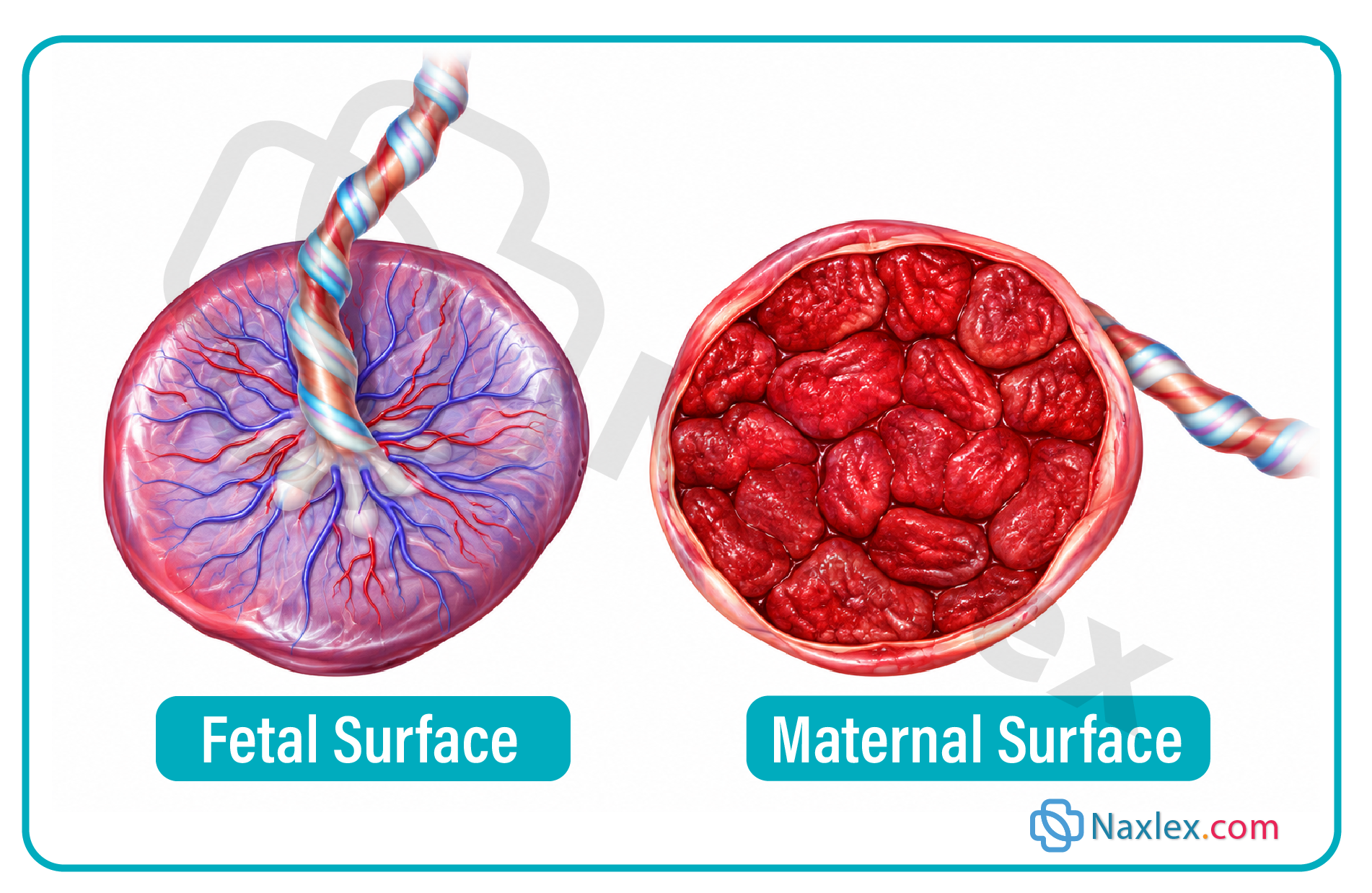
Rationale for correct answers:
1. Inspecting the placenta confirms that all cotyledons and membranes have delivered intact. Examining the maternal surface (lobulated, dark red) and fetal surface (shiny, vascularized) ensures no retained placental fragments remain inside the uterus to trigger delayed hemorrhage or infection.
3. Using quantitative blood loss (QBL) techniques—such as weighing blood-soaked pads and calibrated collection drapes—provides far greater accuracy than visual estimation. Early, precise identification of blood loss allows prompt clinical intervention for obstetric hemorrhage.
4. Administering a prophylactic uterotonic agent (such as oxytocin) immediately after newborn birth is a core component of active management of the third stage. It stimulates strong myometrial contraction, accelerating separation and significantly decreasing blood loss.
Rationale for incorrect answers:
2. Applying continuous fundal pressure prior to confirmed placental separation is strictly contraindicated. Uncontrolled downward force on an unseparated or relaxed uterus can cause acute uterine inversion, leading to severe neurogenic shock and massive hemorrhage.
5. Delaying vital signs is unsafe. Maternal blood pressure, heart rate, and overall responsiveness must be monitored regularly throughout the third stage to detect early physiological signs of hemodynamic instability or hypovolemic shock.
Test-taking strategy:
- Analyze the question: Identify appropriate nursing actions during the third stage of labor (from infant delivery to placental delivery).
- Evaluate choices:
- 1 (Correct): Placental inspection prevents complications from retained tissue.
- 2 (Incorrect): Fundal pressure before separation triggers uterine inversion.
- 3 (Correct): QBL is the standard of care for hemorrhage management.
- 4 (Correct): Prophylactic uterotonics are central to active third-stage management.
- 5 (Incorrect): Vital signs must be monitored continuously to identify instability.
Take home points
- Active management of the third stage includes prophylactic uterotonics, controlled cord traction, and fundal massage post-delivery.
- Quantitative Blood Loss (QBL) provides accurate measurement to detect early postpartum hemorrhage.
- Inspecting both maternal (cotyledons) and fetal (membranes/vessels) surfaces prevents retained placenta complications.
- Fundal pressure prior to separation is strictly avoided to prevent life-threatening uterine inversion.
A nurse caring for a client 25 minutes after newborn birth notes that the placenta has not yet separated despite gentle fundal assessment. Which of the following actions should the nurse take?
Explanation
The third stage of labor encompasses the period from neonatal delivery to placental expulsion, typically taking 5 to 30 minutes. Prolonged retention past 30 minutes signifies a retained placenta, significantly increasing the risk of severe postpartum hemorrhage.
Rationale for correct answer:
2. Normal placental detachment typically occurs within 30 minutes following neonatal birth. The nurse should continue close monitoring while preparing to alert the provider if expulsion exceeds this standard threshold. A delayed expulsion past 30 minutes requires medical intervention to prevent severe maternal complications. This proactive approach ensures timely provider management of potential retained placenta and prevents excess blood loss.
Rationale for incorrect answers:
1. Pulling forcefully on the cord is extremely dangerous and contraindicated. Excessive force can precipitate cord avulsion or catastrophic uterine inversion. Controlled traction should only be gentle during active uterine contraction. The nurse must never apply firm uncoordinated traction.
3. Methylergonovine causes potent vasoconstriction and uterine contraction to manage uterine atony. Administering this medication without assessing blood pressure can trigger severe hypertensive crisis. It is contraindicated in patients with elevated blood pressure or preeclampsia. Medication administration requires a specific provider order rather than an unauthorized repeat.
4. Placental retention approaching 25 minutes requires vigilant observation rather than passive documentation. Dismissing this finding ignores the impending risk of third-stage prolongation. Failure to prepare for intervention exposes the client to unrecognized uterine atony. Proper clinical protocol demands active preparation for potential complications.
Test-taking strategy:
- Analyze the scenario/question: The client is 25 minutes postpartum in the third stage of labor with an unseparated placenta. The prompt asks for the most appropriate nursing intervention given this specific timeframe.
- Apply Obstetrical Principles (Third Stage Management): Normal placental separation occurs within 5 to 30 minutes postpartum through uterine contractions and retroplacental hematoma formation. Expectant management with vigilant monitoring is appropriate until the 30-minute mark is reached, at which point medical intervention for retained placenta is initiated.
- Rule in Choice 2: The duration of 25 minutes is within normal physiological limits but requires preparation for provider notification if placental separation fails to occur by 30 minutes.
- Rule out Choice 1: Excessive cord traction risks uterine inversion and umbilical cord rupture, making aggressive manual pull inappropriate.
- Rule out Choice 3: Ergot alkaloids carry a risk of severe hypertension and must never be administered without vital sign verification or valid prescription.
- Rule out Choice 4: Ignoring the timeline fails to recognize potential postpartum hemorrhage risks as the client approaches the clinical cutoff.
Take home points
- The third stage of labor normally lasts between 5 and 30 minutes after the birth of the neonate.
- Placental separation taking longer than 30 minutes is defined as a retained placenta and requires provider intervention.
- Firm or forceful umbilical cord traction is strictly contraindicated due to the risk of uterine inversion and cord avulsion.
- Methylergonovine requires blood pressure evaluation prior to administration due to its potent vasoconstrictive effects.
A nurse suddenly notes that a client's uterine fundus is no longer palpable abdominally and a reddish mass is visible at the vaginal introitus, accompanied by profound hypotension. Which of the following actions should the nurse take first?
Explanation
An acute uterine inversion occurs when the uterine fundus collapses into the endometrial cavity and prolapses through the cervix into the vagina or beyond the introitus. It represents a catastrophic obstetrical emergency characterized by neurogenic shock and sudden postpartum hemorrhage. Immediate management requires urgent multidisciplinary response and manual replacement before cervical ring constriction occurs, alongside aggressive resuscitative measures.
Rationale for correct answer:
2. Uterine inversion requires an immediate emergency call for additional personnel and anesthesia support. The primary healthcare provider must attempt immediate manual replacement of the inverted fundus before a constriction ring develops around the cervix. Delaying manual replacement drastically reduces success rates and accelerates neurogenic shock. Prompt positioning and rapid response team activation are critical first steps.
Rationale for incorrect answers:
1. Uterotonic agents such as oxytocin cause immediate uterine contraction and cervical ring closure. Administering uterotonics while the uterus is still inverted traps the fundus outside the body and prevents manual repositioning. Oxytocin must only be administered after successful manual replacement is completed to maintain uterine tone.
3. Massaging an inverted or prolapsed uterus causes severe pain, accelerates maternal shock, and worsens active tissue hemorrhage. Direct mechanical manipulation of the exposed inverted fundus disrupts clotting and prevents successful manual reduction. Fundal massage is strictly contraindicated until the organ is completely restored to its anatomical position.
4. Maternal bearing down increases intra-abdominal pressure and forces further extrusion of the inverted organ through the vaginal opening. Instructing the client to push worsens the degree of pelvic prolapse and speeds cardiovascular collapse. The client must be positioned flat or in Trendelenburg position while remaining relaxed.
Test-taking strategy:
- Analyze the scenario/question: The client presents with a non-palpable abdominal fundus, a visible vaginal mass, and sudden profound hypotension, indicating an acute uterine inversion with shock. The question asks for the priority initial nursing action.
- Apply Priority Emergency Principles (Emergency Resuscitation and Reduction): Uterine inversion is a life-threatening emergency requiring rapid stabilization, immediate cessation of uterotonics/massage, and immediate manual replacement of the uterus before cervical trapping occurs.
- Expound further on the Apply knowledge of immediate emergency response: Recognizing the classic triad of a missing abdominal fundus, visible vaginal prolapse, and maternal shock is essential for triggering an emergency code. Early intervention preserves maternal hemodynamic stability and prevents irreversible hemorrhagic collapse.
- Rule in Choice 2: Calling for help mobilizes the necessary anesthesia and surgical team required for emergency manual repositioning and rapid fluid hemodynamic stabilization.
- Rule out Choice 1: Uterotonic administration prior to reduction causes cervical contraction, making subsequent manual replacement of the uterus impossible.
- Rule out Choice 3: External massage on an inverted uterus exacerbates structural injury, accelerates maternal hemorrhage, and increases neurogenic shock.
- Rule out Choice 4: Increased intra-abdominal pressure from pushing leads to complete organ extrusion and worsens acute cardiovascular collapse.
- Expound further on the Apply knowledge of immediate emergency response: Recognizing the classic triad of a missing abdominal fundus, visible vaginal prolapse, and maternal shock is essential for triggering an emergency code. Early intervention preserves maternal hemodynamic stability and prevents irreversible hemorrhagic collapse.
Take home points
- Uterine inversion is characterized by a missing abdominal fundus, a visible introitus mass, severe pain, and rapid hemodynamic shock.
- Calling for immediate assistance is the priority action to prepare for manual uterine replacement under possible relaxation anesthesia.
- Uterotonic medications and fundal massage are strictly contraindicated until the uterus is fully returned to its correct anatomical location.
- Failure to reduce an inverted uterus promptly leads to cervical ring tightening, massive hemorrhage, and profound maternal shock.
A nurse is reviewing causes of postpartum hemorrhage occurring during the third stage of labor. Which of the following causes should the nurse identify as the most common?
Explanation
Uterine atony represents the failure of myometrial muscle fibers to contract effectively following placental separation, allowing open blood vessels at the placental site to bleed profusely. It accounts for up to 80% of all primary postpartum hemorrhage cases globally. Risk factors include uterine overdistension, prolonged labor, and chorioamnionitis.
Rationale for correct answer:
3. Myometrial contraction is the primary physiological mechanism for occluding spiral arteries after placental detachment. When uterine atony occurs, lack of muscle tone leads to rapid and heavy bleeding from the exposed placental site. This condition is universally recognized as the single most common cause of early postpartum hemorrhage.
Rationale for incorrect answers:
1. Lacerations of the cervix, vagina, or perineum account for approximately 20% of postpartum bleeding instances. While genital trauma is the second most common cause, it is typically characterized by continuous trickle bleeding despite a firm, contracted fundus, rather than atony.
2. Retained tissues prevent effective physiological contraction of the myometrium, leading to secondary bleeding. Although retained placenta is a major cause of early and late hemorrhage, it occurs less frequently than primary lack of myometrial tone alone.
4. Coagulopathies account for less than 1% of all postpartum hemorrhage presentations. While disorders like von Willebrand disease or DIC exacerbate bleeding, inherited or acquired clotting defects are relatively rare primary drivers of immediate third-stage hemorrhage.
Test-taking strategy:
- Analyze the scenario/question: The prompt asks the nurse to identify the most frequent underlying etiology of postpartum hemorrhage occurring during the third stage of labor.
- Apply Epidemiological Knowledge (The 4 Ts of Postpartum Hemorrhage): Postpartum hemorrhage etiologies are categorized by Tone (atony), Trauma (lacerations), Tissue (retained placenta), and Thrombin (coagulopathies). Tone issues represent the vast majority of clinical presentations.
- Expound further on the Apply knowledge of postpartum hemorrhage statistics: Understanding the order of frequency among the 4 Ts enables the nurse to anticipate interventions starting with fundal massage and uterotonics. Knowledge of baseline incidence directs rapid, systematic clinical assessment during acute maternal bleeding events.
- Rule in Choice 3: Myometrial failure to contract (uterine atony) is statistically the primary cause, accounting for the vast majority of maternal hemorrhage cases.
- Rule out Choice 1: Perineal and cervical genital trauma represents a significant cause of bleeding but occurs less frequently than atony.
- Rule out Choice 2: Incomplete expulsion of retained fragments contributes significantly to blood loss but is secondary in incidence to tone loss.
- Rule out Choice 4: Underlying maternal coagulopathy defects represent the least common cause among the four major diagnostic categories.
- Expound further on the Apply knowledge of postpartum hemorrhage statistics: Understanding the order of frequency among the 4 Ts enables the nurse to anticipate interventions starting with fundal massage and uterotonics. Knowledge of baseline incidence directs rapid, systematic clinical assessment during acute maternal bleeding events.
Take home points
- Uterine atony is the most common cause of early postpartum hemorrhage, accounting for up to 80% of cases.
- The four main causes of postpartum hemorrhage are remembered using the "4 Ts" mnemonic: Tone, Trauma, Tissue, and Thrombin.
- Genital tract lacerations are suspected when continuous bright red bleeding occurs despite a firm, well-contracted fundus.
- Immediate management of uterine atony begins with firm fundal massage and administration of prescribed uterotonic medications.
Practice Exercise 4
A nurse is caring for a client 30 minutes after vaginal birth. Which of the following findings should the nurse identify as an expected physiologic change during this period?
Explanation
Postpartum puerperal bradycardia (heart rate 50 to 70 beats per minute) is a normal physiological phenomenon occurring within the first 6 to 10 days after birth. It results from a sudden increase in central venous return as autotransfusion occurs following placental delivery, alongside reduced vascular bed capacitance.
Rationale for correct answer:
2. A maternal heart rate of 58 beats per minute represents normal physiological adaptation due to autotransfusion of placental blood into maternal circulation. Increased stroke volume triggers a reflex vagal response, leading to transient bradycardia. In the presence of stable blood pressure, a heart rate of 50 to 70 beats per minute is an expected non-pathological finding requiring standard postpartum monitoring.
Rationale for incorrect answers:
1. A sustained heart rate of 130 beats per minute indicates severe maternal tachycardia requiring urgent assessment. Tachycardia in the early postpartum period is a key indicator of hidden postpartum hemorrhage, hypovolemia, severe pain, or acute infection, and is never an expected change.
3. A maternal temperature of 39.2°C (102.6°F) exceeds the expected mild low-grade temperature elevation seen with exertion and dehydration. While a temperature up to 38.0°C (100.4°F) can be normal in the first 24 hours, a higher spike indicates maternal infection or severe intrapartum chorioamnionitis.
4. A blood pressure reading of 85/50 mmHg with accompanying tachycardia is a classic presentation of severe hypotension and cardiovascular instability. This finding indicates significant hypovolemic shock secondary to excessive blood loss or occult internal bleeding rather than a normal adaptation.
Test-taking strategy:
- Analyze the scenario/question: The prompt asks the nurse to identify an expected, normal physiological change in a maternal client at 30 minutes following a vaginal delivery.
- Apply Normal Postpartum Physiology: Immediate hemodynamic adjustments occur post-delivery, including a surge in central blood volume from autotransfusion, which elevates stroke volume and lowers heart rate. Vital signs must be distinguished from signs of hemorrhage or infection.
- Expound further on the Apply knowledge of normal physiological vs pathological postpartum changes: Differentiating expected cardiovascular adaptations from early subtle signs of instability prevents unnecessary emergency activations while ensuring true complications are caught early. Recognizing transient bradycardia as a compensatory mechanism allows the nurse to safely reassure the client during early recovery.
- Rule in Choice 2: Early puerperal bradycardia (50 to 70 bpm) with normal blood pressure reflects normal cardiovascular autotransfusion and reflex vagal stimulation.
- Rule out Choice 1: Persistent marked maternal tachycardia (>100 bpm) is a primary sign of early blood loss or systemic distress.
- Rule out Choice 3: Marked pyrexia (>38.0°C) points directly toward an infectious etiology rather than transient dehydration.
- Rule out Choice 4: Hypotension accompanied by compensatory tachycardia represents cardiovascular collapse requiring immediate resuscitation.
- Expound further on the Apply knowledge of normal physiological vs pathological postpartum changes: Differentiating expected cardiovascular adaptations from early subtle signs of instability prevents unnecessary emergency activations while ensuring true complications are caught early. Recognizing transient bradycardia as a compensatory mechanism allows the nurse to safely reassure the client during early recovery.
Take home points
- Puerperal bradycardia (50 to 70 beats per minute) is an expected physiological finding in the early postpartum period.
- Maternal temperature up to 38.0°C (100.4°F) within the first 24 hours can occur from dehydration and exertion, but higher spikes indicate infection.
- Tachycardia in the early postpartum period is often the earliest clinical indicator of postpartum hemorrhage or hypovolemia.
- Hypotension combined with tachycardia demands immediate evaluation for hypovolemic shock and acute blood loss.
A nurse palpates a client's uterine fundus 1 hour after birth and finds it soft and difficult to delineate. Which of the following actions should the nurse take first?
Explanation
A soft and poorly delineated fundus indicates uterine atony, the primary cause of early postpartum hemorrhage. The immediate priority is applying fundal massage to stimulate myometrial contractions, contract open spiral arteries, and promote normal uterine muscle tone.
Rationale for correct answer:
2. A soft, uncontracted fundus represents uterine atony and requires immediate mechanical stimulation. The nurse must perform firm, circular massage over the fundus to induce myometrial contraction and prevent severe blood loss. Massaging the fundus directly addresses the underlying cause of bleeding and is the mandatory first-line intervention for early uterine atony.
Rationale for incorrect answers:
1. Delaying intervention for 4 hours when uterine atony is present allows unrecognized, heavy maternal bleeding to continue unchecked. A boggy fundus demands immediate active intervention, and routine documentation without corrective action constitutes negligent nursing during critical postpartum recovery.
3. Delaying immediate bedside intervention to call the primary healthcare provider permits rapid and potentially catastrophic maternal blood loss. The nurse must first initiate fundal massage to stabilize the client before delegating or executing provider notification for persistent refractory atony.
4. Instructing an unstable client with active uterine atony to ambulate increases the risk of syncope, sudden fall injuries, and accelerated hemorrhage. While a full bladder can cause uterine displacement, the nurse should assist with a bedpan or catheterization rather than ordering unassisted ambulation while hemodynamically unassessed.
Test-taking strategy:
- Analyze the scenario/question: The client is 1 hour postpartum with a soft, poorly delineated uterine fundus (uterine atony). The question asks for the priority ("first") nursing intervention.
- Apply Clinical Prioritization Principles (First-Line Emergency Nursing Interventions): Uterine atony requires immediate bedside action to halt bleeding. Direct physical manipulation via fundal massage is the fastest, non-invasive method to stimulate myometrial contraction before seeking secondary orders or performing lower-priority tasks.
- Expound further on the Apply knowledge of immediate nursing action for postpartum uterine atony: Recognizing that fundal massage is a primary, independent nursing action allows for immediate hemorrhage control without waiting for a prescription. Prompt mechanical stimulation compresses open placental site vessels and restores basic myometrial tone.
- Rule in Choice 2: Initiating firm fundal massage provides direct mechanical stimulation to stop active bleeding and restore vital myometrial tone.
- Rule out Choice 1: Postponing care and providing simple passive documentation severely endangers the client by allowing hidden hemorrhage to progress.
- Rule out Choice 3: Postponing bedside physical intervention to make a provider call delays critical life-saving mechanical stabilization.
- Rule out Choice 4: Forcing an unassessed client to engage in immediate ambulation introduces severe orthostatic collapse and trauma risks.
- Expound further on the Apply knowledge of immediate nursing action for postpartum uterine atony: Recognizing that fundal massage is a primary, independent nursing action allows for immediate hemorrhage control without waiting for a prescription. Prompt mechanical stimulation compresses open placental site vessels and restores basic myometrial tone.
Take home points
- A soft, poorly delineated, or boggy fundus is the primary clinical sign of postpartum uterine atony.
- Perform immediate fundal massage as the independent, first-line action to stimulate uterine muscle contraction.
- Never leave a bleeding client with uterine atony unattended to call a provider before initiating basic physical interventions.
- Distended bladders can displace the uterus, but bedpans or catheterization must be used if the client is hemodynamically unstable.
A nurse is assessing a client's lochia on postpartum day 2. Which of the following findings should the nurse document as expected at this time?
Explanation
The normal progression of puerperal lochia spans three distinct stages following placental expulsion, reflecting normal uterine mucosal healing and endometrial regeneration. On postpartum days 1 through 3, the discharge is characterized as lochia rubra, consisting predominantly of erythrocytes, decidual debris, epithelial cells, and mucus.
Rationale for correct answer:
3. Lochia rubra is the expected, normal vaginal discharge for the first 3 to 4 days postpartum. It is dark red in color and may contain small, fleshy clots smaller than a nickel. Documenting lochia rubra on day 2 aligns with standard postpartum involution and indicates appropriate endometrial shedding without abnormal vascular disruption.
Rationale for incorrect answers:
1. Lochia alba represents the final stage of lochia, appearing creamy yellow-to-white starting around postpartum day 10 to 14. Any foul odor present at any stage strongly suggests uterine infection or endometritis rather than an expected finding.
2. Lochia serosa is a pinkish-brown or serosanguineous discharge that typically appears around postpartum day 4 and lasts through day 10. Finding lochia serosa on day 2 is premature and warrants evaluation for accelerated involution or improper stage classification.
4. A complete absence of lochia during the early postpartum period is abnormal and concerning. It may indicate a blocked cervical os secondary to retained tissue or blood clot entrapment, increasing the risk for severe puerperal infection.
Test-taking strategy:
- Analyze the scenario/question: The client is on postpartum day 2, and the nurse needs to identify the physiological stage and normal characteristics of lochia expected for this timeframe.
- Apply Obstetrical Knowledge (Lochia Stages and Timeline): Systematically evaluate the expected chronological progression of lochia: Rubra (days 1 to 3, dark red), Serosa (days 4 to 10, pinkish-brown), and Alba (days 10 to 14+, yellowish-white).
- Expound further on the Apply knowledge of normal lochia progression and clinical timelines: Accurate stage identification allows the nurse to distinguish expected postpartum recovery from early signs of hemorrhage or endometritis. Recognizing that lochia rubra contains normal blood elements on days 1 through 3 prevents false alarms regarding maternal bleeding.
- Rule in Choice 3: Dark red discharge containing small non-pathological clots describes lochia rubra, which is normal for postpartum day 2.
- Rule out Choice 1: Yellowish-white discharge with a foul odor signifies lochia alba and potential endometritis infection.
- Rule out Choice 2: Pinkish-brown serosanguineous discharge corresponds strictly to lochia serosa, typically presenting after day 3.
- Rule out Choice 4: A total lack of lochia indicates cervical obstruction or lochiometra, requiring immediate physical evaluation.
- Expound further on the Apply knowledge of normal lochia progression and clinical timelines: Accurate stage identification allows the nurse to distinguish expected postpartum recovery from early signs of hemorrhage or endometritis. Recognizing that lochia rubra contains normal blood elements on days 1 through 3 prevents false alarms regarding maternal bleeding.
Take home points
- Lochia rubra is dark red with small clots and normally lasts for the first 1 to 3 days postpartum.
- Lochia serosa is pinkish-brown and typically lasts from day 4 through day 10 postpartum.
- Lochia alba is yellowish-white and occurs from roughly day 10 up to 6 weeks postpartum.
- A foul odor or sudden cessation of lochia is abnormal and suggests endometritis or cervical outflow tract obstruction.
A nurse is caring for a client during the first hour postpartum. Which of the following assessment findings should be reported to the primary health care provider immediately?
Explanation
During the first hour postpartum, the client requires close observation for early postpartum hemorrhage. While moderate lochia is anticipated, excessive vaginal bleeding defined as saturating a perineal pad within one hour indicates potential uterine atony or genital tract laceration, requiring immediate provider notification and rapid intervention.
Rationale for correct answer:
2. Saturating a perineal pad in under 60 minutes indicates heavy, excessive lochia and serves as a critical warning sign of early postpartum hemorrhage. The nurse must immediately evaluate for uterine atony, retained tissues, or undetected lacerations. Provider notification is essential to obtain orders for additional uterotonic agents and prevent severe hypovolemic shock.
Rationale for incorrect answers:
1. A firm, midline fundus located at the level of the umbilicus represents expected uterine position and tone during the first hour following birth. This finding indicates effective myometrial contraction and successful placental site homeostasis, which does not require provider notification.
3. Mild afterpains are normal uterine contractions caused by oxytocin release, particularly during breastfeeding or in multiparous clients. Adequate pain relief following prescribed oral analgesia demonstrates expected postpartum recovery rather than a pathological process.
4. Maternal thirst and hunger immediately after birth are common physiological responses due to exertion, fluid restriction, and energy expenditure during labor. Offering fluids and light nutrition supports normal recovery and does not warrant emergency reporting.
Test-taking strategy:
- Analyze the scenario/question: The client is in the first hour postpartum. The prompt asks which assessment finding requires immediate reporting to the primary healthcare provider, implying a significant deviation from normal recovery.
- Apply Priority Nursing Principles (Bleeding Thresholds and Hemorrhage Control): Differentiate between expected early postpartum physiological findings and signs of excessive blood loss. Pad saturation within 1 hour is universally recognized as the clinical threshold for excessive bleeding requiring escalation.
- Expound further on the Apply knowledge of postpartum bleeding assessment and safety thresholds: Monitoring lochia volume against precise time intervals allows early identification of occult or overt hemorrhage. Recognizing that rapid pad saturation indicates active vascular bleeding ensures prompt intervention before cardiovascular decompensation occurs.
- Rule in Choice 2: Rapid saturation of a perineal pad within 45 minutes indicates excessive lochia and potential postpartum hemorrhage.
- Rule out Choice 1: A firm, midline fundus at the umbilicus represents expected myometrial tone and appropriate involution.
- Rule out Choice 3: Mild uterine contractions managed with medication indicate normal involutional afterpains and effective comfort care.
- Rule out Choice 4: Client requests for food and water reflect normal post-exertion recovery following labor.
- Expound further on the Apply knowledge of postpartum bleeding assessment and safety thresholds: Monitoring lochia volume against precise time intervals allows early identification of occult or overt hemorrhage. Recognizing that rapid pad saturation indicates active vascular bleeding ensures prompt intervention before cardiovascular decompensation occurs.
Take home points
- Saturating a perineal pad within 1 hour or less defines excessive lochia and signals potential postpartum hemorrhage.
- A firm, midline fundus at the level of the umbilicus is the normal expected finding in the first hour post-delivery.
- Afterpains are expected uterine contractions that assist with involution and are typically managed with standard analgesics.
- Immediate reporting of excessive bleeding allows for rapid administration of uterotonics and prevention of hypovolemic shock.
A nurse is caring for a client during the fourth stage of labor. Which of the following actions are appropriate components of nursing management? Select all that apply
Explanation
The fourth stage of labor encompasses the critical early recovery phase spanning the first 1 to 4 hours post-delivery, where the risk of primary postpartum hemorrhage is highest. Key nursing interventions focus on maintaining myometrial tone, ensuring effective vesical emptying, stabilizing maternal vital signs, and promoting neonate physiological transition.
Rationale for correct answers:
1. Frequent monitoring of maternal vital signs every 15 minutes during the first hour postpartum is mandatory to detect early signs of hypovolemic shock, internal bleeding, or hemodynamic instability. Rapid assessment of pulse and blood pressure ensures early identification of cardiovascular decompensation.
2. A full, distended bladder displaces the uterus laterally and upward, impairing effective uterine muscle contractions and directly precipitating uterine atony. Assisting the client to void maintains a empty bladder and facilitates appropriate fundal involution.
4. Initiating breastfeeding within the first hour of life takes advantage of the newborn's early quiet alert state and stimulates endogenous maternal oxytocin release. Oxytocin promotes strong myometrial contractions to compress placental site blood vessels, reducing blood loss while establishing infant bonding.
5. Evaluating fundal location, height, and tone concurrently with every vital sign check ensures continuous assessment for uterine atony and hidden bleeding. Simultaneous evaluation prevents missed clinical changes and ensures prompt hemorrhage management.
Rationale for incorrect answers:
3. Delaying skin-to-skin contact interferes with neonatal thermoregulation, cardiorespiratory stabilization, and early breastfeeding success. Direct, uninterrupted skin-to-skin contact should begin immediately after birth in stable mother-infant dyads, rather than waiting for postpartum transfer.
Test-taking strategy:
- Analyze the scenario/question: The client is in the fourth stage of labor (first hour post-delivery). The question asks for appropriate nursing management actions in a Select All That Apply format.
- Apply Postpartum Recovery Principles (The Fourth Stage Protocols): Evaluate each action against evidence-based postpartum guidelines prioritizing maternal hemodynamic monitoring, bladder control, fundal checks, and immediate neonatal bonding.
- Expound further on the Apply knowledge of fourth-stage maternal and neonatal care principles: Mastering the routine assessment schedule and early bonding interventions during the first hour postpartum ensures both maternal physical recovery and optimal newborn transition. Preventing bladder distension and encouraging natural oxytocin release via breastfeeding form the foundation of immediate hemorrhage prevention.
- Rule in Choice 1: Vital sign checks every 15 minutes ensure immediate detection of postpartum hemorrhage and hemodynamic shifts.
- Rule in Choice 2: Promoting maternal voiding prevents bladder distension from inhibiting myometrial contraction.
- Rule out Choice 3: Postponing maternal-infant contact delays crucial thermoregulation adaptation and immediate oxytocin release.
- Rule in Choice 4: Early breastfeeding promotes endogenous oxytocin release and supports infant attachment.
- Rule in Choice 5: Checking the fundus concurrently with vital signs ensures ongoing monitoring for uterine atony.
- Expound further on the Apply knowledge of fourth-stage maternal and neonatal care principles: Mastering the routine assessment schedule and early bonding interventions during the first hour postpartum ensures both maternal physical recovery and optimal newborn transition. Preventing bladder distension and encouraging natural oxytocin release via breastfeeding form the foundation of immediate hemorrhage prevention.
Take home points
- The fourth stage of labor requires maternal vital signs and fundal assessments every 15 minutes for the first hour.
- A full bladder displaces the uterus and is a major preventable cause of postpartum uterine atony.
- Immediate skin-to-skin contact and early breastfeeding stimulate natural oxytocin release to contract the uterus and stabilize the newborn.
- Concurrent assessment of vital signs, fundal tone, and lochia volume forms the core safety protocol of immediate postpartum care.
A client reports severe, unrelieved vaginal and rectal pain 1 hour after a vacuum-assisted vaginal birth, despite a firm fundus and moderate lochia rubra. Which of the following complications should the nurse suspect?
Explanation
A vulvovaginal hematoma occurs following injury to pelvic blood vessels, leading to occult bleeding that collects in soft tissues without visible external lochial changes. It typically arises from operative deliveries like vacuum extraction or tissue trauma, causing intense pain out of proportion to exam findings and severe rectal pressure.
Rationale for correct answer:
3. Severe, unyielding perineal or rectal pain accompanied by feelings of pressure in the presence of a firm fundus and moderate lochia strongly suggests an expanding hematoma. Operative vaginal delivery using vacuum extraction creates significant vascular injury in pelvic tissues. Prompt assessment of the perineum and vaginal wall is essential to identify swelling and prevent hypovolemic shock.
Rationale for incorrect answers:
1. Uterine atony presents primarily as a boggy, soft fundus with heavy lochia rubra and active bleeding. Because the scenario explicitly states that the fundus is firm and lochia is moderate, uterine atony is ruled out as the cause of pain.
2. Retained placental tissues cause uterine subinvolution and persistent bleeding from uncontracted myometrial segments. While retained fragments lead to lochial changes and cramping, they do not produce localized, extreme rectal pressure or severe perineal pain with a firm fundus.
4. Uterine inversion is characterized by sudden profound shock, severe hemorrhage, and a non-palpable abdominal fundus with a visible vaginal mass. A firm abdominal fundus directly excludes the possibility of uterine inversion.
Test-taking strategy:
- Analyze the scenario/question: The client is 1 hour postpartum following a vacuum-assisted delivery, presenting with severe, unrelieved vaginal/rectal pain, a firm fundus, and moderate lochia. The nurse must identify the underlying complication.
- Apply Clinical Prioritization Principles (Differentiating Postpartum Pain Sources): Evaluate the classic signs of hidden tissue bleeding versus open uterine hemorrhage. Intense pain out of proportion to visible blood loss with normal fundal tone points directly to soft tissue vascular trauma.
- Expound further on the Apply knowledge of occult postpartum vascular trauma: Recognizing that operative births increase the risk of soft tissue vessel rupture helps the nurse locate hidden blood accumulation early. Identifying intense pressure symptoms while the fundus remains firm prevents delayed diagnosis of deep pelvic bleeding.
- Rule in Choice 3: Severe rectal pressure and intense pain despite a firm fundus are pathognomonic for an expanding vaginal hematoma.
- Rule out Choice 1: A firm fundal tone directly contradicts a diagnosis of uterine atony.
- Rule out Choice 2: Retained tissue typically presents with uterine bogginess and abnormal bleeding rather than isolated rectal pain.
- Rule out Choice 4: A firm, palpable fundus confirms structural integrity and excludes acute uterine inversion.
- Expound further on the Apply knowledge of occult postpartum vascular trauma: Recognizing that operative births increase the risk of soft tissue vessel rupture helps the nurse locate hidden blood accumulation early. Identifying intense pressure symptoms while the fundus remains firm prevents delayed diagnosis of deep pelvic bleeding.
Take home points
- Severe, unrelieved perineal or rectal pain with a firm fundus and normal lochia indicates a hidden hematoma.
- Operative vaginal deliveries involving vacuum or forceps significantly increase the risk of vulvovaginal hematomas.
- Unlike uterine atony, hematomas cause severe pressure and pain rather than noticeable excessive external bleeding.
- Immediate inspection of the perineum and vaginal vault is required to evaluate for expanding swelling or tissue discoloration.
A nurse is educating a client on the physiologic benefit of early breastfeeding initiation during the fourth stage of labor. Which of the following statements should the nurse include?
Explanation
Initiating breastfeeding within the first hour of birth triggers the release of endogenous oxytocin from the posterior pituitary gland. Oxytocin stimulates potent myometrial contractions, compressing open blood vessels at the placental site and facilitating natural uterine involution to mitigate hemorrhage risk.
Rationale for correct answer:
2. Infant suckling during early breastfeeding stimulates the neurohormonal reflex arc, causing rapid release of endogenous oxytocin into maternal circulation. Oxytocin binds directly to myometrial receptors to promote strong, rhythmic uterine contractions. These contractions effectively clamp down on open placental site blood vessels, decreasing the primary risk of postpartum hemorrhage and accelerating initial fundal involution.
Rationale for incorrect answers:
1. Early breastfeeding accelerates uterine recovery rather than delaying it. The oxytocin surge induced by infant suckling speeds up the process of myometrial contraction and tissue regression, helping the organ return to its pre-pregnancy state far more efficiently.
3. Breastfeeding directly impacts postpartum blood loss by promoting consistent uterine contraction. Claiming that suckling has no effect ignores the critical physiological role of endogenous oxytocin release in preventing early uterine atony.
4. Delaying infant feeding for 4 hours misses the newborn's initial period of quiet alertness and delays essential maternal physiological benefits. Early skin-to-skin contact and suckling within the first hour are recommended standards for maternal recovery and successful lactation onset.
Test-taking strategy:
- Analyze the scenario/question: The prompt asks for the correct educational statement regarding the physiological benefit of early breastfeeding during the fourth stage of labor.
- Apply Endocrine and Postpartum Physiology (Oxytocin Pathway): Evaluate how early lactation interacts with uterine muscle tone. Infant suckling triggers oxytocin release, which stimulates myometrial contraction and decreases early blood loss.
- Expound further on the Apply knowledge of neuroendocrine reflexes in the postpartum period: Educating clients on the physiological connection between nipple stimulation and uterine contractions empowers them to participate actively in hemorrhage prevention. Understanding how endogenous oxytocin prevents uterine atony reinforces the clinical priority of immediate mother-infant contact.
- Rule in Choice 2: Infant suckling stimulates endogenous oxytocin secretion, which maintains essential myometrial tone and prevents bleeding.
- Rule out Choice 1: Oxytocin-induced contractions speed up rather than delay normal fundal involution.
- Rule out Choice 3: Suckling directly reduces maternal bleeding by preventing uterine atony.
- Rule out Choice 4: Postponing breastfeeding misses the crucial early alert state and delays maternal hemorrhage protection.
- Expound further on the Apply knowledge of neuroendocrine reflexes in the postpartum period: Educating clients on the physiological connection between nipple stimulation and uterine contractions empowers them to participate actively in hemorrhage prevention. Understanding how endogenous oxytocin prevents uterine atony reinforces the clinical priority of immediate mother-infant contact.
Take home points
- Early breastfeeding stimulates the release of endogenous oxytocin from the posterior pituitary gland.
- Oxytocin causes strong myometrial contractions that compress placental site blood vessels to prevent postpartum hemorrhage.
- Breastfeeding accelerates fundal involution, helping the uterus return to its pre-pregnancy size more rapidly.
- Initiating skin-to-skin contact and breastfeeding within the first hour postpartum provides optimal physiological benefits for both mother and infant.
A nurse is identifying risk factors for uterine atony in a client following delivery of a 4500-gram (9-pound, 15-ounce) newborn after a prolonged labor. Which of the following additional findings would further increase this client's risk?
Explanation
Uterine atony occurs when myometrial muscle fibers fail to contract adequately, leading to uncompressed placental site blood vessels and rapid hemorrhage. Major predisposing factors include uterine overdistension from fetal macrosomia, myometrial fatigue following prolonged labor, grand multiparity, and a history of retained placental tissue.
Rationale for correct answer:
2. Grand multiparity results in repetitive stretching and fibrous tissue replacement within the myometrium, significantly impairing the muscle's contractility. A history of retained placental fragments further compromises effective placental site separation and involution. Combined with existing risk factors like fetal macrosomia and prolonged labor, these historical findings drastically elevate the risk of severe postpartum hemorrhage due to refractory myometrial fatigue.
Rationale for incorrect answers:
1. Primigravida status with a spontaneous vaginal delivery does not inherently increase the risk for uterine atony. Risk rises with high parity, uterine overdistension, or operative intervention, making a simple first birth a non-contributing baseline factor regarding myometrial tone.
3. A first-degree perineal laceration involves superficial skin and mucosal tissue without muscular disruption. While lacerations are a source of traumatic bleeding, they do not impair myometrial muscle contraction or increase the specific risk of uterine atony.
4. Delayed cord clamping for 45 seconds is an evidence-based practice that enhances neonatal blood volume and iron stores. Brief delays in cord clamping do not compromise placental separation or inhibit subsequent uterine involution.
Test-taking strategy:
- Analyze the scenario/question: The client already has key risk factors for uterine atony (fetal macrosomia of 4500 grams and prolonged labor). The prompt asks to identify an additional finding that further compounds this risk.
- Apply Obstetrical Risk Assessment Principles (Causes of Reduced Myometrial Tone): Evaluate how muscle fatigue, tissue stretching, and historical uterine complications impact post-delivery contractility. Conditions that cause structural muscle changes or previous retained tissue directly compound atony risk.
- Expound further on the Apply knowledge of predisposing factors for uterine atony: Identifying compounding risk factors enables the nurse to prepare active third-stage management protocols before delivery occurs. Recognizing how grand multiparity and past placental retention impair muscle contraction allows the healthcare team to anticipate heavy blood loss proactively.
- Rule in Choice 2: Grand multiparity reduces muscle elasticity, and a history of retained placenta further compromises post-delivery uterine contraction.
- Rule out Choice 1: First-time pregnancy (primigravida status) without operative trauma does not predispose the client to atony.
- Rule out Choice 3: Superficial mucosal trauma like a first-degree laceration causes localized bleeding but does not affect fundal contractility.
- Rule out Choice 4: Brief delayed cord clamping provides neonatal benefits without impairing myometrial contractility or placental separation.
- Expound further on the Apply knowledge of predisposing factors for uterine atony: Identifying compounding risk factors enables the nurse to prepare active third-stage management protocols before delivery occurs. Recognizing how grand multiparity and past placental retention impair muscle contraction allows the healthcare team to anticipate heavy blood loss proactively.
Take home points
- Uterine overdistension (macrosomia, polyhydramnios, multifetal pregnancy) and prolonged labor are major causes of myometrial fatigue and uterine atony.
- Grand multiparity permanently alters myometrial tissue elasticity, greatly increasing the risk of postpartum hemorrhage.
- A history of retained placental tissue increases the risk of recurrent tissue retention and impaired uterine contraction.
- Perineal lacerations cause traumatic postpartum bleeding but do not cause or worsen uterine atony.
Comprehensive Questions
A nurse is caring for a client whose fetus is in an occiput posterior position. The nurse understands that this fetal position may most significantly delay which of the following cardinal movements of labor?
Explanation
An occiput posterior position occurs when the fetal occiput faces the maternal sacrum rather than the pubis. This alignment places the largest transverse diameter of the fetal skull against the rigid pelvic bony architecture, significantly impeding internal rotation. The resulting mechanical friction leads to prolonged second-stage labor, severe maternal back labor, and labor dystocia.
Rationale for correct answer:
2. In an occiput posterior presentation, the fetal head must rotate a longer arc of 135 degrees anteriorly to reach the symphysis pubis, compared to only 45 degrees in an occiput anterior position. This requirement for an extended arc of rotation against the sacral promontory directly delays internal rotation. Failure or delay in completing this rotation leads to prolonged labor and persistent posterior positioning.
Rationale for incorrect answers:
1. Engagement occurs when the widest transverse diameter of the fetal biparietal plane passes through the pelvic inlet. While an occiput posterior head may engage somewhat later due to incomplete flexion, engagement itself is not the primary movement halted by the posterior alignment during active labor progression; the main barrier occurs deeper in the midpelvis during internal rotation.
3. Expulsion is the final cardinal movement involving delivery of the anterior and posterior shoulders and trunk after the head is fully born. Shoulder expulsion relies on anterior-posterior pelvic clearance rather than occiput positioning, meaning it is not the primary mechanism delayed by an occiput posterior orientation.
4. External rotation, or restitution, occurs after the fetal head is completely born and un-twists to align with the shoulders in the pelvic canal. Because external rotation happens entirely outside the maternal birth canal after head delivery, it is not the primary internal movement inhibited by sacral occiput alignment.
Test-taking strategy:
- Analyze the scenario/question: The fetus is in an occiput posterior position. The question asks which cardinal movement of labor is most significantly delayed by this specific malposition.
- Apply Obstetrical Knowledge (Cardinal Movements and Malposition Mechanics): Review the sequence of cardinal movements: Engagement, Descent, Flexion, Internal Rotation, Extension, External Rotation, and Expulsion. Posterior positioning requires the head to execute a much wider arc of movement deep within the midpelvic cavity to orient under the pubic arch.
- Expound further on the Apply knowledge of cardinal movements in malpositioned fetuses: Understanding how the fetal head negotiates pelvic planes helps the nurse anticipate labor dystocia and position changes that facilitate fetal descent. Recognizing that an occiput posterior fetus must rotate 135 degrees explains why the second stage of labor becomes prolonged.
- Rule in Choice 2: The extended 135-degree arc required for the occiput to turn anteriorly directly delays internal rotation and prolongs labor.
- Rule out Choice 1: Initial passage of the biparietal diameter into the pelvic inlet during engagement is not the main movement impaired by sacral fetal alignment.
- Rule out Choice 3: Final body delivery during expulsion depends on shoulder clearance rather than occiput rotation dynamics.
- Rule out Choice 4: Fetal head realignment during external rotation occurs externally after head delivery is already completed.
- Expound further on the Apply knowledge of cardinal movements in malpositioned fetuses: Understanding how the fetal head negotiates pelvic planes helps the nurse anticipate labor dystocia and position changes that facilitate fetal descent. Recognizing that an occiput posterior fetus must rotate 135 degrees explains why the second stage of labor becomes prolonged.
Take home points
- Occiput posterior position requires a longer arc of rotation (135 degrees vs 45 degrees) to complete internal rotation.
- Delayed internal rotation in an occiput posterior position is a primary cause of prolonged second stage labor and labor dystocia.
- Clients with an occiput posterior fetus frequently experience severe "back labor" due to fetal pressure on the maternal sacrum.
- Maternal position changes (such as hands-and-knees or side-lying) can help encourage rotation of an occiput posterior fetus to an occiput anterior position.
A nurse is caring for a client who received epidural analgesia and is now fully dilated but does not yet feel a strong urge to push. Which of the following approaches should the nurse anticipate implementing?
Explanation
Delayed pushing, also known as passive descent or "laboring down," allows uterine contractions to propel the fetus through the birth canal without active maternal bearing down. In clients with epidural analgesia, blunted pelvic floor sensation suppresses the involuntary Ferguson reflex. Delaying active pushing conserves maternal energy, reduces perineal trauma, and decreases operational vaginal delivery rates.
Rationale for correct answer:
2. Allowing a period of passive descent for 1 to 2 hours permits the fetal head to rotate and descend lower into the pelvis using uterine contractions alone. In nulliparous or epidural-anesthetized clients without a pushing urge, delayed pushing leads to higher rates of spontaneous vaginal delivery and less maternal fatigue. The nurse should support passive descent while monitoring fetal status.
Rationale for incorrect answers:
1. Immediate directed pushing using closed-glottis (Valsalva) maneuvers when no urge is present causes rapid maternal exhaustion, increased perineal trauma, and fetal oxygen desaturation. Closed-glottis pushing lowers maternal cardiac output and reduces placental blood flow, making it an inappropriate initial approach for epidural-induced anesthesia.
3. An absent push urge at full dilation in a client with a functioning epidural is an expected physiological finding rather than a sign of labor arrest or fetal distress. Mandating a cesarean birth without allowing a trial of passive descent represents unnecessary surgical intervention and improper labor management.
4. Administering a second epidural bolus increases the sensory block, further dulling pelvic nerve sensation and completely abolishing the Ferguson reflex. Additional local anesthetic delays active labor progress and increases the likelihood of operative vaginal delivery.
Test-taking strategy:
- Analyze the scenario/question: The client is fully dilated (stage two of labor) with epidural analgesia but lacks a natural urge to push. The question asks for the appropriate nursing approach to manage this specific scenario.
- Apply Obstetrical Practice Guidelines (Second Stage Management with Epidurals): Evaluate evidence-based care protocols regarding the second stage of labor. Suppressed sensation due to regional anesthesia is managed by allowing uterine contractions to naturally lower the fetus before initiating voluntary bearing-down efforts.
- Expound further on the Apply knowledge of laboring down and second-stage physiology: Recognizing that passive descent allows pelvic rotation without active maternal effort protects maternal energy reserves and preserves fetal oxygenation. Understanding how regional blockades affect the Ferguson reflex guides the nurse to advocate for evidence-based delayed pushing rather than forced exertion.
- Rule in Choice 2: Practicing delayed pushing utilizes natural uterine contractions to facilitate fetal descent while conserving maternal energy.
- Rule out Choice 1: Forcing closed-glottis pushing without a natural urge increases maternal fatigue and compromises fetal perfusion.
- Rule out Choice 3: Performing an emergency cesarean for a temporary lack of sensation represents premature and non-indicated surgical intervention.
- Rule out Choice 4: Giving an additional epidural bolus deepens pelvic numbness and further eliminates the natural pushing reflex.
- Expound further on the Apply knowledge of laboring down and second-stage physiology: Recognizing that passive descent allows pelvic rotation without active maternal effort protects maternal energy reserves and preserves fetal oxygenation. Understanding how regional blockades affect the Ferguson reflex guides the nurse to advocate for evidence-based delayed pushing rather than forced exertion.
Take home points
- Epidural analgesia often blunts the Ferguson reflex, removing the involuntary urge to push at full dilation.
- Delayed pushing ("laboring down") allows passive fetal descent via uterine contractions, saving maternal energy and reducing operative delivery risk.
- Closed-glottis or forced pushing without an urge increases maternal exhaustion and can compromise fetal oxygenation.
- Passive descent is typically supported for 1 to 2 hours as long as maternal and fetal heart rate patterns remain reassuring.
A nurse is monitoring fetal heart rate during active pushing and observes abrupt, jagged decreases in fetal heart rate that resolve quickly at the end of each contraction. Which of the following underlying causes should the nurse associate with this pattern?
Explanation
Variable decelerations are abrupt, jagged decreases in fetal heart rate defined by a drop from baseline to nadir in less than 30 seconds, lasting at least 15 seconds but less than 2 minutes. They are caused by transient umbilical cord compression, which acutely increases systemic vascular resistance and triggers a compensatory vagal response.
Rationale for correct answer:
2. The rapid, jagged drop in fetal heart rate that aligns with contractions describes variable decelerations, which stem directly from cord entrapment or compression. Umbilical vein and artery occlusion reduces blood flow to the fetus, causing a transient spike in arterial pressure and a rapid reflex decline in heart rate. Relieving cord pressure through maternal position changes is the primary targeted nursing intervention for this specific fetal heart rate pattern.
Rationale for incorrect answers:
1. Uteroplacental insufficiency produces late decelerations, which present as gradual, smooth declines in fetal heart rate starting after the contraction peak and returning to baseline well after the contraction ends. They reflect compromised placental perfusion and fetal hypoxia rather than jagged umbilical vessel occlusion.
3. Fetal head compression causes early decelerations, which are gradual, uniform, mirror-image decreases that coincide perfectly with the onset, peak, and recovery of a contraction. Early decelerations represent a normal vagal reflex during head descent and do not exhibit abrupt, jagged structural waveforms.
4. Maternal pyrexia causes persistent baseline fetal tachycardia (heart rate exceeding 160 beats per minute) rather than episodic, jagged decelerations. Elevated maternal body temperature increases fetal metabolic demand and cardiac workload, resulting in a elevated baseline baseline rate without acute episodic decelerations.
Test-taking strategy:
- Analyze the scenario/question: The nurse observes abrupt, jagged drops in fetal heart rate during pushing that resolve rapidly after contractions. The prompt asks to identify the underlying physiological cause of this specific pattern.
- Apply Fetal Monitoring Principles (Deceleration Morphology and Etiology): Match the waveform characteristics to their physiological origins: Abrupt/jagged drops = Variable decelerations (Cord compression); Gradual drops mirroring contractions = Early decelerations (Head compression); Gradual drops after contraction peaks = Late decelerations (Uteroplacental insufficiency).
- Expound further on the Apply knowledge of fetal heart rate deceleration mechanics: Dissecting the timing, onset speed, and shape of decelerations allows the nurse to execute targeted clinical corrective measures instantly. Identifying that abrupt onset points to mechanical cord occlusion directs the nurse to immediately alter maternal positioning to restore umbilical flow.
- Rule in Choice 2: Abrupt, jagged decelerations represent variable decelerations caused by mechanical umbilical cord compression.
- Rule out Choice 1: Impaired uteroplacental insufficiency causes late decelerations characterized by a gradual, delayed return to baseline.
- Rule out Choice 3: Mechanical fetal head compression produces early, symmetrical decelerations that mirror contractions smoothly.
- Rule out Choice 4: Maternal systemic fever produces continuous baseline fetal tachycardia rather than episodic deceleration patterns.
- Expound further on the Apply knowledge of fetal heart rate deceleration mechanics: Dissecting the timing, onset speed, and shape of decelerations allows the nurse to execute targeted clinical corrective measures instantly. Identifying that abrupt onset points to mechanical cord occlusion directs the nurse to immediately alter maternal positioning to restore umbilical flow.
Take home points
- Variable decelerations are abrupt, jagged drops in fetal heart rate caused by umbilical cord compression.
- The primary nursing intervention for variable decelerations is changing maternal position to relieve pressure on the umbilical cord.
- Early decelerations are caused by fetal head compression, mirror contractions, and are benign.
- Late decelerations indicate uteroplacental insufficiency and require immediate intrauterine resuscitation (oxygen, fluids, stopping oxytocin).
A nurse is preparing a client for an anticipated operative vaginal delivery due to a prolonged second stage. Which of the following findings in the client's labor course most likely contributed to this outcome?
Explanation
A prolonged second stage of labor is frequently caused by mechanical and power factors, most notably a persistent occiput posterior position combined with uterine hypocontraction. Malpositioning requires a larger pelvic circumference for descent, creating significant resistance that slows fetal progress and leads to labor dystocia.
Rationale for correct answer:
2. The persistent occiput posterior position causes the fetal head to present larger diameters to the pelvic canal, creating mechanical friction and preventing smooth descent. When combined with inadequate uterine contractions, the maternal-fetal expulsion force is insufficient to overcome pelvic resistance, resulting in a prolonged second stage. This specific combination is a primary indication for operative vaginal delivery using vacuum extraction or forceps to achieve safe fetal descent.
Rationale for incorrect answers:
1. A rapid fetal descent of 3 cm within 30 minutes reflects efficient labor progress and effective uterine contractility. Rapid progress reduces second-stage duration and eliminates the need for mechanical assistance, directly contradicting the clinical presentation of labor dystocia.
3. Multiparity combined with a history of precipitous delivery strongly correlates with short labor durations and rapid fetal expulsion. Multiparous clients generally experience reduced soft tissue resistance, making a prolonged second stage and subsequent operative delivery highly unlikely.
4. Spontaneous rupture of membranes at complete dilation is a normal physiological occurrence that facilitates fetal descent and enhances uterine contraction efficiency. This event supports normal labor progression rather than contributing to a prolonged second stage.
Test-taking strategy:
- Analyze the scenario/question: The client requires an operative vaginal delivery due to a prolonged second stage of labor. The prompt asks to identify the factor that most likely contributed to this labor arrest.
- Apply Obstetrical Knowledge (Etiology of Second-Stage Labor Dystocia): Evaluate the "3 Ps" of labor (Power, Passenger, Passage). Prolonged labor typically results from ineffective contractions (power) or fetal malpositioning/malpresentation (passenger) failing to navigate the maternal pelvis (passage).
- Expound further on the Apply knowledge of mechanical and power failure in labor dystocia: Analyzing how malpositioning interacts with hypocontraction enables the nurse to anticipate complications long before second-stage limits are reached. Recognizing that occiput posterior presentation requires enhanced uterine work highlights why weak contractions lead directly to operative delivery.
- Rule in Choice 2: The combination of occiput posterior malposition and inadequate contractions directly causes severe descent failure and prolonged labor.
- Rule out Choice 1: Accelerated rapid fetal descent demonstrates effective expulsion forces and rules out labor arrest.
- Rule out Choice 3: Historical precipitous delivery predicts rapid pelvic clearance rather than prolonged second-stage labor.
- Rule out Choice 4: Membrane rupture at complete dilation is a normal milestone that enhances rather than hinders fetal descent.
- Expound further on the Apply knowledge of mechanical and power failure in labor dystocia: Analyzing how malpositioning interacts with hypocontraction enables the nurse to anticipate complications long before second-stage limits are reached. Recognizing that occiput posterior presentation requires enhanced uterine work highlights why weak contractions lead directly to operative delivery.
Take home points
- Occiput posterior position and uterine hypocontraction are primary causes of a prolonged second stage of labor.
- Operative vaginal delivery (vacuum or forceps) is indicated when the second stage is prolonged and maternal-fetal risks emerge.
- Maternal exhaustion, regional anesthesia, and fetal malpositioning significantly increase the need for mechanical delivery assistance.
- Multiparous status and precipitous labor histories are protective against second-stage labor dystocia.
A nurse is assisting with delivery when the fetal head retracts tightly against the maternal perineum immediately after birth of the head. Suprapubic pressure and the McRoberts maneuver fail to resolve the impaction. Which of the following complications should the nurse anticipate as the primary concern for the newborn?
Explanation
Shoulder dystocia is an obstetrical emergency where the anterior fetal shoulder becomes impacted behind the maternal symphysis pubis following delivery of the head. When initial maneuvers such as the McRoberts maneuver and suprapubic pressure fail to dislodge the shoulder, severe mechanical traction on the fetal head or prolonged pressure in the brachial plexus trunk risks traction injuries to the nerve roots, leading to Erb-Duchenne palsy or Klumpke palsy.
Rationale for correct answer:
2. Failure of first-line maneuvers during a shoulder dystocia event increases the risk of fetal structural and neurological trauma. Excessive lateral traction exerted on the fetal head during attempt to free the impacted anterior shoulder stretches the spinal nerves of the brachial plexus, making nerve paralysis and clavicular fractures the most common direct structural complications in the neonate.
Rationale for incorrect answers:
1. Neonatal hyperbilirubinemia can occur as a secondary complication if significant birth trauma leads to extensive cephalhematoma or bruising. However, it develops over several days postpartum due to erythrocyte breakdown rather than presenting as the immediate, primary mechanical hazard during acute shoulder dystocia.
3. Congenital diaphragmatic hernia is a structural developmental defect of the diaphragm occurring during early fetal organogenesis. It is an intrinsic malformation unrelated to intrapartum mechanical birth trauma or shoulder impaction.
4. Neonatal hypoglycemia is a metabolic condition commonly associated with maternal diabetes or fetal intrauterine growth restriction. While infant of a diabetic mother is a major risk factor for macrosomia and subsequent shoulder dystocia, hypoglycemia itself is a metabolic issue rather than the direct mechanical complication of shoulder dystocia.
Test-taking strategy:
- Analyze the scenario/question: The scenario describes the classic "turtle sign" (fetal head retracting against the perineum) indicating shoulder dystocia, which remains unresolved after McRoberts maneuver and suprapubic pressure. The question asks for the primary newborn complication anticipated from this specific emergency.
- Apply Obstetrical Emergency Principles (Complications of Shoulder Dystocia): Distinguish direct mechanical birth injuries caused by shoulder impaction from unrelated metabolic or congenital disorders. Excessive lateral traction on the fetal head during shoulder extraction places acute tension on the cervical nerve roots.
- Expound further on the Apply knowledge of mechanical traction hazards during delivery: Understanding the anatomical vulnerability of the brachial plexus during difficult shoulder extractions guides the nurse to avoid applying downward fundal pressure. Recognizing that traction forces damage C5-T1 nerve roots allows the neonatal team to perform immediate post-delivery neurological and clavicular assessments.
- Rule in Choice 2: Mechanical force applied to free the impacted shoulder directly stretches cervical nerve roots, leading to brachial plexus paralysis.
- Rule out Choice 1: Secondary hyperbilirubinemia may develop later from tissue bruising but is not the primary acute mechanical danger.
- Rule out Choice 3: Structural diaphragmatic hernia is an embryological defect completely unrelated to physical delivery mechanics.
- Rule out Choice 4: Metabolic hypoglycemia is a secondary condition related to underlying risk factors like maternal diabetes rather than the mechanical injury itself.
- Expound further on the Apply knowledge of mechanical traction hazards during delivery: Understanding the anatomical vulnerability of the brachial plexus during difficult shoulder extractions guides the nurse to avoid applying downward fundal pressure. Recognizing that traction forces damage C5-T1 nerve roots allows the neonatal team to perform immediate post-delivery neurological and clavicular assessments.
Take home points
- The "turtle sign" (head retracting against the perineum) is the classic clinical indicator of shoulder dystocia.
- McRoberts maneuver (hyperflexing maternal legs) and suprapubic pressure are first-line actions to resolve shoulder impaction.
- Brachial plexus injury (Erb palsy) and clavicle fractures are the primary physical complications in neonates following shoulder dystocia.
- Fundal massage or pressure is strictly contraindicated during shoulder dystocia because it further impacts the shoulder behind the symphysis pubis.
A nurse notes that a client's placenta has not separated 20 minutes after birth of the newborn, and no active bleeding is present. Which of the following actions is most appropriate at this time?
Explanation
The third stage of labor spans from the delivery of the neonate to the total placental expulsion, typically taking 5 to 30 minutes to complete. When active maternal hemorrhage is absent, simple observation during this window is appropriate while waiting for spontaneous placental detachment.
Rationale for correct answer:
2. Placental separation within 20 minutes is an expected normal physiological variation. In the absence of heavy bleeding, the nurse should continue gentle fundal observation and monitor for classic separation signs, as retained placenta is only formally diagnosed after 30 minutes elapse. Patient monitoring avoids unnecessary invasive interventions while preserving maternal safety and supporting normal third-stage progress.
Rationale for incorrect answers:
1. Pulling forcefully on the cord before detachment occurs is extremely dangerous. Strong traction risks snapping the cord, inducing severe cord avulsion, or causing life-threatening uterine inversion. Controlled cord traction should only be applied gently during active uterine contractions after separation signs appear.
3. Preparing for invasive manual removal at 20 minutes is premature and increases risks for uterine trauma and puerperal infection. Manual extraction is reserved for true retained placenta cases exceeding 30 minutes or cases complicated by uncontrolled hemorrhage.
4. Administering unprescribed uterotonics outside established clinical protocols compromises patient safety. While oxytocin is routinely given after neonatal birth, giving an extra dose without provider direction or clinical indication risks hyperstimulation risks and severe side effects.
Test-taking strategy:
- Analyze the scenario/question: The client is 20 minutes postpartum with an unseparated placenta and no active bleeding. The prompt asks for the most appropriate nursing intervention for this timeframe and clinical presentation.
- Apply Obstetrical Knowledge (Third Stage Timing and Physiology): Normal placental delivery requires up to 30 minutes. If the client remains hemodynamically stable without active hemorrhage, expectant management and close monitoring represent the standard of care.
- Expound further on the Apply knowledge of normal third-stage labor management: Differentiating normal physiological delays from true third-stage complications prevents premature, high-risk interventions. Understanding that 20 minutes falls within the safe 30-minute window guides the nurse to maintain patient safety through passive observation.
- Rule in Choice 2: Observation is appropriate because placental delivery normally takes up to 30 minutes, confirming safe expected timeframe care without active bleeding.
- Rule out Choice 1: Aggressive pull on the umbilical cord risks cord avulsion and dangerous uterine inversion.
- Rule out Choice 3: Initiating invasive manual removal before 30 minutes increases maternal infection and tissue trauma.
- Rule out Choice 4: Giving extra medication without orders introduces protocol deviation and unwanted medication toxicity.
- Expound further on the Apply knowledge of normal third-stage labor management: Differentiating normal physiological delays from true third-stage complications prevents premature, high-risk interventions. Understanding that 20 minutes falls within the safe 30-minute window guides the nurse to maintain patient safety through passive observation.
Take home points
- The third stage of labor normally lasts between 5 and 30 minutes postpartum.
- Retained placenta is defined as failure of the placenta to deliver within 30 minutes following birth.
- Umbilical cord traction should never be forced or firm prior to clear signs of placental separation.
- Expectant monitoring is appropriate when no active bleeding is present during the initial 30 minutes post-delivery.
A nurse is caring for a client who received carboprost tromethamine for management of postpartum hemorrhage. Which of the following pre-existing conditions in the client's history should have prompted the nurse to question this order?
Explanation
Carboprost tromethamine (Hemabate) is a potent synthetic prostaglandin F2-alpha analog used to treat refractory postpartum hemorrhage secondary to uterine atony. Prostaglandin F2-alpha stimulates smooth muscle contraction throughout the body, inducing severe bronchospasm and airway constriction, making it strictly contraindicated in clients with bronchial asthma.
Rationale for correct answer:
2. Carboprost tromethamine causes intense smooth muscle constriction in the respiratory tract alongside its intended action on the myometrium. Administering this medication to a client with a history of asthma can trigger life-threatening bronchospasm and acute respiratory distress. The nurse must recognize this critical contraindication and advocate for alternative uterotonic agents like oxytocin or misoprostol.
Rationale for incorrect answers:
1. Type 2 diabetes mellitus is not a contraindication for carboprost tromethamine. While prostaglandins can influence metabolic pathways during prolonged use, a single emergency dose for uterine atony does not pose an immediate danger to glucose regulation or exacerbate underlying diabetic vasculopathy.
3. Mild gestational hypertension is not a direct contraindication for carboprost tromethamine, though caution is warranted. The uterotonic agent that is strictly contraindicated in hypertensive disorders (due to severe vasoconstriction) is methylergonovine, whereas carboprost primarily affects smooth muscle in the bronchi.
4. Iron deficiency anemia is extremely common among postpartum clients experiencing excessive blood loss and is not a contraindication for carboprost tromethamine. Treating atony rapidly stops further depletion of maternal hemoglobin and prevents severe hypovolemic shock.
Test-taking strategy:
- Analyze the scenario/question: The client received carboprost tromethamine (Hemabate) for postpartum hemorrhage. The question asks to identify the pre-existing condition in the medical history that serves as a contraindication and should have prompted questioning the order.
- Apply Pharmacological Knowledge (Uterotonic Contraindications): Match specific postpartum hemorrhage medications with their absolute contraindications: Methylergonovine = Hypertension/Preeclampsia; Carboprost tromethamine = Asthma/Reactive Airway Disease; Misoprostol/Oxytocin = Generally safe across these conditions.
- Expound further on the Apply knowledge of uterotonic contraindications and smooth muscle selectivity: Recognizing the target organ effects of prostaglandins helps the nurse protect clients with underlying pulmonary disease. Differentiating the pulmonary smooth muscle impact of carboprost from the vascular smooth muscle impact of methylergonovine is essential for safe emergency medication administration.
- Rule in Choice 2: Carboprost tromethamine induces severe airway smooth muscle contraction, directly exacerbating bronchial asthma and triggering acute bronchospasm.
- Rule out Choice 1: Controlled diabetes mellitus does not restrict the acute emergency use of prostaglandin uterotonics.
- Rule out Choice 3: Elevated blood pressure strictly contraindicates methylergonovine rather than gestational hypertension limiting carboprost use.
- Rule out Choice 4: Underlying iron deficiency requires aggressive bleeding control and does not limit uterotonic therapy.
- Expound further on the Apply knowledge of uterotonic contraindications and smooth muscle selectivity: Recognizing the target organ effects of prostaglandins helps the nurse protect clients with underlying pulmonary disease. Differentiating the pulmonary smooth muscle impact of carboprost from the vascular smooth muscle impact of methylergonovine is essential for safe emergency medication administration.
Take home points
- Carboprost tromethamine (Hemabate) is strictly contraindicated in clients with asthma due to severe bronchospasm risk.
- Methylergonovine (Methergine) is strictly contraindicated in clients with hypertension or preeclampsia due to vasoconstrictive effects.
- Oxytocin and misoprostol are generally safe first- and second-line options for clients with hypertension or asthma.
- Nurses must screen client histories for respiratory and cardiovascular conditions prior to administering secondary uterotonic medications.
A nurse palpates a client's uterine fundus 2 hours postpartum and finds it displaced above the umbilicus and deviated toward the right side of the abdomen. Which of the following actions should the nurse take first?
Explanation
A fundus that is elevated above the umbilicus and deviated laterally—most commonly to the right—is the classic clinical presentation of urinary bladder distension. A full bladder mechanically displaces the uterus, preventing effective myometrial contraction and increasing the risk of uterine atony and postpartum hemorrhage. The immediate priority is facilitating vesical evacuation.
Rationale for correct answer:
2. Bladder distension physically pushes the uterus upward and laterally, impairing the muscle fibers from contracting tightly around placental site blood vessels. Assisting the client to void or using a bedside bladder scanner to confirm retention directly addresses the underlying cause of uterine displacement. Emptying the bladder allows the fundus to return to the midline position and regain adequate myometrial tone.
Rationale for incorrect answers:
1. Massaging a uterus that is displaced by a full bladder will not resolve the underlying structural obstruction and causes unnecessary pain. While fundal massage is the primary treatment for un-displaced atony, the nurse must first empty the bladder to allow effective, natural uterine involution.
3. Administering uterotonic medications like oxytocin is ineffective if the uterus is physically prevented from contracting due to urinary retention. Pharmacological intervention without addressing anatomical displacement leads to medication misuse and fails to control bleeding risks caused by vesical pressure.
4. Uterine inversion presents as a non-palpable abdominal fundus and a visible or palpable mass at the vaginal introitus accompanied by severe shock. Reporting uterine inversion in this scenario represents a significant diagnostic error, as a elevated, deviated fundus indicates simple urinary retention.
Test-taking strategy:
- Analyze the scenario/question: The client is 2 hours postpartum with a fundus displaced above the umbilicus and deviated to the right. The prompt asks for the initial nursing action.
- Apply Obstetrical Knowledge (Anatomical Causes of Fundal Displacement): Recognize the classic clinical presentation of bladder distension in the early postpartum period: elevation and lateral deviation of the fundus. Interventions must target the primary root cause before secondary pharmacological or physical manipulations are attempted.
- Expound further on the Apply knowledge of postpartum bladder management and fundal position: Identifying how full urinary bladders alter pelvic anatomy enables the nurse to quickly reverse uterine bogginess caused by mechanical displacement. Prompt voiding or catheterization restores the uterus to its natural midline position, reducing blood loss without invasive procedures.
- Rule in Choice 2: Assisting the client to void addresses urinary retention, removing mechanical displacement so the fundus can return midline.
- Rule out Choice 1: Massaging a displaced uterus fails to address the underlying bladder pressure preventing contraction.
- Rule out Choice 3: Giving uterotonics like oxytocin is useless until vesical evacuation allows the myometrium to contract fully.
- Rule out Choice 4: Suspecting structural uterine inversion is incorrect because the fundus is clearly palpable abdominally.
- Expound further on the Apply knowledge of postpartum bladder management and fundal position: Identifying how full urinary bladders alter pelvic anatomy enables the nurse to quickly reverse uterine bogginess caused by mechanical displacement. Prompt voiding or catheterization restores the uterus to its natural midline position, reducing blood loss without invasive procedures.
Take home points
- A fundus elevated above the umbilicus and deviated to the right is a pathognomonic sign of a distended bladder.
- Bladder distension prevents the uterus from contracting, leading to uterine atony and postpartum hemorrhage.
- Assisting the client to void or performing catheterization is the first-line action for a displaced uterus.
- Once the bladder is emptied, the fundus should be reassessed to ensure it returns to a firm, midline position.
A nurse is caring for a client whose umbilical cord, upon inspection after placental delivery, is found to contain only two vessels. Which of the following actions should the nurse take?
Explanation
A normal umbilical cord contains three vessels: two umbilical arteries and one umbilical vein. The presence of a single umbilical artery (a two-vessel cord) is a congenital structural variation resulting from primary vascular agenesis or secondary atrophy of one artery. This finding is strongly associated with underlying congenital anomalies, particularly affecting the cardiovascular, renal, and gastrointestinal systems, requiring prompt provider notification and further diagnostic evaluation.
Rationale for correct answer:
2. Identifying a two-vessel umbilical cord requires immediate reporting to the primary healthcare provider because it serves as an early clinical marker for internal organ malformations. Approximately 20% to 30% of infants with a single umbilical artery present with concurrent structural birth defects, most commonly within the genitourinary or cardiac systems. Early notification ensures the neonate receives targeted renal ultrasonography and pediatric evaluation.
Rationale for incorrect answers:
1. Dismissing a two-vessel cord as a completely benign anatomical variant ignores its well-documented clinical association with major fetal organ malformations. Passive documentation without provider notification delays necessary screening for potentially life-threatening occult anomalies and compromises standard neonatal care.
3. A two-vessel cord is a anatomical finding in the neonate's umbilical cord, not an indicator of acute maternal trauma or retained intra-abdominal tissue. Preparing the maternal client for emergency surgical exploration is entirely non-indicated, medically inappropriate, and reflects a failure to understand maternal-fetal anatomy.
4. Prophylactic uterotonic agents are administered to the maternal client during the third stage of labor to stimulate myometrial contractions and prevent uterine atony. Umbilical cord vessel variations in the neonate have no bearing on maternal bleeding risks or the safety profile of uterotonic administration.
Test-taking strategy:
- Analyze the scenario/question: The nurse inspects the delivered umbilical cord and identifies only two vessels instead of the expected three. The prompt asks for the appropriate nursing action based on this specific clinical finding.
- Apply Fetal and Neonatal Physical Assessment Principles (Umbilical Cord Vessel Anomalies): Recall that standard umbilical anatomy consists of 2 arteries and 1 vein. A single umbilical artery is associated with structural organ defects, requiring provider notification for specialized neonatal follow-up.
- Expound further on the Apply knowledge of two-vessel umbilical cord implications: Recognizing that umbilical vessel abnormalities correlate with organogenesis disruption allows the nurse to initiate essential neonatal diagnostic pathways early. Understanding the difference between maternal third-stage protocols and newborn structural screenings prevents inappropriate interventions.
- Rule in Choice 2: Reporting a two-vessel cord allows the provider to order specialized diagnostic workups for associated congenital anomalies.
- Rule out Choice 1: Documenting it as a harmless variant fails to account for high rates of hidden genitourinary defects.
- Rule out Choice 3: Preparing the mother for surgical exploration is completely non-indicated for a fetal cord vessel variation.
- Rule out Choice 4: Withholding maternal prophylactic uterotonics based on a fetal umbilical finding endangers maternal hemodynamic stability.
- Expound further on the Apply knowledge of two-vessel umbilical cord implications: Recognizing that umbilical vessel abnormalities correlate with organogenesis disruption allows the nurse to initiate essential neonatal diagnostic pathways early. Understanding the difference between maternal third-stage protocols and newborn structural screenings prevents inappropriate interventions.
Take home points
- A normal umbilical cord contains three vessels: two umbilical arteries and one umbilical vein (AVA).
- A two-vessel cord (single umbilical artery) is significantly associated with congenital renal, cardiac, and gastrointestinal anomalies.
- The primary nursing action upon discovering a two-vessel cord is to notify the primary healthcare provider and document the finding for neonatal follow-up.
- Fetal umbilical cord anomalies do not alter standard maternal third-stage management or uterotonic administration protocols.
A nurse is caring for a client 3 hours postpartum who reports feeling generalized chills and mild shaking without fever. Which of the following nursing actions is most appropriate?
Explanation
Immediate postpartum chills ("postpartum tremors") are a normal physiological phenomenon occurring within the first few hours after birth due to rapid nervous system changes, vasomotor instability, and fluid shifts. In the absence of a fever, this self-limiting response requires warm blankets and reassurance rather than medical escalation.
Rationale for correct answer:
3. Postpartum shaking or chills without fever is a harmless, transient physiological reaction caused by the release of pressure on pelvic nerves, sudden hormonal drops, and maternal physical exhaustion. Providing a warm blanket and reassuring the client addresses comfort directly and alleviates anxiety without unnecessary interventions.
Rationale for incorrect answers:
1. Antipyretic medications like acetaminophen are indicated for fever reduction. Administering an antipyretic to an afebrile client experiencing non-febrile vasomotor chills is clinically inappropriate and violates safe medication administration practices.
2. Chills accompanied by fever or foul-smelling lochia warrant infection screening. However, isolated, immediate postpartum shaking without fever is a standard physiological response rather than an indicator of puerperal sepsis or infection.
4. Obtaining blood cultures is an invasive procedure indicated for suspected systemic sepsis or bacteremia accompanied by high fever and hemodynamic instability. Ordering blood cultures for normal physiological postpartum tremors is unnecessary.
Test-taking strategy:
- Analyze the scenario/question: The client is 3 hours postpartum experiencing generalized chills and mild shaking with no fever. The prompt asks for the most appropriate nursing action.
- Apply Postpartum Physiology Principles: Differentiate between harmless physiological adaptation mechanisms (postpartum chills) and signs of infection or sepsis (fever, purulent lochia, tachycardia).
- Assess key presentation markers: Lack of fever rules out acute infection; timing (3 hours postpartum) aligns with expected vasomotor instability.
- Rule in Choice 3: Offering a warm blanket and reassurance directly manages non-febrile postpartum tremors.
- Rule out Choice 1: Antipyretics are not indicated when body temperature is within normal limits.
- Rule out Choice 2: Infection reporting requires systemic fever or clinical signs of sepsis.
- Rule out Choice 4: Invasive diagnostic testing like blood cultures is non-indicated without signs of sepsis.
- Assess key presentation markers: Lack of fever rules out acute infection; timing (3 hours postpartum) aligns with expected vasomotor instability.
Take home points
- Postpartum chills and shaking without fever are normal physiological reactions occurring in the first few hours after birth.
- Primary nursing interventions focus on maternal comfort, such as providing warm blankets and offering reassurance.
- Infection workups (blood cultures, antipyretics) are reserved for clients presenting with fever, uterine tenderness, or purulent lochia.
A nurse is assessing a client's lochia on postpartum day 12 and notes a yellowish-white discharge with a mild, non-foul odor. Which of the following should the nurse document this finding as?
Explanation
Lochia alba is the final stage of postpartum uterine discharge, occurring approximately days 10 to 14 (and up to 6 weeks) postpartum. It is characterized by a yellowish-white color composed primarily of leukocytes, decidual cells, epithelial cells, mucus, and bacteria, with a normal non-foul odor.
Rationale for correct answer:
3. Yellowish-white discharge on postpartum day 12 fits the expected timeframe for lochia alba. Documenting this as a normal physiological finding is correct because fundal involution and endometrial shedding transition through predictable stages post-birth.
Rationale for incorrect answers:
1. Lochia rubra is dark red, consists mainly of blood and decidual debris, and is present during the first 1 to 3 days postpartum. Red lochia on day 12 would indicate late postpartum hemorrhage or retained placental fragments.
2. Lochia serosa is brownish-pink or serosanguineous, consisting of red blood cells, mucus, and tissue exudate, occurring typically from days 4 to 10 postpartum. By day 12, the color shifts from pinkish-brown to yellowish-white.
4. A yellowish-white discharge with a normal, non-foul odor on day 12 is expected. Provider notification is only necessary if the lochia contains large clots, reverts to bright red, or presents with a foul odor indicative of endomyometritis.
Test-taking strategy:
- Analyze the scenario/question: The client is on postpartum day 12 with yellowish-white discharge and a mild, non-foul odor. Determine the stage of lochia and whether it is expected.
- Apply Knowledge of Lochia Stages and Timelines:
- Lochia rubra: Days 1–3 (Dark red)
- Lochia serosa: Days 4–10 (Pinkish-brown)
- Lochia alba: Days 10–14+ (Yellowish-white)
- Rule in Choice 3: Yellowish-white lochia on day 12 is expected lochia alba.
- Rule out Choice 1: Lochia rubra occurs only in the immediate postpartum period (days 1–3).
- Rule out Choice 2: Lochia serosa typically transitions to lochia alba around day 10.
- Rule out Choice 4: Non-foul yellowish-white lochia is physiological, not abnormal.
Take home points
- Lochia progresses through three distinct stages: rubra (days 1–3), serosa (days 4–10), and alba (days 10–14+).
- Lochia alba consists of leukocytes, mucus, and epithelial cells and is yellowish-white in color.
- A foul odor or a reversal back to bright red bleeding indicates potential infection or secondary postpartum hemorrhage and requires further clinical evaluation.
A nurse is caring for a client with a known history of grand multiparity and a macrosomic newborn weighing 4300 grams (9 pounds, 8 ounces). Which of the following postpartum complications should the nurse most closely anticipate?
Explanation
Uterine atony—the inability of the myometrium to contract effectively around placental site blood vessels—is the leading cause of early postpartum hemorrhage. Fetal macrosomia (>4000 g) and grand multiparity stretch the myometrial muscle fibers beyond their normal limits, impairing their contractility post-delivery and drastically increasing the risk of uterine atony.
Rationale for correct answer:
1. Grand multiparity causes permanent loss of muscle tone and increased fibrous tissue within the myometrium due to repeated pregnancies, while a macrosomic fetus physically overdistends the uterine cavity. This combined overstretching impairs the smooth muscle fibers' ability to contract tightly after placental separation, leading directly to uterine atony and potential postpartum hemorrhage. The nurse must monitor fundal tone and lochia closely.
Rationale for incorrect answers:
2. Retained placental fragments result from incomplete separation of the placenta or abnormal adherent placentation (such as placenta accreta). While grand multiparity is a minor risk factor for placenta previa, fetal macrosomia itself does not cause placenta previa or retained tissue.
3. Perineal hematomas stem from vascular trauma during delivery (such as from operative birth or rapid descent). While macrosomia increases the risk of perineal lacerations, it is a secondary concern compared to the life-threatening risk of uterine atony caused by overdistension.
4. Delayed lactation can occur due to retained placental fragments, severe blood loss, or hormonal imbalances, but it is not the primary physiological complication directly anticipated from myometrial overdistension and grand multiparity.
Test-taking strategy:
- Analyze the scenario/question: The client has two major risk factors: grand multiparity and a macrosomic infant (4300 g). Identify the primary postpartum complication associated with these factors.
- Apply Obstetrical Knowledge (Causes of Uterine Atony): Recall the main causes of myometrial fatigue and overdistension: polyhydramnios, multifetal gestation, fetal macrosomia, prolonged labor, and high parity.
- Evaluate options for primary clinical priority: Muscle overstretching directly leads to loss of uterine tone (atony).
- Rule in Choice 1: Overdistension from macrosomia combined with grand multiparity directly impairs myometrial contractility, causing uterine atony.
- Rule out Choice 2: Retained placental fragments stem from abnormal adherence or incomplete separation, not directly from muscle overdistension.
- Rule out Choice 3: Hematomas cause localized pain and bleeding but are not the primary systemic risk associated with an overdistended uterus.
- Rule out Choice 4: Delayed lactation is a secondary issue and not an immediate life-threatening physiological complication.
- Evaluate options for primary clinical priority: Muscle overstretching directly leads to loss of uterine tone (atony).
Take home points
- Uterine overdistension (macrosomia, polyhydramnios, multiple gestation) and grand multiparity are major risk factors for uterine atony.
- Uterine atony is the most common cause of early postpartum hemorrhage.
- Nurses must prioritize frequent fundal assessments and maintain preparedness for atony management in clients with overdistended uteri.
A nurse is caring for a client 45 minutes postpartum whose vital signs show a heart rate increasing from 88 to 112 beats per minute while blood pressure remains within normal limits. Which of the following interpretations by the nurse is most accurate?
Explanation
Postpartum hemorrhage involves blood loss ≥ 1000 mL or bleeding accompanied by signs of hypovolemia within 24 hours of delivery. Tachycardia represents an early compensatory response to hypovolemia, maintaining cardiac output before systolic blood pressure drops. Additional clinical indicators include uterine atony, vaginal bleeding, and diaphoresis.
Rationale for correct answer:
2. An elevated heart rate of 112 beats per minute reflects early compensatory tachycardia secondary to occult intravascular volume depletion. Compensatory mechanisms preserve arterial pressure despite significant early blood loss. Immediate uterine tone and lochia evaluation is indicated.
Rationale for incorrect answers:
1. A persistent heart rate above 100 beats per minute is abnormal postpartum and warrants immediate intervention. Dismissing tachycardia ignores key physiological markers of early hemodynamic instability. Ongoing maternal assessment remains mandatory to identify hemorrhage early.
3. Postpartum bradycardia typically manifests as a resting heart rate between 50 and 70 beats per minute due to decreased cardiac workload. A pulse of 112 beats per minute represents clear tachycardia rather than a physiological pulse rate reduction.
4. Attributing sustained maternal tachycardia solely to psychological stress or anxiety risks delaying necessary clinical interventions. While anxiety can elevate heart rate, hypovolemia must always be systematically excluded first. Maternal hemodynamic stability requires priority objective diagnostic evaluation.
Test-taking strategy:
- Analyze the scenario/question: The client is 45 minutes postpartum with a heart rate increase from 88 to 112 beats per minute, while blood pressure remains normal. The nurse must identify the most accurate clinical interpretation of these physiological vital sign changes.
- Apply the ABCs and Normal Physiology: Evaluate Circulation principles to detect early signs of cardiovascular compromise. Blood pressure often remains stable initially due to systemic vascular resistance adjustments, making heart rate changes the primary indicator of volume status.
- Apply knowledge of maternal cardiovascular adaptation following delivery. Autotransfusion of uteroplacental blood temporarily maintains blood pressure, hiding early blood loss. Tachycardia is often the earliest detectable sign of impending hypovolemic shock before hypotension manifests.
- Rule in Choice 2: An increasing pulse rate to 112 beats per minute indicates early compensatory tachycardia driven by sympathetic stimulation to maintain cardiac output.
- Rule out Choice 1: Normal postpartum adjustments do not include persistent tachycardia, making this option unsafe and incorrect.
- Rule out Choice 3: Physiological bradycardia involves pulse rates between 50 and 70 beats per minute, contradicting the client's current heart rate.
- Rule out Choice 4: Attributing vital sign changes to anxiety without ruling out hemorrhage ignores life-threatening hypovolemia.
- Apply knowledge of maternal cardiovascular adaptation following delivery. Autotransfusion of uteroplacental blood temporarily maintains blood pressure, hiding early blood loss. Tachycardia is often the earliest detectable sign of impending hypovolemic shock before hypotension manifests.
- Select Choice 2 as the most accurate interpretation of the clinical findings.
Take home points
- Compensatory tachycardia is an early sign of postpartum hemorrhage that precedes hypotension.
- Maternal blood pressure can remain within normal limits despite substantial intravascular volume loss.
- Postpartum bradycardia is a normal physiological finding, whereas tachycardia always warrants immediate investigation.
- Uterine tone and lochia volume must be assessed promptly whenever maternal heart rate increases.
A nurse is reviewing risk factors associated with postpartum hemorrhage during the third and fourth stages of labor. Which of the following should the nurse identify as risk factors? Select all that apply
Explanation
Postpartum hemorrhage primary etiology involves uterine atony, trauma, tissue retention, and thrombin defects. Uterine overdistension compromises myometrial contractility, while retained tissue prevents efficient vascular occlusion. Maternal risk factors include grand multiparity, prolonged labor, and precipitous delivery.
Rationale for correct answers:
1. Uterine overdistension stretches myometrial muscle fibers beyond normal limits, impairing post-delivery contraction. Conditions like multiple gestation reduce the uterus's ability to achieve effective hemostasis following placental separation.
2. Prolonged labor depletes uterine glycogen reserves, resulting in myometrial fatigue and post-delivery atony. Previous uterine surgery increases the risk of abnormal placental adherence or uterine rupture.
4. Retained placental fragments physically disrupt normal uterine involution and vascular constriction. Unoccluded maternal spiral arteries continue to bleed until the retained tissue is completely removed.
5. Grand multiparity leads to increased fibrotic tissue accumulation in the uterine wall, diminishing contractile strength. Precipitous labor causes rapid uterine exhaustion, significantly predisposing the client to hemorrhage.
Rationale for incorrect answers:
3. Spontaneous unassisted vaginal delivery with an intact perineum minimizes soft tissue trauma and lacrimation risk. An intact perineum preserves physiological anatomical barriers without creating vascular lacerations. Uncomplicated deliveries without instrumentation present a lower baseline risk for third and fourth stage hemorrhage. Normal labor mechanics reduce the likelihood of severe maternal morbidity.
Test-taking strategy:
- Analyze the scenario/question: The nurse is reviewing risk factors for postpartum hemorrhage during the third and fourth stages of labor. The question requires identifying all conditions that increase the risk of maternal hemorrhage.
- Apply Obstetrical Knowledge (4 Ts of Hemorrhage): Evaluate each choice using the framework of Tone (atony), Tissue (retained placenta), Trauma (lacerations), and Thrombin (coagulopathies).
- Apply knowledge of uterine physiology and labor dynamics. Any factor that overstretches myometrial fibers, causes muscular fatigue, damages tissue, or prevents complete uterine clamping increases bleeding risk. Identifying predisposing clinical conditions is essential for early hemorrhage prevention and management.
- Rule in Choice 1: Excessive stretching from multiple gestation impairs post-delivery myometrial contraction, leading directly to uterine atony.
- Rule in Choice 2: Protracted labor exhausts uterine muscle, while prior surgery increases risk for placental adherence anomalies.
- Rule out Choice 3: An unassisted delivery with an intact perineum represents low tissue trauma and normal physiological progression.
- Rule in Choice 4: Adherent tissue prevents complete spiral artery occlusion, causing persistent intravascular blood loss.
- Rule in Choice 5: High parity creates myometrial dysfunction, while rapid delivery precipitates acute muscle fatigue.
- Apply knowledge of uterine physiology and labor dynamics. Any factor that overstretches myometrial fibers, causes muscular fatigue, damages tissue, or prevents complete uterine clamping increases bleeding risk. Identifying predisposing clinical conditions is essential for early hemorrhage prevention and management.
- Select Choices 1, 2, 4, and 5 as the correct risk factors for postpartum hemorrhage.
Take home points
- Uterine atony secondary to myometrial fatigue or overdistension is the leading cause of postpartum hemorrhage.
- Retained placental fragments prevent proper mechanical occlusion of maternal uterine blood vessels.
- Grand multiparity and precipitous labor significantly impair post-delivery uterine contractile efficiency.
- Traumatic risk factors include lacerations, whereas intact perineal structures carry a low risk of hemorrhage.
A nurse is teaching a client about expected findings during the fourth stage of labor. Which of the following statements by the client indicate an accurate understanding? Select all that apply
Explanation
The fourth stage of labor represents the immediate two-hour postpartum recovery phase where maternal physiology shifts toward stabilization. Normal findings include a firm fundus at the umbilicus, physiologic bradycardia, and moderate lochia without rapid saturation. Thermoregulation via skin-to-skin and rehydration address fluid loss.
Rationale for correct answers:
1. Immediately following delivery, the uterine fundus contracts firmly to occlude maternal spiral arteries. A midline location at or near the umbilicus level confirms proper involution and effective hemostasis.
2. Diaphoresis, fluid shifts, and physical exertion during labor induce intense postpartum thirst and caloric depletion. Rapid provision of oral oral fluids and nutritional intake supports early maternal metabolic recovery.
4. Direct contact promotes maternal-infant thermoregulation while stimulating endogenous oxytocin release. Intercutaneous thermal transfer stabilizes the newborn's physiological parameters and prevents cold stress.
5. Maternal blood volume shifting from the uteroplacental bed into systemic circulation increases cardiac stroke volume. This physiological adaptation triggers vagal response, resulting in transient benign bradycardia without hemodynamic compromise.
Rationale for incorrect answers:
3. Saturating a perineal pad within one hour signifies excessive maternal blood loss requiring urgent assessment. Normal lochia rubra should be moderate, whereas rapid saturation suggests hidden uterine atony. Immediate clinical evaluation of uterine tone and lochia accumulation is mandatory. Uncontrolled blood loss quickly leads to hypovolemic shock if left unmanaged.
Test-taking strategy:
- Analyze the scenario/question: The nurse is evaluating client understanding regarding normal physiological adaptations and expected clinical findings during the fourth stage of labor. The question requires identifying all accurate client statements.
- Apply Obstetrical Knowledge (Fourth Stage Recovery): Evaluate each choice against expected immediate postpartum baseline physiological norms versus signs of early maternal or neonatal complications.
- Apply knowledge of early postpartum maternal adaptations and newborn transitional physiology. Maternal physiological shifts involve autotransfusion, fluid redistribution, uterine involution, and metabolic depletion. Recognizing expected maternal findings prevents unnecessary interventions while ensuring rapid identification of abnormal bleeding patterns.
- Rule in Choice 1: A firmly contracted uterus located at the umbilicus level confirms normal uterine involution and active bleeding control.
- Rule in Choice 2: Caloric expenditure and fluid loss during active labor make maternal rehydration and hunger normal recovery responses.
- Rule out Choice 3: Saturating a perineal pad within one hour indicates potential postpartum hemorrhage rather than an expected finding.
- Rule in Choice 4: Direct contact facilitates neonatal temperature regulation and maternal oxytocin production to assist recovery.
- Rule in Choice 5: Autotransfusion increases stroke volume, resulting in physiological maternal bradycardia during early recovery.
- Apply knowledge of early postpartum maternal adaptations and newborn transitional physiology. Maternal physiological shifts involve autotransfusion, fluid redistribution, uterine involution, and metabolic depletion. Recognizing expected maternal findings prevents unnecessary interventions while ensuring rapid identification of abnormal bleeding patterns.
- Select Choices 1, 2, 4, and 5 as the accurate understandings of the fourth stage of labor.
Take home points
- The fundus should remain firm, midline, and at or slightly below the level of the umbilicus during early recovery.
- Saturating a perineal pad in one hour or less is a sign of excessive bleeding that requires immediate intervention.
- Postpartum bradycardia (50 to 60 bpm) is a normal physiological finding caused by increased autotransfusion and stroke volume.
- Early skin-to-skin contact supports neonatal thermoregulation and stimulates endogenous oxytocin release to assist uterine contraction.
A nurse is caring for a client during active management of the third stage of labor. Which of the following interventions are components of this approach? Select all that apply
Explanation
Active management of the third stage of labor reduces postpartum hemorrhage risk by accelerating placental delivery. Key components include immediate oxytocin administration, continuous controlled traction, and post-delivery fundal massage. Routine management prevents myometrial atony and reduces overall maternal blood loss.
Rationale for correct answers:
1. Administering a prophylactic uterotonic immediately after newborn birth stimulates myometrial contraction. Oxytocin promotes rapid placental separation and occludes maternal spiral arteries, effectively minimizing early postpartum blood loss.
2. Controlled cord traction applied with counter-pressure prevents uterine inversion while facilitating placental descent. Upward abdominal pressure stabilizes the lower uterine segment as gentle traction guides placental expulsion.
4. Fundal massage following placental delivery stimulates continued uterine contractility. Massaging the fundus prevents blood clot accumulation inside the uterine cavity and maintains myometrial tone.
Rationale for incorrect answers:
3. Withholding uterine massage for 24 hours allows uterine relaxation, significantly increasing the risk of uterine atony. Immediate fundal massage is critical for sustaining uterine contraction and preventing acute postpartum hemorrhage.
5. Delaying uterotonic administration until complete spontaneous placental separation contradicts active management protocols. Prompt uterotonic delivery immediately after infant birth expedites placental detachment and prevents unnecessary intravascular bleeding.
Test-taking strategy:
- Analyze the scenario/question: The nurse is identifying the specific evidence-based nursing interventions that comprise the active management of the third stage of labor (AMTSL) to prevent postpartum hemorrhage.
- Apply Obstetrical Knowledge (AMTSL Guidelines): Evaluate each choice against established clinical protocols designed to shorten placental delivery time and reduce maternal bleeding risk.
- Apply knowledge of third stage labor management protocols. AMTSL combines pharmacological and mechanical techniques to contract the myometrium and safely deliver the placenta. Identifying correct components ensures standardized care that directly lowers maternal morbidity and mortality associated with hemorrhage.
- Rule in Choice 1: Administering a uterotonic like oxytocin immediately following infant delivery promotes swift myometrial contraction.
- Rule in Choice 2: Combining traction with suprapubic counter-pressure ensures safe placental expulsion while safeguarding against uterine inversion.
- Rule out Choice 3: Delaying uterine massage for 24 hours leads to uterine relaxation and severe blood loss.
- Rule in Choice 4: Post-delivery fundal massage maintains uterine firmness and evacuates accumulated intrauterine blood clots.
- Rule out Choice 5: Waiting for spontaneous separation delays uterotonic action, increasing the duration of third stage bleeding.
- Apply knowledge of third stage labor management protocols. AMTSL combines pharmacological and mechanical techniques to contract the myometrium and safely deliver the placenta. Identifying correct components ensures standardized care that directly lowers maternal morbidity and mortality associated with hemorrhage.
- Select Choices 1, 2, and 4 as the correct interventions for active management of the third stage of labor.
Take home points
- Active management of the third stage of labor reduces the incidence of postpartum hemorrhage.
- Prophylactic uterotonic administration (e.g., oxytocin) should occur immediately after the birth of the infant.
- Controlled cord traction must always be paired with counter-pressure above the symphysis pubis to protect against uterine inversion.
- Fundal massage immediately following placental delivery ensures sustained uterine firmness and prevents blood clot pooling.
A nurse is assessing a client for signs of uterine inversion following delivery. Which of the following findings should alert the nurse to this obstetric emergency? Select all that apply
Explanation
Uterine inversion is a life-threatening obstetric emergency where the uterine fundus collapses into the endometrial cavity and turns inside out. Severe pelvic pain, profound neurogenic shock, and sudden hypotension occur immediately. Examination reveals fundal absence abdominally and a dark reddish mass at the introitus.
Rationale for correct answers:
1. Traction on the inverted uterine fundus exerts intense mechanical pressure on pelvic nerves and ligaments, provoking acute severe pain. Sudden stretching of the broad ligament initiates a severe visceral nerve reaction and immediate pelvic distress.
3. Profound hypotension results from combined neurogenic shock caused by parasympathetic stimulation and acute intravascular blood loss. Sudden stretching of the pelvic peritoneal structures triggers vagal stimulation, causing rapid hemodynamic collapse.
4. When the fundus inverts and descends into the lower uterine segment or vagina, the normal abdominal contour collapses completely. Abdominal palpation reveals a distinct fundal depression or total absence of the expected uterine structure.
5. In complete or prolapsed inversion, the inverted fundal wall extends entirely through the cervical os into the vagina. A smooth, dark reddish mass protrudes beyond the introitus, representing the exposed endometrial lining.
Rationale for incorrect answers:
2. A firm fundus located at the umbilicus represents normal physiological postpartum contraction and rules out inversion. Uterine inversion manifests as an abdominal void rather than a firm, properly positioned fundus. Finding a firm fundus indicates proper myometrial tone and expected involution. Persistent midline firmness ensures that the uterine walls have not inverted into the uterine cavity.
Test-taking strategy:
- Analyze the scenario/question: The nurse is assessing a client for clinical signs of uterine inversion following placental delivery. The question requires selecting all findings that indicate this specific obstetric emergency.
- Apply Obstetrical Knowledge (Uterine Inversion Pathophysiology): Evaluate each choice based on the anatomical displacement, neurogenic response, and hemorrhagic complications associated with uterine inversion.
- Apply knowledge of maternal emergency assessment and uterine anatomy. Inversion causes the fundus to turn inside out and pull downward, triggering visceral pain, severe vagal responses, and sudden blood loss. Recognizing the classic triad of hemorrhage, shock, and absent abdominal fundus is critical for emergency management.
- Rule in Choice 1: Mechanical traction on broad ligaments and visceral nerves causes sudden, excruciating pelvic pain.
- Rule out Choice 2: A firm, umbilical fundus demonstrates normal uterine tone and excludes structural inversion.
- Rule in Choice 3: Sudden pelvic ligament stretching induces acute vagal stimulation, leading to rapid hypotension and neurogenic shock.
- Rule in Choice 4: Downward displacement of the inverted fundal wall leaves an abdominal cavity void upon manual palpation.
- Rule in Choice 5: An inverted uterine body descending past the vagina presents as a protruding vascular mass.
- Apply knowledge of maternal emergency assessment and uterine anatomy. Inversion causes the fundus to turn inside out and pull downward, triggering visceral pain, severe vagal responses, and sudden blood loss. Recognizing the classic triad of hemorrhage, shock, and absent abdominal fundus is critical for emergency management.
- Select Choices 1, 3, 4, and 5 as the findings indicating uterine inversion.
Take home points
- Uterine inversion is characterized by a missing abdominal fundus, severe pelvic pain, and a visible vaginal mass.
- Hypotension in uterine inversion is often disproportionate to blood loss due to severe vagal nerve stimulation and neurogenic shock.
- A firm, palpable fundus at the umbilicus confirms normal uterine position and rules out uterine inversion.
- Prompt recognition and immediate emergency resuscitation are required to manual replace the inverted uterus and restore hemodynamic stability.
A nurse is educating a client on warning signs of postpartum hemorrhage to report immediately after discharge. Which of the following signs should the nurse include in this education? Select all that apply
Explanation
Late postpartum hemorrhage typically occurs secondary to subinvolution, retained placental fragments, or infection after discharge. Excessive bleeding manifests as rapid pad saturation, passage of large clots, and secondary hemodynamic instability. Clinical indicators include orthostatic lightheadedness, weakness, persistent maternal tachycardia, and foul-smelling lochia.
Rationale for correct answers:
1. Saturating a perineal pad in less than one hour indicates excessive maternal bleeding requiring urgent medical evaluation. Heavy lochia flow signals compromised uterine involution or retained placental fragments, risking rapid intravascular depletion.
2. While small lochia clots can occur normally, clots exceeding golf-ball size indicate significant intrauterine blood accumulation. Large clots suggest impaired myometrial contraction and ongoing active bleeding within the uterine cavity.
4. Sudden lightheadedness upon rising indicates orthostatic hypotension secondary to reduced circulating blood volume. Intravascular volume loss decreases cerebral perfusion, signaling potential early hypovolemic shock.
5. A persistently elevated resting heart rate combined with generalized weakness reflects compensatory sympathetic activation during volume loss. Tachycardia helps maintain cardiac output when stroke volume drops from ongoing hemorrhage.
Rationale for incorrect answers:
3. The transition of lochia from dark red (rubra) to pinkish-brown (serosa) over three to four days reflects expected physiological healing. Normal lochia progression demonstrates effective endometrial regeneration and progressive uterine healing. Reversion from serosa back to heavy rubra would indicate delayed postpartum bleeding, whereas normal color progression indicates an expected, healthy postpartum recovery course.
Test-taking strategy:
- Analyze the scenario/question: The nurse is identifying client discharge education topics regarding red-flag warnings for late postpartum hemorrhage. The question asks to select all accurate warning signs requiring immediate client reporting.
- Apply Obstetrical Knowledge (Signs of Postpartum Hemorrhage): Evaluate each choice to distinguish between physiological lochia progression and clinical manifestations of excessive uterine bleeding or hemodynamic collapse.
- Apply knowledge of postpartum discharge assessment and maternal physiological recovery. Maternal education focuses on recognizing abnormal bleeding patterns and early systemic signs of hypovolemia after leaving the hospital. Prompt identification of secondary hemorrhage prevents severe complications such as severe anemia, hypovolemic shock, and delayed emergency interventions.
- Rule in Choice 1: Soaking more than one pad an hour demonstrates heavy active bleeding that exceeds normal lochial discharge limits.
- Rule in Choice 2: Excreting blood clots larger than a golf ball signals poolings of blood caused by inefficient uterine contractility.
- Rule out Choice 3: Progression of lochia rubra to serosa represents normal expected endometrial involution and healing.
- Rule in Choice 4: Sudden postural lightheadedness signals acute orthostatic hypotension caused by diminished circulating blood volume.
- Rule in Choice 5: Unresolved tachycardia accompanied by weakness indicates systemic compensation for intravascular volume loss.
- Apply knowledge of postpartum discharge assessment and maternal physiological recovery. Maternal education focuses on recognizing abnormal bleeding patterns and early systemic signs of hypovolemia after leaving the hospital. Prompt identification of secondary hemorrhage prevents severe complications such as severe anemia, hypovolemic shock, and delayed emergency interventions.
- Select Choices 1, 2, 4, and 5 as the critical warning signs to report immediately after discharge.
Take home points
- Saturating more than one perineal pad per hour or passing blood clots larger than a golf ball requires immediate medical evaluation.
- Normal lochia expectedly transitions from rubra to serosa to alba; a reversal in this pattern indicates potential hemorrhage or infection.
- Dizziness, lightheadedness, and weakness are early functional indicators of orthostatic hypotension and hypovolemia.
- Persistent tachycardia is a key compensatory mechanism reflecting significant intravascular volume depletion from ongoing blood loss.
Lessons
 Naxlex
Just Now
Naxlex
Just Now
- Objectives
- Introduction
- Stage 2 Of Labor: Onset, Mechanism, And Maternal Fetal Assessment
- Practice Exercise 1
- Stage 2 Of Labor: Management And Complications
- Practice Exercise 2
- Stage 3 Of Labor: Placental Separation And Delivery
- Practice Exercise 3
- Stage 4 Of Labor: Immediate Postpartum Recovery
- Practice Exercise 4
- Summary
- Comprehensive Questions
Notes Highlighting is available once you sign in. Login Here.
Objectives
By the end of this section, the learner will be able to:
- Define the second stage of labor and identify its onset and expected duration in nulliparous and multiparous clients.
- Describe the seven cardinal movements of labor and explain the biomechanical rationale behind each movement.
- Explain the maternal physiologic changes that occur during the second stage of labor, including cardiovascular, respiratory, and musculoskeletal adaptations.
- Differentiate fetal station, fetal position, and fetal descent patterns using the Friedman labor curve and current evidence-based labor progress standards.
- Interpret fetal heart rate patterns during the second stage of labor and correlate findings with maternal pushing efforts.
- Identify appropriate nursing assessments and interventions to promote maternal-fetal wellbeing throughout the second stage of labor.
Introduction
Labor is a continuous physiologic process traditionally divided into four distinct stages, each characterized by specific maternal and fetal events.
- The second stage of labor begins with complete cervical dilation and ends with the birth of the newborn.
- The third stage of labor begins immediately after newborn birth and ends with delivery of the placenta and membranes.
- The fourth stage of labor encompasses the first 1 to 4 hours following placental delivery, during which the maternal body begins hemodynamic stabilization.
Nurses play a pivotal role during these stages by conducting continuous assessment, anticipating complications, and providing supportive, evidence-based interventions. A thorough scientific understanding of the mechanisms, physiologic adaptations, and potential complications of each stage is essential to promote a safe outcome for both the client and the newborn.
Stage 2 Of Labor: Onset, Mechanism, And Maternal Fetal Assessment
3.1 Definition, Onset, and Duration
- The second stage of labor begins at full cervical dilation of 10 cm and complete cervical effacement of 100%.
- This stage ends with the complete expulsion of the newborn from the maternal birth canal.
- The second stage is subdivided clinically into two phases.
- The latent (passive) phase occurs when the cervix is fully dilated but the client has not yet felt a strong urge to push.
- The active (descent) phase occurs when the client experiences a strong, involuntary urge to bear down as the fetal presenting part descends.
- Duration varies significantly based on parity, epidural analgesia use, and fetal position.
- Nulliparous clients without epidural anesthesia typically complete this stage within 3 hours.
- Nulliparous clients with epidural anesthesia may take up to 4 hours.
- Multiparous clients without epidural anesthesia typically complete this stage within 2 hours.
- Multiparous clients with epidural anesthesia may take up to 3 hours.
- A prolonged second stage is diagnosed when these time limits are exceeded without adequate fetal descent.
Nursing Insights
- Document the exact time of complete dilation, as this marks the official start of the second stage for legal and clinical records.
- Reassure clients that a longer second stage with epidural analgesia is expected and does not automatically indicate a complication.
- Monitor the client closely for a sudden urge to push, which signals transition into the active descent phase.
3.2 Cardinal Movements (Mechanism of Labor)
The cardinal movements describe the sequential positional changes the fetus undergoes to navigate the maternal pelvis during descent. These movements often overlap rather than occurring in strict isolation.
- Engagement
- Occurs when the biparietal diameter of the fetal head passes through the pelvic inlet.
- Confirmed clinically when the fetal station reaches 0 station.
- Descent
- Refers to the continuous downward progression of the fetal presenting part through the pelvis.
- Facilitated by uterine contractions, maternal pushing efforts, and gravity.
- Flexion
- The fetal chin flexes onto the fetal chest as resistance from the pelvic floor and cervix increases.
- Allows the smallest fetal head diameter, the suboccipitobregmatic diameter, to present to the maternal pelvis.
- Internal Rotation
- The fetal occiput rotates from a transverse position toward the anterior maternal pelvis.
- Aligns the fetal head with the anteroposterior diameter of the maternal pelvic outlet.
- Extension
- Occurs as the fetal head passes beneath the maternal symphysis pubis.
- The fetal head extends as it emerges, delivering the occiput, face, and chin sequentially.
- External Rotation (Restitution)
- The fetal head rotates externally to realign with the fetal shoulders following delivery of the head.
- Facilitates the shoulders' descent into the anteroposterior diameter of the pelvic outlet.
- Expulsion
- The anterior shoulder delivers first beneath the symphysis pubis, followed by the posterior shoulder.
- The remainder of the fetal body follows rapidly thereafter.
Nursing Insights
- Use the mnemonic "Every Darn Fetus Involves Extensive Rotational Efforts" to recall the sequence: Engagement, Descent, Flexion, Internal rotation, Extension, Restitution (external rotation), Expulsion.
- Recognize that failure of internal rotation may indicate a persistent occiput posterior position, which can prolong the second stage.
- Explain to the client that fetal head extension beneath the symphysis pubis is often accompanied by an intense burning or stretching sensation, historically termed the "ring of fire."
3.3 Maternal Physiologic Changes During the Second Stage
- Cardiovascular Changes
- Maternal cardiac output increases substantially due to catecholamine release and Valsalva maneuvers during pushing.
- Blood pressure may fluctuate, rising during contractions and pushing efforts, then normalizing between contractions.
- Respiratory Changes
- Sustained breath-holding during pushing (Valsalva maneuver) can cause transient maternal hypoxia and fetal hypoxia if prolonged excessively.
- Open-glottis pushing techniques are increasingly recommended to minimize these respiratory compromises.
- Musculoskeletal Changes
- Maternal fatigue increases as voluntary and involuntary muscle groups, including abdominal and pelvic floor muscles, work continuously.
- Diaphoresis, trembling, and leg cramps commonly occur secondary to intense muscular exertion.
- Gastrointestinal Changes
- Nausea and vomiting may occur due to increased intra-abdominal pressure and vagal stimulation.
- Rectal pressure sensation intensifies as the fetal presenting part compresses the rectum during descent.
- Perineal Changes
- The perineum progressively thins, distends, and becomes edematous as the fetal head crowns.
- Increased vaginal and vulvar vascularity may cause visible bulging and bluish discoloration during pushing.
Nursing Insights
- Monitor maternal blood pressure and heart rate between contractions rather than solely during pushing efforts, as intra-contraction values are transiently elevated.
- Encourage intermittent, spontaneous, open-glottis pushing rather than prolonged breath-holding to preserve maternal-fetal oxygenation.
- Offer cool cloths and positioning changes to address maternal fatigue and discomfort during prolonged pushing efforts.
3.4 Fetal Descent, Station, and Fetal Heart Rate Monitoring
- Fetal Station
- Station is measured in centimeters relative to the maternal ischial spines, ranging from -5 to +5.
- A station of 0 indicates the fetal presenting part is level with the ischial spines, confirming engagement.
- Positive stations (+1 to +5) indicate progressive descent below the ischial spines toward the perineum.
- Rate of Descent
- Nulliparous clients are expected to descend at a minimum rate of 1 cm per hour.
- Multiparous clients are expected to descend at a minimum rate of 2 cm per hour.
- A protraction disorder is diagnosed when descent occurs slower than these established minimums.
- Fetal Heart Rate (FHR) Monitoring
- Continuous electronic fetal monitoring is recommended throughout the second stage due to increased risk of fetal compromise.
- Baseline FHR should remain between 110 and 160 beats per minute.
- Early decelerations are gradual, mirror-image decreases in FHR corresponding to fetal head compression and are considered benign.
- Variable decelerations are abrupt decreases in FHR associated with umbilical cord compression, common during active pushing.
- Late decelerations are gradual decreases in FHR occurring after the peak of a contraction, indicating uteroplacental insufficiency and requiring immediate intervention.
- Persistent or worsening variable or late decelerations during pushing warrant a temporary halt in pushing, maternal repositioning, and administration of supplemental oxygen as indicated.
Nursing Insights
- Correlate FHR deceleration patterns with the timing of maternal pushing efforts to distinguish benign from concerning patterns.
- Reposition the client to a lateral position if variable or late decelerations occur, as this can relieve cord or uteroplacental compression.
- Notify the primary health care provider immediately if late decelerations persist despite corrective interventions, as this may necessitate an operative or assisted delivery.
Stage 2 Of Labor: Management And Complications
5.1 Maternal Positioning and Pushing Techniques
- Maternal Positioning
- Upright positions, including squatting, sitting, and kneeling, may widen the maternal pelvic outlet by up to 28%.
- Lateral (side-lying) positioning is beneficial for clients with epidural analgesia or fetal heart rate abnormalities.
- Hands-and-knees positioning may facilitate rotation of a fetus in an occiput posterior position.
- Supine positioning should generally be avoided due to the risk of aortocaval compression and resultant maternal hypotension.
- Frequent position changes every 30 to 60 minutes may enhance fetal descent and rotation.
- Pushing Techniques
- Directed (closed-glottis) pushing involves sustained breath-holding for a count of 10 seconds, repeated 3 times per contraction.
- This technique is associated with increased risk of maternal fatigue and transient fetal hypoxia.
- Spontaneous (open-glottis) pushing allows the client to push instinctively, exhaling or vocalizing during exertion.
- This technique is associated with improved maternal-fetal oxygenation and reduced pelvic floor trauma.
- Delayed pushing, also known as "laboring down," allows passive fetal descent for 1 to 2 hours after complete dilation before active pushing begins.
- This approach may reduce total pushing time and maternal exhaustion, particularly with epidural analgesia.
- Directed (closed-glottis) pushing involves sustained breath-holding for a count of 10 seconds, repeated 3 times per contraction.
Nursing Insights
- Encourage upright or lateral positioning rather than supine positioning to optimize pelvic diameters and prevent aortocaval compression.
- Support spontaneous, open-glottis pushing over prolonged directed breath-holding whenever feasible to protect fetal oxygenation.
- Offer delayed pushing to clients with epidural analgesia who do not yet feel a strong urge to bear down.
5.2 Perineal Management and Episiotomy
- Perineal Massage and Warm Compresses
- Application of warm compresses to the perineum during the second stage may reduce the incidence of third- and fourth-degree lacerations.
- Perineal massage with lubricant during the late second stage may improve tissue elasticity.
- Episiotomy
- An episiotomy is a surgical incision of the perineum performed to enlarge the vaginal opening during delivery.
- Midline episiotomy extends from the vaginal opening directly toward the rectum.
- Associated with easier repair and less blood loss.
- Associated with higher risk of extension into a third- or fourth-degree laceration.
- Mediolateral episiotomy extends at a 45-degree angle away from the rectum.
- Associated with lower risk of rectal extension.
- Associated with increased blood loss, pain, and repair difficulty.
- Routine episiotomy is no longer recommended; restrictive use is favored based on current evidence.
- Perineal Lacerations
- First-degree laceration involves the vaginal mucosa and skin only.
- Second-degree laceration extends into the perineal muscles.
- Third-degree laceration extends into the anal sphincter.
- Fourth-degree laceration extends through the anal sphincter into the rectal mucosa.
Nursing Insights
- Apply warm compresses to the perineum during crowning to reduce laceration risk, as supported by current evidence-based practice.
- Recognize that a restrictive approach to episiotomy is preferred over routine use due to associated tissue trauma.
- Document the specific degree of any laceration or episiotomy extension for accurate postpartum wound assessment.
5.3 Signs of Imminent Delivery and Immediate Newborn Care
- Signs of Imminent Delivery
- Increased bloody show with thick mucus discharge.
- Involuntary bearing-down efforts with an uncontrollable urge to push.
- Bulging of the perineum and visible crowning of the fetal head.
- Client statements of rectal pressure or a sensation of needing to have a bowel movement.
- Spontaneous rupture of membranes if not previously ruptured.
- Immediate Newborn Care
- Dry the newborn immediately after birth to prevent evaporative heat loss.
- Position the newborn skin-to-skin on the maternal chest when possible to promote thermoregulation and bonding.
- Suction the mouth before the nose only if visible secretions obstruct the airway.
- Clamp and cut the umbilical cord, with delayed cord clamping of 30 to 60 seconds recommended for term, vigorous newborns.
- Assign Apgar scores at 1 minute and 5 minutes after birth to evaluate newborn transition.
- Apply an identification band to both the newborn and the client immediately following birth.
Nursing Insights
- Recognize an uncontrollable urge to push accompanied by perineal bulging as an indication that delivery is imminent and additional support personnel should be summoned.
- Prioritize immediate drying and skin-to-skin contact to prevent newborn heat loss through evaporation.
- Support delayed cord clamping for vigorous term newborns unless immediate resuscitation is required.
5.4 Nursing Management During the Second Stage
- Assess maternal vital signs, including blood pressure, heart rate, and temperature, at least every 30 minutes.
- Auscultate or continuously monitor fetal heart rate per institutional protocol, typically every 5 to 15 minutes.
- Evaluate uterine contraction frequency, duration, and intensity throughout active pushing.
- Provide continuous emotional support, encouragement, and clear coaching during pushing efforts.
- Prepare the delivery area and ensure availability of emergency equipment, including resuscitation supplies.
- Facilitate maternal position changes to enhance comfort and promote fetal descent.
- Monitor for bladder distension, as a full bladder can impede fetal descent and increase risk of postpartum hemorrhage.
- Notify the primary health care provider or certified nurse-midwife when crowning is observed.
Nursing Insights
- Encourage the client to void prior to active pushing, as bladder distension may obstruct fetal descent.
- Provide continuous, individualized coaching rather than generic instructions to enhance maternal confidence and pushing effectiveness.
- Ensure the neonatal resuscitation cart and warmer are prepared and functioning prior to imminent delivery.
5.5 Complications of the Second Stage
- Shoulder Dystocia
- Defined as impaction of the fetal anterior shoulder against the maternal symphysis pubis after delivery of the fetal head.
- Recognized clinically by the "turtle sign," in which the fetal head retracts against the perineum after delivery.
- Risk factors include fetal macrosomia, maternal diabetes, and prolonged second stage.
- Management involves the McRoberts maneuver, in which maternal hips are hyperflexed toward the abdomen.
- Suprapubic pressure may be applied to dislodge the impacted anterior shoulder.
- Shoulder dystocia is considered an obstetric emergency due to risk of brachial plexus injury and neonatal asphyxia.
- Precipitous Labor
- Defined as labor and delivery occurring within less than 3 hours from onset of contractions to birth.
- Associated with risk of uterine rupture, cervical or vaginal lacerations, and neonatal intracranial hemorrhage.
- Risk factors include grand multiparity and a history of prior precipitous labor.
- Prolonged Second Stage
- Diagnosed when pushing exceeds established time limits based on parity and epidural use, as outlined previously.
- Associated with increased risk of maternal exhaustion, chorioamnionitis, postpartum hemorrhage, and operative delivery.
- Contributing factors include fetal malposition, cephalopelvic disproportion, and inadequate uterine contractions.
Nursing Insights
- Recognize the turtle sign immediately, as prompt McRoberts maneuver and suprapubic pressure can reduce neonatal injury risk in shoulder dystocia.
- Prepare for a precipitous delivery by remaining at the bedside continuously once rapid cervical change or pushing urgency is identified.
- Anticipate the need for possible operative vaginal delivery or cesarean birth when second stage time limits are exceeded without adequate descent.
Stage 3 Of Labor: Placental Separation And Delivery
7.1 Definition and Duration
- The third stage of labor begins immediately after the complete birth of the newborn and ends with the complete delivery of the placenta and membranes.
- This stage encompasses two physiologic processes.
- Placental separation from the uterine wall.
- Placental expulsion through the birth canal.
- Average duration ranges from 5 to 30 minutes following newborn birth.
- A third stage lasting longer than 30 minutes is classified as a retained placenta and requires intervention.
- Duration is influenced by uterine contractility, placental location, and the management approach utilized.
Nursing Insights
- Begin timing the third stage immediately after the newborn's complete birth, as this determines when retained placenta should be suspected.
- Recognize that spontaneous placental delivery beyond 30 minutes significantly increases the risk of postpartum hemorrhage.
- Continue fundal assessment throughout this stage even while awaiting placental separation.
7.2 Signs of Placental Separation
- Classic Signs of Placental Separation
- A sudden gush of dark blood from the vaginal introitus.
- Lengthening or protrusion of the umbilical cord outside the vaginal opening.
- The uterine fundus rises upward and becomes globular and firm, often described as changing from a discoid to a globular shape.
- The uterus rebounds upward toward the umbilicus as the placenta descends into the lower uterine segment.
- Confirmatory Assessment
- Gentle upward pressure on the fundus while observing the umbilical cord for further lengthening confirms separation.
- Absence of cord retraction with fundal elevation suggests the placenta has not yet fully separated.
Nursing Insights
- Observe for all four classic signs together rather than relying on a single isolated finding to confirm placental separation.
- Avoid excessive traction on the umbilical cord before confirming separation, as this may cause uterine inversion.
- Document the time of placental separation signs separately from the time of complete placental expulsion.
7.3 Active Management versus Physiologic (Expectant) Management
- Active Management of the Third Stage
- Involves administration of a prophylactic uterotonic agent immediately after birth of the anterior shoulder or newborn.
- Includes controlled cord traction combined with counter-pressure applied above the symphysis pubis.
- Includes uterine massage performed after placental delivery to promote sustained contraction.
- Associated with significantly reduced incidence of postpartum hemorrhage and shortened third stage duration.
- Physiologic (Expectant) Management
- Allows spontaneous placental separation and expulsion without pharmacologic or mechanical intervention.
- Relies on maternal pushing efforts and gravity to expel the placenta.
- Associated with a longer third stage duration and increased risk of postpartum hemorrhage compared to active management.
- Current evidence-based guidelines recommend active management as the standard of care for most vaginal deliveries to minimize hemorrhage risk.
Nursing Insights
- Advocate for active management as the standard evidence-based approach, as it substantially reduces postpartum hemorrhage rates.
- Explain to the client that a prophylactic uterotonic injection immediately after birth is a routine safety measure, not an emergency intervention.
- Recognize that physiologic management may be client-preferred in low-risk settings but requires vigilant hemorrhage monitoring.
7.4 Techniques of Placental Delivery
- Controlled Cord Traction
- Gentle, steady traction is applied to the umbilical cord while counter-pressure is applied above the symphysis pubis to prevent uterine inversion.
- Traction should only be applied during a uterine contraction and after confirmed placental separation.
- Maternal Pushing
- The client may be instructed to push the placenta out spontaneously once separation is confirmed.
- Placental Inspection
- The placenta is inspected immediately after delivery for completeness of both the maternal (fetal-facing) and fetal (shiny) surfaces.
- The membranes are inspected to ensure complete removal without retained fragments.
- The umbilical cord is inspected for the presence of three vessels, two arteries and one vein.
Nursing Insights
- Always maintain counter-pressure above the symphysis pubis during controlled cord traction to prevent iatrogenic uterine inversion.
- Inspect the placenta thoroughly for missing cotyledons or ragged membrane edges, as retained fragments increase postpartum hemorrhage risk.
- Report a two-vessel umbilical cord to the primary health care provider, as it may be associated with congenital anomalies.
7.5 Pharmacologic Management (Uterotonic Agents)
|
Uterotonic Agent |
Route |
Key Nursing Consideration |
|
Oxytocin |
Intravenous or intramuscular |
First-line agent; monitor for water intoxication with prolonged high-dose infusion |
|
Methylergonovine |
Intramuscular |
Contraindicated in clients with hypertension |
|
Carboprost tromethamine |
Intramuscular |
Contraindicated in clients with asthma; may cause diarrhea and fever |
|
Misoprostol |
Oral, sublingual, or rectal |
Useful when other agents are contraindicated or unavailable |
- Oxytocin is administered routinely as first-line prophylaxis immediately following birth of the newborn or placenta.
- Methylergonovine causes sustained uterine contraction but elevates maternal blood pressure significantly.
- Carboprost tromethamine, a prostaglandin analog, is avoided in clients with active bronchospastic disease due to bronchoconstriction risk.
Nursing Insights
- Always assess maternal blood pressure before administering methylergonovine, as it is contraindicated in hypertensive clients.
- Assess for a history of asthma before administering carboprost tromethamine due to risk of severe bronchospasm.
- Monitor intake and output closely during prolonged high-dose oxytocin infusion due to its antidiuretic effect at high concentrations.
7.6 Nursing Management During the Third Stage
- Continue continuous maternal vital sign monitoring, focusing on early signs of hemorrhage such as tachycardia and hypotension.
- Palpate the uterine fundus gently to assess for firmness without applying excessive pressure before placental separation.
- Administer prophylactic uterotonic medication per institutional protocol and primary health care provider order.
- Inspect the placenta, membranes, and umbilical cord immediately upon delivery for completeness.
- Estimate and document blood loss accurately using visual estimation or quantitative blood loss measurement techniques.
- Prepare for potential manual placental removal if spontaneous separation does not occur within the expected timeframe.
Nursing Insights
- Use quantitative blood loss measurement, such as weighing blood-soaked materials, rather than visual estimation alone, as visual methods often underestimate true blood loss.
- Maintain a calm, reassuring presence, as the client may experience anxiety during the transition immediately following birth.
- Notify the primary health care provider promptly if the placenta has not delivered within 30 minutes of newborn birth.
7.7 Complications of the Third Stage
- Retained Placenta
- Defined as failure of the placenta to deliver within 30 minutes following newborn birth.
- Risk factors include placenta accreta spectrum disorders, uterine anomalies, and prior uterine surgery.
- Management may require manual removal of the placenta under anesthesia or surgical intervention.
- Postpartum Hemorrhage
- Defined as cumulative blood loss of ≥1000 mL or blood loss accompanied by signs and symptoms of hypovolemia within 24 hours of birth.
- The most common cause during the third stage is uterine atony, accounting for approximately 70% of cases.
- Other causes include retained placental fragments, genital tract trauma, and coagulopathy.
- Uterine Inversion
- Defined as the uterine fundus collapsing and turning inside out, protruding into or through the cervix.
- Often caused by excessive cord traction before placental separation or fundal pressure applied inappropriately.
- Presents with sudden severe pain, profound hypotension, and a palpable mass at the vaginal introitus or absence of a palpable fundus abdominally.
- Considered an obstetric emergency requiring immediate manual repositioning and discontinuation of any uterotonic agents until the uterus is repositioned.
Nursing Insights
- Recognize that uterine atony is the leading cause of postpartum hemorrhage and requires prompt fundal massage and uterotonic administration.
- Identify uterine inversion immediately by the absence of a palpable fundus and profound sudden hypotension, and call for emergency assistance.
- Withhold uterotonic agents temporarily during active uterine inversion management, as they can worsen entrapment of the inverted uterus.
Stage 4 Of Labor: Immediate Postpartum Recovery
9.1 Definition and Duration
- The fourth stage of labor begins immediately after complete delivery of the placenta and membranes and extends through the first 1 to 4 hours postpartum.
- This stage represents the period of initial maternal physiologic stabilization following the hemodynamic stress of labor and birth.
- The greatest risk of postpartum hemorrhage occurs within this timeframe, making this stage critical for close observation.
- Some institutions extend fourth stage monitoring protocols up to the first 24 hours postpartum for high-risk clients.
Nursing Insights
- Recognize the first 1 to 2 hours postpartum as the period of highest hemorrhage risk, warranting the most frequent assessments.
- Maintain the client in the delivery or recovery area under direct nursing observation throughout this stage.
- Communicate a clear plan of assessment frequency to the client and support persons to reduce anxiety during this transition.
9.2 Maternal Physiologic Changes and Recovery
- Cardiovascular Changes
- Maternal blood volume shifts as approximately 500 to 1000 mL of blood is auto-transfused from the uteroplacental circulation back into systemic circulation.
- Cardiac output remains elevated for the first 30 to 60 minutes postpartum before gradually declining.
- Bradycardia, with maternal heart rates as low as 50 to 70 beats per minute, may be a normal postpartum finding due to increased stroke volume.
- Thermoregulatory Changes
- Postpartum chills or shaking, unrelated to fever, commonly occur within the first hour due to vasomotor instability and fetal-maternal cell transfer.
- A mild temperature elevation up to 38.0°C (100.4°F) within the first 24 hours may occur secondary to dehydration and exertion.
- Gastrointestinal and Renal Changes
- Maternal thirst and hunger frequently increase due to caloric expenditure during labor.
- Bladder tone is often diminished secondary to trauma or regional anesthesia, increasing risk of urinary retention.
- Musculoskeletal Changes
- Maternal fatigue and generalized muscle soreness are expected due to the physical exertion of the second stage.
Nursing Insights
- Differentiate normal postpartum chills, which resolve spontaneously, from chills accompanied by fever, which may indicate infection.
- Offer warm blankets for postpartum shaking, as this is a normal vasomotor response rather than a sign of complication.
- Assess bladder distension frequently, as urinary retention can displace the uterus and predispose to uterine atony.
9.3 Fundal Assessment and Lochia Evaluation
- Fundal Assessment
- Immediately postpartum, the uterine fundus should be palpable at the level of the umbilicus, firm, and midline.
- A boggy or soft fundus indicates uterine atony and requires immediate fundal massage.
- The fundus descends approximately 1 cm, or one fingerbreadth, per day following delivery.
- A fundus palpated above the umbilicus or deviated laterally, often to the right, suggests bladder distension.
- Lochia Evaluation
- Lochia rubra is dark red in color, consists primarily of blood, and occurs from delivery through approximately day 3 postpartum.
- Lochia serosa is pinkish-brown, consists of blood, mucus, and leukocytes, and occurs from approximately day 4 through day 10 postpartum.
- Lochia alba is yellowish-white, consists primarily of leukocytes and mucus, and occurs from approximately day 11 through day 14, and may persist up to 6 weeks postpartum.
- Lochia amount is quantified as scant, light, moderate, or heavy based on saturation of the perineal pad.
- Saturation of one perineal pad within 1 hour, or passage of large clots, indicates excessive bleeding requiring immediate evaluation.
- A foul odor to lochia suggests possible endometritis and warrants further assessment.
Nursing Insights
- Palpate the fundus gently while supporting the lower uterine segment with the opposite hand to prevent uterine inversion.
- Report saturation of a perineal pad within 1 hour or passage of clots larger than a golf ball as signs of excessive hemorrhage.
- Recognize a return of lochia rubra after progression to serosa or alba as an abnormal finding requiring further assessment.
9.4 Vital Sign and Hemodynamic Monitoring
- Assess maternal blood pressure, heart rate, and respiratory rate every 15 minutes for the first hour postpartum.
- Assess vital signs every 30 minutes during the second hour postpartum if the client remains stable.
- Monitor temperature at least once during the fourth stage, with elevations above 38.0°C (100.4°F) warranting further investigation.
- Evaluate for early signs of hypovolemic shock, including tachycardia, tachypnea, hypotension, and altered mental status.
- Assess perineal and vaginal areas for signs of hematoma formation, including asymmetric swelling and severe, unrelieved pain.
Nursing Insights
- Recognize that maternal hypotension is often a late sign of hemorrhage, as compensatory tachycardia typically precedes blood pressure decline.
- Correlate rising pulse rate with falling or unchanged blood pressure as an early warning sign of hypovolemia.
- Report unrelieved, severe perineal or rectal pain unresponsive to analgesia as a possible indicator of hematoma formation.
9.5 Maternal-Newborn Bonding and Breastfeeding Initiation
- Bonding
- Uninterrupted skin-to-skin contact between the client and newborn during the first hour postpartum, often termed the "sensitive period," promotes attachment.
- Skin-to-skin contact supports newborn thermoregulation, glucose stability, and respiratory and cardiac stabilization.
- Breastfeeding Initiation
- Early initiation of breastfeeding within the first hour of life is associated with improved breastfeeding success and maternal oxytocin release.
- Maternal oxytocin release during breastfeeding promotes uterine contraction, thereby reducing postpartum blood loss.
- The newborn's innate rooting and sucking reflexes are typically most pronounced during this first hour, often termed the "quiet alert stage."
Nursing Insights
- Facilitate uninterrupted skin-to-skin contact immediately after birth whenever the maternal-newborn condition allows, delaying nonessential procedures.
- Educate the client that early breastfeeding stimulates oxytocin release, which assists in maintaining uterine tone and reducing hemorrhage risk.
- Recognize the first hour of life as the optimal window for breastfeeding initiation due to heightened newborn alertness.
9.6 Nursing Management During the Fourth Stage
- Perform fundal assessment and massage as needed at each vital sign interval.
- Assess and document lochia amount, color, consistency, and odor at each assessment interval.
- Monitor perineal condition for edema, hematoma, or excessive bruising.
- Encourage voiding within the first few hours postpartum to prevent bladder distension and resultant uterine displacement.
- Provide analgesia for afterpains and perineal discomfort per primary health care provider orders.
- Support and facilitate early breastfeeding initiation and skin-to-skin contact when appropriate.
- Provide warm blankets and reassurance for postpartum chills and shaking.
- Educate the client on signs of hemorrhage to report immediately, including heavy bleeding, dizziness, or passage of large clots.
Nursing Insights
- Combine fundal assessment with vital sign checks to minimize unnecessary maternal disturbance while ensuring thorough monitoring.
- Encourage early ambulation to the bathroom with assistance to promote voiding and reduce risk of bladder-related uterine atony.
- Provide clear, simple hemorrhage warning signs in client education to empower early self-reporting of concerning symptoms.
9.7 Complications of the Fourth Stage
- Postpartum Hemorrhage
- Defined as cumulative blood loss of ≥1000 mL or blood loss accompanied by hypovolemic signs within 24 hours of birth.
- Early postpartum hemorrhage occurs within the first 24 hours; the fourth stage represents the peak risk window.
- Immediate interventions include fundal massage, bladder emptying, and administration of uterotonic medications.
- Hematoma Formation
- Results from bleeding into the perineal, vaginal, or vulvar soft tissue without external visible bleeding.
- Presents with severe, localized, unrelieved pain disproportionate to the visible findings, along with asymmetric swelling or discoloration.
- May require surgical evacuation and ligation of the bleeding vessel if large or expanding.
- Uterine Atony
- Defined as failure of the myometrium to contract adequately following delivery, leading to continued bleeding from the placental implantation site.
- The most common cause of early postpartum hemorrhage during the fourth stage.
- Risk factors include uterine overdistension from macrosomia or multiple gestation, prolonged labor, grand multiparity, and retained placental fragments.
- Management includes bimanual fundal massage, bladder decompression, and administration of uterotonic agents such as oxytocin, methylergonovine, or carboprost tromethamine.
Nursing Insights
- Perform fundal massage immediately upon identifying a boggy uterus, as prompt intervention can prevent progression to severe hemorrhage.
- Suspect hematoma formation in a client reporting severe, unrelieved perineal or rectal pain despite a firm fundus and normal lochia flow.
- Recognize grand multiparity, macrosomia, and prolonged labor as key risk factors warranting heightened vigilance for uterine atony.
Summary
- The second stage of labor extends from complete cervical dilation of 10 cm to complete expulsion of the newborn, encompassing the seven cardinal movements of engagement, descent, flexion, internal rotation, extension, external rotation, and expulsion.
- Maternal physiologic adaptations during the second stage include increased cardiac output, transient respiratory compromise with sustained Valsalva pushing, and progressive perineal thinning as the fetal head descends and crowns.
- Fetal wellbeing during the second stage is evaluated through station, rate of descent, and continuous fetal heart rate monitoring, with late decelerations signaling uteroplacental insufficiency requiring prompt intervention.
- Evidence-based second stage management favors upright or lateral maternal positioning, spontaneous open-glottis pushing, restrictive episiotomy use, and warm perineal compresses to minimize laceration risk.
- Key second stage complications include shoulder dystocia, identified by the turtle sign and managed with the McRoberts maneuver, precipitous labor, and prolonged second stage related to malposition or cephalopelvic disproportion.
- The third stage of labor begins after newborn birth and ends with complete placental and membrane delivery, confirmed by the classic signs of separation: cord lengthening, a gush of dark blood, and a firm, globular, rising fundus.
- Active management of the third stage, involving prophylactic uterotonic administration, controlled cord traction, and uterine massage, is the evidence-based standard that significantly reduces postpartum hemorrhage risk compared to physiologic management.
- Third stage complications include retained placenta beyond 30 minutes, postpartum hemorrhage predominantly caused by uterine atony, and uterine inversion, an obstetric emergency requiring immediate manual repositioning.
- The fourth stage encompasses the first 1 to 4 hours postpartum, representing the period of highest hemorrhage risk, requiring frequent fundal assessment, lochia evaluation, and vital sign monitoring at 15-minute intervals during the first hour.
- Fourth stage priorities include promoting maternal-newborn bonding and early breastfeeding initiation to stimulate endogenous oxytocin release, alongside vigilant monitoring for uterine atony, hematoma formation, and postpartum hemorrhage.