





 Video Library


Please set your exam date
Fetal Monitoring
Study Questions
Practice Exercise 1
A nurse is applying external fetal monitoring equipment to a client at 38 weeks gestation in early labor. Which of the following actions should the nurse perform first?
Explanation
External fetal monitoring utilizes ultrasound and tocodynamometer transducers to assess fetal heart rate and uterine contractions. Accurate placement relies on identifying the fetal lie and position to optimize acoustic coupling between the transducer and the maternal abdomen.
Prior to applying any electronic monitoring equipment, clinicians must determine fetal orientation to ensure the ultrasound transducer is positioned precisely over the fetal heart. Leopold maneuvers involve systematic abdominal palpation to identify the fetal back, cephalic prominence, and presentation, which directly dictates where the signal will be strongest. Failure to locate the fetal heart leads to signal loss, requiring unnecessary repositioning and causing maternal anxiety. Nurses must also assess the uterine fundus for the tocodynamometer, which detects intrauterine pressure changes through abdominal wall distention. Accurate transducer placement minimizes maternal discomfort while providing continuous monitoring essential for evaluating fetal oxygenation status and labor progression.
Rationale for correct answer:
2. Performing Leopold maneuvers allows the nurse to determine the fetal position, specifically the location of the fetal back where heart sounds are best heard. Proper identification ensures the transducer is placed accurately, preventing signal loss during monitoring. This initial step is clinically essential for assessment.
Rationale for incorrect answers:
1. Applying ultrasound transmission gel to the tocodynamometer is technically incorrect because that device measures mechanical pressure. Gel is only required for the ultrasound transducer to conduct sound waves through the skin. This action would result in unnecessary mess and equipment contamination.
3. Placing a laboring client in the supine position is contraindicated due to the risk of supine hypotension. The weight of the uterus compresses the inferior vena cava, decreasing venous return and cardiac output. Lateral positioning is the standard of care for safety.
4. Securing the ultrasound transducer over the fundus is incorrect as the fetal heart is not located there. The tocodynamometer is placed over the fundus to measure contraction intensity, while the ultrasound transducer is placed over the area of the fetal back.
Test-taking strategy:
- Analyze the scenario/question: The nurse is preparing to initiate external fetal monitoring for a patient in labor. The question asks for the first action, requiring the application of clinical protocols for equipment placement and patient safety.
- Apply knowledge of nursing procedures: The nurse must utilize systematic assessment techniques to determine fetal position before applying devices. Understanding the specific function of each transducer ensures accurate data collection and patient safety during the labor process. This foundational assessment prevents time-wasting errors and ensures the physiological stability of the mother and fetus by avoiding incorrect device placement or poor patient positioning.
- Rule out Choice 1: The tocodynamometer detects mechanical pressure and does not require conductive gel.
- Rule in Choice 2: Leopold maneuvers are the prerequisite assessment to locate the fetal back for accurate heart rate monitoring.
- Rule out Choice 3: Supine positioning causes vena cava compression, leading to maternal hypotension and fetal distress.
- Rule out Choice 4: Incorrect anatomical placement of the ultrasound transducer over the fundus will fail to capture the fetal heart rate.
Take home points
- Leopold maneuvers are essential for identifying fetal position before placing electronic monitoring devices.
- Ultrasound transducers require conductive gel for signal transmission, whereas tocodynamometers measure mechanical pressure changes.
- Supine positioning is avoided in laboring patients to prevent aortocaval compression and potential fetal hypoxia.
- Accurate transducer placement is vital for continuous assessment of fetal well-being and uterine contraction patterns.
A client in active labor is receiving continuous external fetal monitoring. The primary health care provider asks the nurse to report the frequency of uterine contractions. Which of the following best describes how the nurse should measure frequency using the tocodynamometer?
Explanation
External fetal monitoring utilizes a tocodynamometer to track uterine activity by measuring changes in abdominal wall contour. Precise assessment of labor progress requires standardized methods for quantifying the temporal and mechanical characteristics of contractions.
The frequency of uterine contractions is defined as the elapsed time measured in minutes from the beginning of one contraction to the beginning of the next. Clinicians must observe the tocodynamometer tracing on the monitor strip to identify the specific onset point where the uterine muscle begins to contract and the curve deviates from the baseline. Measuring intervals between peaks or utilizing rest periods does not accurately reflect the physiological pace of labor. Consistent monitoring of these time intervals is essential for evaluating the progression of cervical dilation and determining if the contraction pattern meets the criteria for adequate labor.
Rationale for correct answer:
2. Frequency is measured from the onset of one contraction to the onset of the next. This standardized measurement identifies the interval between contractions. Consistent application of this clinical definition ensures accurate reporting of labor progress to the provider.
Rationale for incorrect answers:
1. Measuring from the peak of one contraction to the peak of another is incorrect because it focuses on the acme rather than the start. This method fails to represent the true contraction cycle and timing necessary for assessment.
3. Measuring from the end of one contraction to the onset of the next is incorrect as this calculates the resting period. This duration is known as the interval and does not represent the actual frequency of contractions.
4. Counting the total number of contractions within a 30-minute period measures the contraction count rather than the frequency. Frequency specifically refers to the time interval between the start of consecutive contractions during labor.
Test-taking strategy:
- Analyze the scenario/question: The nurse must identify the correct clinical definition of contraction frequency. The question tests the ability to distinguish between various temporal metrics used to assess uterine activity during labor.
- Apply knowledge of obstetrical monitoring: The nurse must understand that frequency describes the recurring timing of contractions. Distinguishing between onset-to-onset, peak-to-peak, and resting intervals is crucial for accurate charting and communication with the healthcare team. This prevents misinterpretation of labor progression and ensures that clinicians are using a standardized, evidence-based approach to monitor uterine activity in accordance with established fetal monitoring protocols.
- Rule out Choice 1: This describes the interval between contraction peaks, which does not accurately define the initiation of new contractions.
- Rule in Choice 2: The onset-to-onset measurement is the gold standard for defining contraction frequency in obstetric practice.
- Rule out Choice 3: This describes the resting tone interval, which does not account for the duration of the contractions themselves.
- Rule out Choice 4: This describes contraction quantity, which provides an estimate of activity but fails to calculate the specific temporal frequency.
Take home points
- Contraction frequency is measured from the onset of one contraction to the onset of the next.
- Duration is the time from the beginning to the end of a single contraction.
- The interval represents the resting period between the end of one contraction and the start of the next.
- Standardized measurement of contraction patterns is essential for identifying labor progress and potential uterine tachysystole.
A nurse notes that the external fetal monitor tracing shows frequent signal loss and artifact. Which of the following client factors is the most likely cause of this finding?
Explanation
External fetal monitoring relies on the transmission of ultrasound waves and mechanical pressure to capture physiological data through maternal tissues. When the integrity of this transmission is compromised, nurses frequently encounter signal loss and artifact on the tracing.
The primary challenge in maintaining consistent fetal monitoring for patients with an elevated body mass index (BMI) is the increased amount of adipose tissue between the transducer and the fetus. This thick layer of subcutaneous fat acts as a barrier that dissipates acoustic energy, leading to signal attenuation and difficulty in maintaining stable ultrasound coupling. While clinicians often attempt to adjust transducer placement, the physical density of the maternal abdomen remains a significant barrier to signal clarity. In such cases, the equipment often fails to distinguish between fetal heart sounds and maternal signals or background noise, resulting in the characteristic signal loss or erratic tracing artifacts commonly documented in clinical practice.
Rationale for correct answer:
1. A BMI of 35 kg/m² indicates obesity, which increases adipose tissue thickness over the abdomen. This adipose layer scatters ultrasound waves, causing frequent signal loss and making it difficult to maintain continuous, clear fetal heart rate monitoring tracings.
Rationale for incorrect answers:
2. An anterior placenta is a common finding and does not typically cause signal loss. While it may occasionally dampen sound, experienced nurses can usually locate a clear signal by adjusting the transducer position on the maternal abdomen.
3. Epidural anesthesia involves the administration of medication into the epidural space and does not interfere with ultrasound technology. It may change the contraction pattern or fetal heart rate, but it is not a cause of monitor artifacts.
4. Regular contractions are a normal finding during labor and indicate active progress. While contractions may cause minor movement, they do not cause persistent signal loss or chronic artifact on an external monitor tracing if the equipment is positioned correctly.
Test-taking strategy:
- Analyze the scenario/question: The nurse is encountering technical failure with external monitoring equipment. The question asks to identify the most likely maternal factor contributing to poor signal acquisition and trace artifacts.
- Apply knowledge of physiological barriers to monitoring: The nurse must evaluate how maternal body composition and anatomical features affect the transmission of ultrasound waves through the abdominal wall. Adipose tissue is a well-documented physical impediment to the acoustic coupling required for external fetal heart rate sensors. This foundational understanding allows the nurse to prioritize clinical interventions, such as repositioning the client or considering internal monitoring, when external methods are compromised by the patient's body habitus. This ensures that the fetal heart rate is accurately assessed, minimizing the risk of misinterpreting signal loss for fetal distress and improving the quality of clinical documentation during labor.
- Rule in Choice 1: Increased adipose tissue (BMI 35) is the most significant clinical barrier to consistent acoustic signal transmission.
- Rule out Choice 2: An anterior placenta is a standard anatomical variation that rarely precludes accurate ultrasound signal acquisition.
- Rule out Choice 3: Epidural anesthesia affects physiological parameters but does not mechanically interfere with external transducer sensor technology.
- Rule out Choice 4: Normal uterine activity is expected in labor and does not inherently disrupt the functioning of properly placed monitoring sensors.
Take home points
- Increased maternal adipose tissue significantly hinders ultrasound wave transmission and signal clarity.
- External fetal monitoring requires direct contact between the transducer and the maternal skin to minimize air gaps.
- Signal loss in obese patients often necessitates more frequent transducer adjustments or the consideration of fetal scalp electrodes.
- Clinicians must distinguish between true signal loss due to fetal distress and technical artifact caused by maternal body habitus.
A nurse is caring for a client undergoing external electronic fetal monitoring who suddenly becomes hypotensive with a blood pressure of 88/52 mmHg. Which of the following nursing actions is the priority?
Explanation
Supine hypotension syndrome in pregnancy results from the gravid uterus compressing the inferior vena cava and aorta, which significantly reduces maternal venous return and cardiac output. This physiological shift compromises placental perfusion and necessitates immediate hemodynamic intervention.
The priority action for a pregnant client experiencing acute hypotension is to relieve the mechanical obstruction of the great vessels. Repositioning the client into a lateral tilt or side-lying position immediately alleviates aortocaval compression, allowing for rapid restoration of blood flow to the maternal heart and the fetus. Relying on secondary interventions, such as fluid boluses or oxygen administration, delays the definitive correction of the underlying hemodynamic impairment. Nurses must recognize that swift repositioning is the most effective and fastest method to reverse maternal hypotension and restore stable perfusion, thereby preventing prolonged fetal hypoxia and potential compromise during labor.
Rationale for correct answer:
2. Repositioning to a lateral position is the priority action to relieve compression on the inferior vena cava. This maneuver promptly improves maternal circulation and restores cardiac output, effectively addressing the root cause of the observed acute hypotension.
Rationale for incorrect answers:
1. Increasing intravenous fluids is a secondary intervention for hypotension. While often necessary, it does not address the mechanical cause of supine hypotension and should not delay the immediate repositioning required to restore venous return to the heart.
3. Notifying the provider is a necessary step but is not the first priority during a hemodynamic emergency. The nurse must first implement life-saving nursing interventions that stabilize the client before initiating the standard communication protocol for abnormal findings.
4. Administering oxygen is indicated for fetal distress but is not the primary treatment for maternal hypotension. This action does not fix the underlying obstruction and is considered a supportive measure rather than the essential intervention required for this patient.
Test-taking strategy:
- Analyze the scenario/question: The client is exhibiting signs of supine hypotension, a common complication in late pregnancy caused by aortocaval compression. The question requires the application of the nursing process to prioritize the most effective, immediate intervention for a hemodynamically unstable pregnant patient.
- Apply knowledge of obstetrical physiology: The nurse must utilize the ABC principle alongside the specific physiological requirements of the pregnant client to address the compression of major vessels. By identifying that the gravid uterus is the cause of the hypotension, the nurse can deduce that mechanical relief—rather than medical or pharmacological support—is the critical first step. This ensures that the underlying obstruction is resolved instantly, maximizing the time available for reperfusion and preventing further compromise to the fetal-maternal unit. This systematic approach allows the nurse to act decisively in an emergency to restore maternal stability and fetal oxygenation.
- Rule in Choice 2: Lateral repositioning is the definitive mechanical correction for aortocaval compression and the priority nursing intervention.
- Rule out Choice 1: Fluid resuscitation is a secondary supportive measure and does not address the mechanical blockage causing the hypotension.
- Rule out Choice 3: Notification of the provider should occur only after or concurrently with the implementation of immediate stabilizing interventions.
- Rule out Choice 4: Oxygen administration addresses potential hypoxemia but does not correct the causative factor of maternal hypotension.
Take home points
- Supine hypotension is caused by the gravid uterus compressing the inferior vena cava and aorta.
- Lateral positioning is the immediate priority intervention to relieve aortocaval compression in pregnancy.
- Nursing interventions for maternal hypotension must focus on restoring venous return before secondary measures.
- Prompt correction of maternal hypotension is essential to maintain adequate fetal placental perfusion and prevent fetal distress.
A nurse is preparing to apply external fetal monitoring devices to a laboring client. Which of the following statements accurately describe the tocodynamometer? Select all that apply
Explanation
The tocodynamometer is a primary tool in external electronic fetal monitoring, functioning as a pressure-sensitive device to track uterine activity. Understanding the mechanism and limitations of this equipment is crucial for accurate clinical assessment during labor.
The tocodynamometer operates by detecting mechanical abdominal wall tension created by the hardening of the uterus during a contraction. Unlike internal pressure catheters, this device measures relative changes rather than absolute intensity in millimeters of mercury. Nurses must accurately place the pressure-sensitive sensor over the uterine fundus, where the muscle is most active and contractile, to capture reliable data on the contraction cycle. Because it records both the start and end of these physical shifts, it effectively tracks the frequency and duration of labor events. Clinicians must periodically assess the equipment to ensure it remains positioned over the optimal location as the uterus shifts.
Rationale for correct answer:
2. The device utilizes a pressure-sensing element to detect changes in abdominal wall tension. This mechanical sensitivity allows the nurse to monitor the onset and offset of uterine activity throughout the labor process in a noninvasive manner.
3. Because it consistently captures the mechanical hardening of the abdomen, the device provides accurate data regarding contraction frequency and duration. This allows the nurse to establish a reliable baseline for labor progress and uterine activity assessment throughout the client's care.
Rationale for incorrect answers:
1. It does not measure exact intrauterine pressure in millimeters of mercury. That data requires an intrauterine pressure catheter (IUPC), which must be inserted into the uterus. The tocodynamometer provides only a qualitative tracing of uterine activity.
4. Ultrasound transmission gel is exclusively required for the ultrasound transducer, not the tocodynamometer. Using gel on this device is technically unnecessary and will likely interfere with the mechanical contact required between the sensor and the maternal skin.
5. The tocodynamometer is placed over the fundus, but it does not need to be moved as the fundal height changes. While the fundal position may fluctuate slightly, the device is secured with a belt to maintain constant contact over the most active area.
Test-taking strategy:
- Analyze the scenario/question: The nurse needs to identify accurate clinical characteristics and functional parameters of the tocodynamometer. The question requires differentiating between the capabilities of external pressure sensors and internal monitoring devices.
- Apply knowledge of monitoring equipment: The nurse must apply an understanding of how external monitors function versus internal ones. By recognizing that the tocodynamometer is a pressure-sensitive tool—not a sound-wave or high-fidelity fluid-pressure device—the nurse can eliminate options that describe inaccurate measurement techniques. This ensures the nurse selects features that align with noninvasive, external mechanical monitoring. This methodical selection process prevents the misapplication of equipment, such as the unnecessary use of gel, and ensures that the data obtained—frequency and duration—is interpreted within the limitations of external technology, thereby maintaining the standard of care for fetal and maternal monitoring.
- Rule out Choice 1: This describes an IUPC, which is the only tool capable of measuring exact intrauterine pressure in mmHg.
- Rule in Choice 2: The device is specifically designed to detect mechanical abdominal wall tension caused by uterine contraction.
- Rule in Choice 3: It is clinically validated to provide accurate timing for contraction frequency and duration during external monitoring.
- Rule out Choice 4: Gel is only required for ultrasound sensors; it would impede the function of the pressure-sensing tocodynamometer.
- Rule out Choice 5: The tocodynamometer is secured over the fundus and remains in place throughout labor, requiring only minor adjustment rather than constant repositioning.
Take home points
- The tocodynamometer measures contraction frequency and duration by detecting changes in abdominal wall tension.
- External monitoring cannot provide quantitative measurement of contraction intensity in mmHg.
- Ultrasound transmission gel is for ultrasound transducers only and should not be used with tocodynamometers.
- Intrauterine pressure catheters are required for the absolute measurement of intrauterine pressure and contraction strength.
A nurse is teaching a nursing student about the ultrasound transducer used in external fetal monitoring. Which of the following statements by the student indicates a need for further teaching?
Explanation
The ultrasound transducer is a critical component of external electronic fetal monitoring, utilizing Doppler technology to assess fetal status. Understanding the specific functionality and limitations of this device is essential for nurses to obtain clear, actionable data and avoid diagnostic errors during labor.
The ultrasound transducer functions by emitting high-frequency sound waves that reflect off the fetal heart valves, converting mechanical movement into an audible signal and a visual tracing. To achieve this, the device must be placed over the area where the fetal heart is closest to the maternal abdomen, a location typically identified through Leopold maneuvers. Because air gaps significantly impede sound wave transmission, ultrasound gel is applied to ensure a continuous acoustic path between the skin and the transducer. Unlike the tocodynamometer, this device is strictly for heart rate assessment and has no capability to evaluate myometrial activity or the mechanical intensity of contractions, which are distinct physiologic processes.
Rationale for correct answer:
3. The statement that the transducer measures contraction strength is incorrect. This device is exclusively for fetal heart rate monitoring, not uterine activity. Contraction intensity is either assessed by manual palpation or measured via an intrauterine pressure catheter.
Rationale for incorrect answers:
1. Placement over the area of maximum intensity is the standard protocol. This ensures that the Doppler signal is captured clearly, minimizing the risk of signal loss and ensuring an accurate heart rate tracing for the clinician.
2. Ultrasound gel is essential for acoustic coupling. It eliminates air, which would otherwise block transmission of sound waves, and ensures the continuous, clear reception of the fetal cardiac signals through the dense maternal tissues.
4. Movement often results in signal interruption, which manifests as vertical spikes or chaotic patterns on the monitor. Recognizing this artifact is vital so the nurse does not mistake technical noise for actual fetal heart rate decelerations or distress.
Test-taking strategy:
- Analyze the scenario/question: The nurse is assessing the student's understanding of external fetal monitoring equipment. The question asks for the statement indicating a need for further teaching, which requires identifying the incorrect assertion regarding transducer function.
- Apply knowledge of obstetrical technology: The nurse must differentiate between the two primary sensors used in external monitoring: the ultrasound transducer and the tocodynamometer. By knowing that the former measures cardiac activity while the latter measures mechanical pressure, the nurse can identify the statement that conflates these two distinct functions. This foundational knowledge is critical for ensuring the correct equipment is used for its intended purpose, preventing clinical errors, and ensuring that nursing documentation remains accurate. This process of elimination allows the nurse to confirm that the student understands both the mechanical requirements, such as gel usage, and the technical limitations of the monitoring equipment.
- Rule out Choice 1: This is a correct clinical practice; therefore, it does not indicate a need for further teaching.
- Rule out Choice 2: This accurately describes the purpose of conductive gel; the student understands the technical requirement for signal acquisition.
- Rule in Choice 3: This statement is false because the ultrasound transducer is incapable of measuring uterine contraction strength, signaling a clear knowledge deficit.
- Rule out Choice 4: This is a correct statement regarding technical interference; the student understands common monitor limitations.
Take home points
- Ultrasound transducers assess fetal heart rate using Doppler technology and do not monitor uterine contractions.
- Conductive gel is mandatory for ultrasound transducers to provide the acoustic medium required for signal transmission.
- Accurate transducer placement relies on systematic assessment to find the location of maximum fetal heart intensity.
- Signal artifact is common during maternal or fetal movement and must be distinguished from true fetal heart rate changes.
A nurse is caring for a client at 40 weeks gestation with an intact amniotic sac who requires continuous fetal monitoring during labor induction with oxytocin. Which of the following is the most appropriate monitoring method for this client?
Explanation
Continuous electronic fetal monitoring is the standard of care for clients undergoing labor induction due to the increased risk of uterine tachysystole and fetal compromise associated with oxytocin infusion. Selecting the appropriate monitoring modality depends on patient safety, cervical status, and the integrity of the amniotic membranes.
The noninvasive nature of external monitoring makes it the primary choice for clients with intact membranes. The tocodynamometer tracks contraction patterns, while the ultrasound transducer captures fetal heart rate data via Doppler technology. Invasive methods like fetal scalp electrodes or intrauterine pressure catheters require artificial membrane rupture, which is contraindicated as a routine first step in patients with intact sacs to prevent unnecessary infection risks. By utilizing external devices, the nurse provides continuous assessment of fetal well-being and labor progress while respecting the integrity of the protective amniotic environment until rupture becomes clinically indicated.
Rationale for correct answer:
3. External monitoring is the most appropriate method for clients with intact membranes. It provides the required continuous assessment for induction without the risks associated with invasive device placement or premature artificial rupture of membranes.
Rationale for incorrect answers:
1. Fetal scalp electrodes require membrane rupture for attachment to the fetal presenting part. Placing these on a client with an intact sac is not possible and would introduce unnecessary risks of infection and maternal-fetal injury.
2. An intrauterine pressure catheter is a highly invasive device that also necessitates membrane rupture for insertion. This device is not indicated as an initial monitoring method for a client whose amniotic sac is currently intact.
4. Intermittent auscultation is insufficient for a client undergoing oxytocin induction. Due to the high risk of uterine hyperstimulation, continuous monitoring is required to detect early signs of fetal distress that auscultation might miss.
Test-taking strategy:
- Analyze the scenario/question: The client is at 40 weeks with an intact amniotic sac and is undergoing induction. The nurse must identify the safest and most appropriate monitoring method that complies with standard clinical protocols for labor management.
- Apply knowledge of obstetrical monitoring and safety: The nurse must apply the principle of least invasive practice while ensuring adequate safety during high-risk procedures like oxytocin induction. Induction agents increase the risk of uterine tachysystole, necessitating continuous surveillance. However, invasive monitoring tools (IUPC/FSE) are clinically inappropriate when the amniotic sac is intact because they require membrane rupture. This ensures the nurse avoids unnecessary surgical or invasive procedures, reducing the risk of chorioamnionitis and fetal injury. This methodology prioritizes patient safety by aligning the technology with the current anatomical status of the laboring client.
- Rule out Choice 1: Scalp electrodes are invasive and cannot be placed while the amniotic membranes are intact.
- Rule out Choice 2: An IUPC is an invasive tool that requires membrane rupture and is not indicated for initial monitoring.
- Rule in Choice 3: External monitoring is noninvasive, safe, and meets the standard of care for continuous monitoring during induction.
- Rule out Choice 4: Intermittent monitoring is contraindicated for induced labor due to the inability to continuously evaluate for uterine tachysystole.
Take home points
- Continuous electronic fetal monitoring is mandatory for all clients undergoing oxytocin induction.
- External monitoring is the gold standard for clients with intact membranes to avoid unnecessary invasive risks.
- Invasive monitoring tools such as scalp electrodes and intrauterine pressure catheters require artificial membrane rupture.
- Oxytocin induction increases the risk of uterine hyperstimulation, necessitating precise and continuous assessment of contraction patterns.
A nurse is documenting fetal monitoring findings for a client in active labor. The external ultrasound transducer tracing is difficult to interpret due to poor signal quality. Which of the following nursing interventions should be implemented first?
Explanation
External electronic fetal monitoring often requires frequent adjustment to maintain accurate signal acquisition, especially as labor progresses and fetal position changes. The nurse must employ systematic troubleshooting techniques to optimize the acoustic window between the maternal abdomen and the fetal heart.
When the fetal heart rate tracing is unclear, the priority is to re-establish a high-quality signal through noninvasive corrective measures. Repositioning the ultrasound transducer ensures it is located precisely over the fetal back, where the heart sounds are most audible, while the addition of ultrasound gel provides the necessary medium for sound wave transmission. Failure to maintain a clear tracing obscures the clinical picture, potentially leading to diagnostic delays. By performing these simple, immediate interventions first, the nurse avoids unnecessary invasive procedures while ensuring the continuous surveillance required for safety during active labor.
Rationale for correct answer:
2. Repositioning the transducer and adding gel is the priority intervention. This simple, noninvasive action restores acoustic coupling, which is the most common reason for signal loss, ensuring the nurse obtains a clear tracing without resorting to invasive methods.
Rationale for incorrect answers:
1. Requesting a fetal scalp electrode is an invasive procedure that requires membrane rupture. This should only be considered after noninvasive troubleshooting has failed, as it introduces risks of infection and potential fetal scalp injury during application.
3. Documenting that the heart rate cannot be obtained is clinically negligent until all troubleshooting measures have been exhausted. The nurse has a professional obligation to actively resolve technical issues to ensure the fetus is adequately monitored during labor.
4. Increasing intravenous fluids is indicated for suspected fetal distress, not for equipment signal loss. This action does not address the mechanical cause of the poor tracing and is inappropriate when the primary issue is a technical equipment failure.
Test-taking strategy:
- Analyze the scenario/question: The nurse is facing an equipment-related technical issue involving a poor fetal heart rate signal. The question requires prioritizing interventions based on the least invasive, most effective clinical practice.
- Apply knowledge of equipment troubleshooting: The nurse must apply the "least invasive first" principle. Because external monitoring is noninvasive, the initial response to signal loss must involve basic equipment adjustments. Only when these interventions are unsuccessful—and if the clinical situation dictates—should the nurse progress to invasive options like the fetal scalp electrode. This ensures that the nurse provides safe, evidence-based care that minimizes patient risk while maximizing diagnostic accuracy. This methodology helps the nurse filter out actions that are either premature, inappropriate for the context, or indicative of failure to meet professional standards of care.
- Rule out Choice 1: This is an invasive step reserved for when noninvasive attempts to acquire a signal have failed.
- Rule in Choice 2: Repositioning and gel application are the immediate, noninvasive gold-standard interventions for signal loss.
- Rule out Choice 3: This action demonstrates a failure to provide necessary patient care and violates nursing documentation standards.
- Rule out Choice 4: Increasing fluids is a physiological intervention for fetal distress and does not resolve mechanical signal artifact.
Take home points
- Always attempt noninvasive troubleshooting of equipment before considering invasive monitoring methods.
- Ultrasound transducers require proper positioning over the fetal back and sufficient conductive gel for signal clarity.
- Maintain a continuous attempt to re-acquire the fetal heart rate signal before documenting an inability to obtain data.
- Invasive monitoring, such as fetal scalp electrodes, is reserved for situations where external methods fail to provide adequate surveillance.
Practice Exercise 2
A nurse is assisting the primary health care provider with insertion of a fetal scalp electrode. Which of the following findings would be a contraindication to this procedure?
Explanation
A fetal scalp electrode (FSE) is an invasive monitoring tool used to provide an accurate, continuous fetal heart rate tracing when external methods are insufficient. The procedure involves attaching a small bipolar electrode directly to the fetal scalp, which creates a direct route for blood-borne and viral pathogens to enter the fetal circulatory system.
The presence of maternal genital herpes simplex virus (HSV) is a significant contraindication for FSE placement due to the high risk of vertical transmission. Inserting an electrode into the fetal scalp creates a portal of entry that could facilitate the inoculation of the fetus with the virus, potentially leading to severe neonatal infection, encephalitis, or disseminated disease. Clinicians must strictly adhere to safety guidelines and avoid any invasive procedures that could compromise fetal skin integrity in the presence of active maternal infections. Ensuring patient safety involves identifying these contraindications during the admission assessment to prevent preventable neonatal morbidity.
Rationale for correct answer:
2. Active herpes simplex virus lesions are an absolute contraindication to FSE placement. The procedure creates a puncture site on the fetal scalp, significantly increasing the risk of neonatal transmission of the virus and subsequent severe clinical complications.
Rationale for incorrect answers:
1. Cervical dilation of 3 cm is a standard clinical requirement for the safe and successful placement of an FSE. While the membranes must be ruptured, this finding indicates the cervix is sufficiently open to allow for procedure access.
3. A vertex presentation with the fetal head well applied to the cervix is the ideal position for electrode placement. This finding confirms that the presenting part is accessible, making the procedure technically feasible and safe to perform.
4. Clear amniotic fluid is a normal clinical finding and does not impact the safety or advisability of the procedure. Absence of meconium suggests that the fetus is not currently experiencing meconium staining, which is not a factor in FSE placement.
Test-taking strategy:
- Analyze the scenario/question: The nurse must identify contraindications for an invasive fetal monitoring procedure. The question focuses on maternal-fetal safety protocols regarding infectious disease transmission during labor.
- Apply knowledge of contraindications for invasive monitoring: The nurse must utilize the principle of risk reduction to prevent neonatal infection. When evaluating the safety of an FSE, the nurse must consider the risks of breaking the fetal skin barrier, especially in the presence of maternal infections like HSV, human immunodeficiency virus (HIV), or hepatitis. By recognizing that invasive monitoring is a direct pathway for infection, the nurse can rule out situations where the benefit of monitoring does not outweigh the risk of pathogen transmission. This ensures that the nurse acts as a safety barrier, protecting the fetus from unnecessary medical interventions that could lead to life-altering health outcomes, thereby upholding the standard of clinical practice.
- Rule out Choice 1: This is a favorable finding for FSE placement and does not preclude the procedure.
- Rule in Choice 2: Maternal HSV infection is a direct contraindication to invasive procedures that breach fetal skin integrity.
- Rule out Choice 3: Optimal fetal presentation is required for the procedure and does not represent a risk factor.
- Rule out Choice 4: Clear fluid is expected and does not impact the decision-making process for electrode application.
Take home points
- Fetal scalp electrodes provide continuous heart rate data but involve a breach of the fetal skin barrier.
- Maternal infections such as genital herpes simplex virus are absolute contraindications to invasive fetal monitoring.
- Proper application of a fetal scalp electrode requires cervical dilation and a well-applied vertex presentation.
- Invasive monitoring should be avoided if the potential for neonatal infection outweighs the diagnostic benefit of the procedure.
A nurse notes that a client's intrauterine pressure catheter tracing shows a resting tone of 6 mmHg between contractions. Which of the following actions should the nurse take?
Explanation
An intrauterine pressure catheter (IUPC) is a high-fidelity device used to measure the precise mechanical force of uterine contractions in millimeters of mercury. Evaluating the resting tone is a standard component of assessing labor adequacy and monitoring for complications like uterine hypertonus.
The normal resting tone of the uterus during labor, as measured by an IUPC, typically ranges between 5 mmHg and 20 mmHg. A value of 6 mmHg is well within these expected physiologic limits, indicating that the uterine muscle is properly relaxing between contractions and that the fetal perfusion is likely optimal. When the resting tone is stable and within this range, the nurse does not need to intervene or alert the provider, as it confirms that the uterus is not experiencing pathologic hypertonicity. Continuous documentation of these normal findings ensures accurate clinical records and ongoing assessment of labor progression.
Rationale for correct answer:
2. A resting tone of 6 mmHg is within the normal range for a laboring uterus. Because this value signifies adequate uterine relaxation, the nurse should document the finding as expected and continue routine monitoring.
Rationale for incorrect answers:
1. Notifying the provider of a normal finding is unnecessary and wastes clinical time. The nurse must be able to distinguish between normal physiologic measurements and those that indicate potential complications, such as uterine hypertonus.
3. Repositioning the catheter is not indicated because the device is functioning correctly and providing a valid reading. This action would cause unnecessary discomfort to the client and disrupt the continuity of the data being collected.
4. Amnioinfusion is a treatment for repetitive variable decelerations or thick meconium, not for a normal resting tone. This procedure is inappropriate in this context and would be considered an unnecessary invasive intervention.
Test-taking strategy:
- Analyze the scenario/question: The nurse is evaluating intrauterine pressure catheter (IUPC) data during labor. The question requires the application of clinical knowledge regarding normal hemodynamic ranges for uterine activity.
- Apply knowledge of uterine monitoring ranges: The nurse must utilize the standard IUPC reference ranges to interpret the tracing. By identifying that 6 mmHg is a normal physiological resting tone, the nurse can recognize that no intervention or escalation of care is needed. This ensures the nurse remains confident in their clinical assessment and prevents the unnecessary disruption of care or improper escalation of findings to the provider. This approach relies on the ability to differentiate between expected baseline values and abnormal findings, such as elevated tone or tachysystole, ensuring that care is focused on actual patient needs rather than normal, documented clinical variations.
- Rule out Choice 1: The nurse should only notify the provider of findings that fall outside of the normal physiological range.
- Rule in Choice 2: A resting tone of 6 mmHg is a normal clinical finding that requires standard documentation.
- Rule out Choice 3: There is no evidence of device malfunction; the reading is within established clinical parameters.
- Rule out Choice 4: Amnioinfusion is not the treatment for a normal, healthy resting tone measured during labor.
Take home points
- The normal resting tone of the uterus as measured by an intrauterine pressure catheter is 5 to 20 mmHg.
- Uterine hypertonus is typically defined as a resting tone greater than 20 to 25 mmHg.
- Nurses must distinguish between expected clinical findings and abnormal values to avoid unnecessary interventions.
- Documentation of normal fetal monitoring findings supports the ongoing assessment of labor progress and fetal well-being.
A client's Montevideo unit calculation from intrauterine pressure catheter data equals 130 MVUs after 4 hours of oxytocin augmentation. Which of the following is the priority nursing action?
Explanation
Montevideo units (MVUs) are a quantitative measure of cumulative uterine activity during labor, calculated by summing the intensity of contractions in mmHg over a 10-minute window. This metric helps clinicians assess the adequacy of labor progression and determine if intervention, such as oxytocin dose adjustment, is necessary.
The standard threshold for adequate labor is typically defined as 200 to 250 MVUs within a 10-minute period. A calculation of 130 MVUs indicates that the current uterine activity is insufficient to facilitate effective cervical dilation. Because the client is undergoing oxytocin augmentation, the primary health care provider often requires an increase in the infusion rate to reach the desired contraction threshold. Nurses must collaborate with the clinical team to optimize labor patterns while continuously monitoring the fetus for signs of distress, ensuring that the myometrial activity is safely titrated to achieve the progress necessary for a vaginal birth.
Rationale for correct answer:
2. An MVU value of 130 is below the threshold of 200 required for adequate labor progress. Increasing the oxytocin rate is the standard nursing action to strengthen contraction patterns and achieve the necessary uterine force for cervical change.
Rationale for incorrect answers:
1. Discontinuing oxytocin is only indicated for uterine tachysystole or fetal distress. This finding shows hypotonic labor, not hyperstimulation, so stopping the infusion would unnecessarily arrest the progress of labor at this stage.
3. Simply documenting and continuing routine monitoring is insufficient because the client is not achieving the necessary labor progress. The nurse must actively facilitate the augmentation protocol as ordered to ensure that the birth process remains moving forward.
4. An emergency cesarean birth is only indicated for fetal compromise or maternal instability. A low MVU calculation is a common clinical finding that requires pharmacologic titration rather than surgical intervention, assuming the fetal heart rate remains reassuring.
Test-taking strategy:
- Analyze the scenario/question: The nurse is evaluating labor progress using Montevideo units during an oxytocin induction. The question asks for the priority action when the calculated value falls below the standard target for adequate labor.
- Apply knowledge of labor management: The nurse must utilize the standard clinical definition of adequate labor (200-250 MVUs) to determine the next step in care. By identifying that the current value (130 MVUs) is inadequate, the nurse can deduce that the oxytocin infusion needs to be adjusted. This application of physiological benchmarks allows the nurse to move from passive observation to active participation in the labor augmentation process. This ensures that the client's care remains proactive, reducing the time spent in suboptimal labor patterns while maintaining the safety of the mother and fetus through cautious medication management.
- Rule out Choice 1: The MVU value is low, not high, so tachysystole is not present.
- Rule in Choice 2: The current MVU is below the therapeutic goal, requiring an increase in oxytocin stimulation.
- Rule out Choice 3: Active management is required because labor is not progressing according to established numerical targets.
- Rule out Choice 4: There is no indication of fetal or maternal crisis that would necessitate an emergent surgical delivery.
Take home points
- Adequate labor is generally defined as 200 to 250 Montevideo units (MVUs) within a 10-minute window.
- Montevideo units are calculated by summing the amplitude of contractions in mmHg over a 10-minute period.
- Insufficient MVUs in a laboring client on oxytocin typically necessitate titration of the medication to increase contraction intensity.
- Continuous monitoring of fetal heart rate is essential whenever the oxytocin infusion rate is adjusted to ensure fetal tolerance.
A nurse is caring for a client undergoing amnioinfusion through an intrauterine pressure catheter for management of variable decelerations. Which of the following findings indicates the therapy is effective?
Explanation
Amnioinfusion involves the instillation of sterile isotonic fluid into the uterine cavity to increase amniotic fluid volume. This technique is clinically indicated to alleviate umbilical cord compression, which manifests as variable decelerations on the electronic fetal monitoring tracing.
The therapeutic goal of amnioinfusion is to restore the amniotic cushion, which provides mechanical support and prevents the umbilical cord from becoming compressed against the fetal presenting part or the uterine wall. When the therapy is effective, the mechanical force acting on the cord is relieved, leading to the resolution of the characteristic variable decelerations previously noted during contractions. Nurses must vigilantly monitor the tracing for this improvement in the fetal heart rate pattern, as the restoration of blood flow ensures adequate oxygenation. Maintaining this cushion-like environment throughout the remainder of the labor process is crucial for preventing the recurrence of fetal distress.
Rationale for correct answer:
1. The primary purpose of amnioinfusion is to resolve variable decelerations caused by cord compression. Improvement in the fetal heart rate tracing indicates that the procedure has successfully achieved its intended therapeutic goal for the fetus.
Rationale for incorrect answers:
2. Increased frequency of contractions is not a desired outcome and may indicate uterine hypertonus. Excessive uterine activity is a potential complication of amnioinfusion that could lead to fetal distress or compromised perfusion, requiring immediate reassessment.
3. Pelvic pressure is a normal sensation during the descent of the fetus in labor. It is not an indicator that the amnioinfusion therapy has successfully addressed the underlying issue of umbilical cord compression or fetal distress.
4. Clear fluid return confirms the absence of meconium but does not assess the success of the fetal heart rate stabilization. The return of fluid is a technical aspect of the procedure, not a marker of therapeutic effectiveness regarding oxygenation.
Test-taking strategy:
- Analyze the scenario/question: The nurse is managing a client receiving amnioinfusion for variable decelerations. The question asks for the clinical indicator that demonstrates the procedure has achieved its objective of improving fetal heart rate patterns.
- Apply knowledge of obstetrical interventions: The nurse must utilize the principle of physiological correction to evaluate the procedure. Amnioinfusion is a specific intervention for variable decelerations caused by cord compression. By recognizing that the resolution of these specific patterns is the target outcome, the nurse can differentiate between therapeutic success and other findings, such as fluid character or maternal sensation, which are irrelevant to fetal oxygenation. This focus on the FHR tracing ensures the nurse remains centered on the fetus's status. This methodology allows for quick verification of treatment success and enables the nurse to report findings to the provider accurately, ensuring that the intervention is appropriately managed until delivery.
- Rule in Choice 1: The goal of amnioinfusion is the resolution of cord compression-related variable decelerations.
- Rule out Choice 2: Increased contraction frequency suggests uterine irritation or hyperstimulation, which is an adverse event.
- Rule out Choice 3: Maternal sensations are not objective measures of fetal oxygenation or treatment efficacy.
- Rule out Choice 4: The appearance of return fluid is a diagnostic indicator for meconium and does not define the success of the amnioinfusion.
Take home points
- Amnioinfusion is primarily used to relieve umbilical cord compression and resolve variable decelerations.
- The success of amnioinfusion is directly measured by the improvement of the fetal heart rate tracing.
- Monitoring for uterine hypertonus is a critical nursing responsibility during the administration of an amnioinfusion.
- Variable decelerations are classic signs of cord compression, which can be mitigated by increasing the amniotic fluid volume.
A nurse is reviewing prerequisites for internal fetal monitoring insertion with a nursing student. Which of the following are required before an internal device can be placed? Select all that apply.
Explanation
Internal fetal monitoring utilizes specialized equipment, including the fetal scalp electrode and the intrauterine pressure catheter, to provide high-fidelity data when external methods prove inadequate. The insertion of these devices is a clinical procedure that requires specific anatomical and physiological conditions to ensure safety and accuracy.
Before any internal tool can be inserted into the uterine cavity, the amniotic membranes must be ruptured to allow for direct access to the fetus and the uterine environment. Clinicians must also confirm that the cervical dilation is sufficient—typically at least 2 cm—to allow the provider to safely navigate and place the device. Furthermore, the fetal presenting part must be engaged within the maternal pelvis, ensuring the sensor can be securely attached to the scalp or that the pressure catheter can be placed without risking injury to the fetal face or maternal structures. These strict prerequisites minimize the risk of infection, fetal trauma, and procedural failure during the transition to invasive monitoring.
Rationale for correct answer:
1. Ruptured membranes are a mandatory prerequisite because the device must be placed directly onto the fetal skin or within the uterine cavity. This rupture allows for the direct contact required for high-fidelity signal acquisition and pressure measurement.
2. Cervical dilation of at least 2 cm is required to provide the clinician with adequate space for manual access. This anatomical opening ensures that the device can be placed precisely without causing trauma to the cervix.
3. An engaged presenting part is necessary to stabilize the device during and after placement. This fetal position prevents the electrode from slipping or causing injury, ensuring the sensor remains securely attached for continuous data collection during labor.
Rationale for incorrect answers:
4. Negative hepatitis B and HIV status are not prerequisites for invasive monitoring. While clinicians must be aware of maternal infections to assess the risk of vertical transmission, these conditions are not absolute barriers to the use of internal devices if clinical necessity dictates their use.
5. The lithotomy position is not required throughout labor for the patient. Clients are encouraged to move into various positions for comfort and labor progression; internal devices are designed to remain secure during these maternal movements and changes in posture.
Test-taking strategy:
- Analyze the scenario/question: The nurse is assessing the student's knowledge regarding the requirements for invasive internal fetal monitoring. The question requires identifying the mandatory physiological and mechanical conditions that must be met before performing these procedures.
- Apply knowledge of obstetrical protocols: The nurse must utilize standard obstetric safety guidelines. By identifying that membrane rupture, cervical access, and fetal engagement are mechanical necessities for placement, the nurse can isolate the correct requirements. This ensures the nurse understands that invasive procedures are limited by specific anatomical benchmarks rather than general maternal health status or labor positioning. This methodical approach to selecting prerequisites ensures the nurse can safely guide the student through clinical expectations, maintaining a focus on protecting fetal and maternal integrity through adherence to strict, evidence-based procedural standards.
- Rule in Choice 1: Membrane rupture is a mandatory mechanical step for access to the uterus and fetal scalp.
- Rule in Choice 2: Sufficient cervical dilation is the primary anatomical requirement for safe device insertion.
- Rule in Choice 3: An engaged presenting part is essential for the secure, safe attachment of the fetal scalp electrode.
- Rule out Choice 4: Infectious disease status is an assessment factor, not a technical prerequisite for the placement of monitoring equipment.
- Rule out Choice 5: Patient positioning is flexible during labor and does not need to be restricted to lithotomy for the monitoring to function.
Take home points
- Internal fetal monitoring requires ruptured membranes and sufficient cervical dilation for safe insertion.
- Engagement of the fetal presenting part is necessary to ensure the scalp electrode can be safely and securely attached.
- Invasive monitoring is not contingent upon the absence of maternal infectious diseases, though those factors impact clinical risk assessment.
- Patients do not need to remain in a fixed lithotomy position once internal devices are securely in place during labor.
A nurse is caring for a client with a fetal scalp electrode in place. Which of the following findings at the insertion site 2 hours after birth requires immediate nursing notification to the primary health care provider?
Explanation
A fetal scalp electrode (FSE) is an invasive monitoring tool that breaches the skin integrity of the fetal presenting part. While minor skin changes are expected following its use, signs of localized infection or systemic inflammatory response require prompt clinical investigation and intervention.
The presence of purulent drainage at the site, combined with induration (hardening of the underlying tissue), is a classic clinical indicator of a localized bacterial infection at the electrode insertion site. This suggests that pathogens, potentially including flora from the maternal genital tract, have infiltrated the fetal scalp tissues, creating an abscess or cellulitis. The nurse must recognize that this finding is not a benign consequence of monitoring but a potential neonatal complication that necessitates immediate medical evaluation. Early recognition and reporting allow for the timely initiation of antibiotic therapy, which is critical to preventing the progression of the infection to more severe neonatal sepsis.
Rationale for correct answer:
2. Purulent drainage and induration are clear signs of an active scalp infection. This finding indicates that the invasive procedure has resulted in a localized pathological process, requiring immediate notification to the provider for potential antibiotic treatment.
Rationale for incorrect answers:
1. Mild erythema is a common finding following the attachment of a scalp electrode due to the localized pressure. It is typically a transient response that resolves on its own and does not indicate an infectious process.
3. A small superficial abrasion is a minor skin injury resulting from the mechanical attachment of the electrode. It is considered an expected, non-threatening result of invasive monitoring and does not require provider notification.
4. A slight residual imprint is a normal result of the spiral electrode being secured to the scalp. This mark is purely mechanical in nature and will fade naturally as the skin recovers, requiring no specific medical follow-up.
Test-taking strategy:
- Analyze the scenario/question: The nurse is performing a post-birth assessment of a neonate who had an FSE placed during labor. The question asks to identify the finding that represents a clinical complication requiring immediate escalation of care.
- Apply knowledge of neonatal assessment and infection control: The nurse must utilize the principles of wound assessment to distinguish between normal postpartum recovery and signs of infection. By focusing on descriptors like "purulent" and "induration," the nurse can recognize the hallmarks of a developing infectious process. This ensures that the nurse does not confuse benign mechanical markers (imprints or abrasions) with serious clinical findings. This methodical approach to assessment protects the newborn by ensuring that any signs of infection are reported and managed before they can escalate into a systemic illness. This clinical judgment is a vital component of providing safe, high-quality neonatal care following invasive obstetric procedures.
- Rule out Choice 1: Mild redness is a normal, expected response to localized pressure and does not signify infection.
- Rule in Choice 2: Purulence and induration are clinical red flags that indicate a localized bacterial infection requiring medical intervention.
- Rule out Choice 3: Superficial abrasions are minor, expected mechanical trauma and are not indicative of serious infection.
- Rule out Choice 4: A simple imprint left by the electrode device is a normal, expected finding following internal monitoring.
Take home points
- The site of a fetal scalp electrode must be assessed thoroughly for signs of infection following birth.
- Purulent drainage and induration are clinical indicators of a localized bacterial infection at the insertion site.
- Mild erythema, superficial abrasions, and electrode imprints are benign, expected mechanical findings.
- Immediate notification of the provider is required for any sign of infection to initiate early antibiotic therapy if necessary.
A nurse is caring for a laboring client who is a candidate for internal fetal monitoring. Which of the following client conditions would the nurse identify as an appropriate indication for placing an intrauterine pressure catheter rather than continuing external monitoring alone?
Explanation
Internal fetal monitoring with an intrauterine pressure catheter (IUPC) provides precise, high-fidelity measurement of uterine contraction intensity and frequency. This invasive modality is indicated when external methods fail to provide the objective data necessary for clinical decision-making during labor management.
When a client’s body mass index (BMI) significantly interferes with the tocodynamometer’s ability to detect abdominal wall tension, the nurse often lacks a reliable metric for contraction adequacy. In scenarios involving oxytocin augmentation, this lack of data complicates the provider's ability to assess labor progression. By placing an IUPC, the nurse provides the clinical team with absolute values in millimeters of mercury, enabling accurate calculation of Montevideo units and informed titration of labor-inducing medications. This objective monitoring is critical for ensuring that labor progresses safely and effectively, particularly when maternal habitus creates significant barriers to standard, noninvasive external monitoring technology.
Rationale for correct answer:
1. Obesity creates a physical barrier that prevents the tocodynamometer from accurately capturing uterine activity. Placing an IUPC allows the nurse to obtain precise, quantitative data on uterine contraction patterns necessary to manage labor progression effectively.
Rationale for incorrect answers:
2. Intact membranes are a contraindication for invasive monitoring, as placement requires artificial rupture. The client is not currently a candidate for an IUPC until the amniotic sac has been ruptured, either spontaneously or by the provider.
3. A footling breech presentation with an unengaged head is a major contraindication for invasive monitoring. The device could cause fetal injury, and the risk of prolapsed cord is significantly increased with invasive procedures in this presentation.
4. Placenta previa is an absolute contraindication for any invasive monitoring, including IUPC placement. The insertion could cause severe hemorrhage by damaging the placental tissue, which lies over or near the internal cervical os.
Test-taking strategy:
- Analyze the scenario/question: The nurse must identify when an intrauterine pressure catheter (IUPC) is both clinically indicated and safe to use. The question requires distinguishing between technical limitations (like poor external signals) and clinical contraindications (like placental location or fetal presentation).
- Apply knowledge of obstetrical monitoring indications and contraindications: The nurse must utilize the principle of risk-benefit analysis. While an IUPC is a useful diagnostic tool for quantifying contractions, its invasive nature carries specific risks that prohibit its use in high-risk scenarios, such as placenta previa or malpresentation. By filtering out choices where the procedure would be dangerous, the nurse can identify that the only appropriate use is to overcome technical difficulties caused by maternal habitus. This ensures that invasive monitoring is only employed when external means are insufficient and the safety of the mother and fetus can be maintained throughout the procedure.
- Rule in Choice 1: The IUPC is specifically designed to overcome signal acquisition challenges related to maternal body mass.
- Rule out Choice 2: Invasive procedures cannot be performed until the amniotic membranes have been ruptured.
- Rule out Choice 3: Breech presentation and unengaged fetal parts increase the risk of procedural injury and cord prolapse.
- Rule out Choice 4: Placenta previa makes any invasive cervical procedure life-threatening due to the risk of hemorrhage.
Take home points
- Intrauterine pressure catheters provide quantitative measurement of contraction intensity in mmHg.
- Invasive monitoring is indicated when maternal habitus precludes reliable external monitoring of labor progress.
- Membrane rupture is a mandatory prerequisite for the insertion of any internal fetal monitoring device.
- Placenta previa and breech presentations are critical contraindications to the use of internal monitoring tools.
A nurse is preparing to assist with insertion of an intrauterine pressure catheter. Which of the following client history findings should the nurse report to the primary health care provider before the procedure?
Explanation
An intrauterine pressure catheter (IUPC) is a specialized diagnostic device inserted into the uterine cavity to provide continuous, high-fidelity monitoring of contraction intensity. The procedure is invasive and requires strict adherence to anatomical and pathological safety standards.
The presence of placenta previa, which is the attachment of the placenta over or near the internal cervical os, serves as an absolute contraindication to the insertion of an IUPC or any invasive cervical instrumentation. During the placement process, the catheter must pass through the cervix and into the amniotic space; if the placenta is covering the cervix, this manipulation can disrupt the placental attachment, leading to severe hemorrhage that poses a life-threatening risk to both the mother and the fetus. Nurses must thoroughly review the patient's prenatal history and ultrasound findings prior to any invasive procedure to ensure that such risks are identified and avoided, thereby maintaining the clinical safety of the laboring client.
Rationale for correct answer:
3. Placenta previa is a contraindication for the insertion of an IUPC. Mechanical disruption of the placenta by the catheter can cause profuse hemorrhage, making it a vital history finding that must be reported to the provider immediately.
Rationale for incorrect answers:
1. A cervical dilation of 4 cm is well beyond the minimum requirement of 2 cm for safe IUPC insertion. This finding confirms the client has met the anatomical criteria necessary to proceed with the instrumentation safely.
2. Spontaneous rupture of membranes is a necessary prerequisite for the placement of any internal monitoring device. Clear amniotic fluid is a reassuring finding that indicates the patient is technically cleared for the procedure.
4. Epidural anesthesia does not interfere with the insertion of an intrauterine pressure catheter. As long as the patient is hemodynamically stable, the presence of an epidural is irrelevant to the procedural safety of placing the internal monitoring device.
Test-taking strategy:
- Analyze the scenario/question: The nurse must identify critical contraindications for an invasive obstetric procedure. The question focuses on maternal anatomy and pathology that would render intrauterine monitoring hazardous.
- Apply knowledge of procedural safety and contraindications: The nurse must utilize the principle of "do no harm" by screening for conditions that make invasive instrumentation inherently dangerous. Placenta previa is a well-documented hazard for any procedure involving the cervix or uterus. By identifying this condition in the client's history, the nurse can prevent a potentially catastrophic event. This screening process ensures that the nurse acts as a primary safety officer, preventing the initiation of procedures that conflict with the client's established obstetric pathology, thereby prioritizing maternal and fetal well-being over the need for additional monitoring data.
- Rule out Choice 1: This is a favorable finding that indicates the client meets the minimum requirements for the procedure.
- Rule out Choice 2: Ruptured membranes are a mandatory mechanical requirement for the insertion of internal monitoring tools.
- Rule in Choice 3: Placenta previa is a definitive contraindication due to the extreme risk of severe placental hemorrhage.
- Rule out Choice 4: Epidural anesthesia is a supportive measure for labor management and does not impact the safety of device placement.
Take home points
- Placenta previa is an absolute contraindication for the insertion of an intrauterine pressure catheter due to the risk of hemorrhage.
- Before placing any invasive monitoring device, the nurse must confirm the integrity of the amniotic sac is compromised (ruptured).
- Sufficient cervical dilation (at least 2 cm) is a mandatory mechanical requirement for the safe insertion of internal equipment.
- Maternal history review is a critical nursing responsibility to prevent the initiation of dangerous procedures in high-risk pregnancies.
Practice Exercise 3
A nurse is reviewing a fetal heart rate tracing and identifies a baseline of 165 bpm sustained for 15 minutes with minimal variability. Which of the following maternal findings would the nurse anticipate as a possible cause?
Explanation
Fetal tachycardia is defined as a baseline fetal heart rate exceeding 160 beats per minute (bpm) for a duration of at least 10 minutes. This clinical finding often reflects fetal stress or compensatory mechanisms in response to the maternal environment.
Maternal fever, evidenced by an elevated temperature, is a frequent cause of fetal tachycardia due to the direct transfer of heat across the placenta, which increases the fetal metabolic rate. As the fetus attempts to compensate for the higher temperature, the heart rate rises to meet the increased oxygen demands of the tissues. This phenomenon is often associated with conditions like chorioamnionitis, which necessitates thorough assessment of the mother for signs of infection. Recognizing that an elevated maternal temperature is the likely etiology allows the nurse to initiate appropriate interventions, such as antipyretics or cooling measures, while continuously monitoring the fetus for signs of further hemodynamic compromise during the labor process.
Rationale for correct answer:
1. A temperature of 38.4°C indicates a febrile state, which is a leading cause of fetal tachycardia. The maternal fever directly accelerates fetal metabolism, causing the fetal heart rate to increase as a standard physiological response to thermal stress.
Rationale for incorrect answers:
2. A blood pressure of 110/70 mmHg is within the normal range for an adult. This stable reading indicates adequate maternal perfusion and is not associated with the pathological shift in the fetal heart rate baseline.
3. A pulse oximetry reading of 98% is a normal oxygen saturation level. This finding indicates that the mother has adequate respiratory function and is not experiencing maternal hypoxia, which would be a separate clinical concern.
4. Recent oral intake of clear fluids is a standard practice that does not impact fetal heart rate physiology. This action is unrelated to the development of tachycardia and does not indicate any pathological change in the fetal status.
Test-taking strategy:
- Analyze the scenario/question: The nurse is assessing a persistent fetal tachycardia (165 bpm) with minimal variability. The question requires identifying the maternal factor most likely responsible for this elevated heart rate baseline.
- Apply knowledge of fetal-maternal physiology: The nurse must utilize the principle of cause-and-effect in obstetric monitoring. By understanding that fetal heart rate is sensitive to the maternal thermal environment, the nurse can link the fetal response to the maternal temperature. This application of physiological principles allows the nurse to screen maternal assessment data for the most probable source of the tachycardia. This methodology ensures that the nurse evaluates the patient holistically, prioritizing the detection of infection (via fever) over irrelevant findings like normal vital signs or oral intake, thereby facilitating timely clinical action.
- Rule in Choice 1: Maternal fever is a direct, clinically documented cause of increased fetal metabolic rate and subsequent tachycardia.
- Rule out Choice 2: Normal maternal blood pressure does not explain an increase in the fetal heart rate baseline.
- Rule out Choice 3: Normal oxygen saturation levels rule out maternal hypoxia as a causative factor for fetal tachycardia.
- Rule out Choice 4: Oral intake is a benign, expected behavior that does not cause fetal cardiac changes.
Take home points
- Fetal tachycardia is defined as a baseline fetal heart rate above 160 bpm for at least 10 minutes.
- Maternal fever is the most common cause of fetal tachycardia due to increased fetal metabolic demand.
- Assessment of fetal tachycardia must always include a check for maternal fever and clinical signs of infection.
- Persistent tachycardia with minimal variability is a non-reassuring sign that warrants prompt maternal assessment and medical intervention.
A nurse notes moderate baseline variability on a client's fetal heart rate tracing. Which of the following statements best reflects the significance of this finding?
Explanation
Fetal heart rate variability refers to the fluctuations in the baseline heart rate caused by the continuous interaction between the sympathetic and parasympathetic branches of the autonomic nervous system. Assessing variability is a fundamental component of electronic fetal monitoring and provides essential data on fetal well-being.
Moderate variability, defined as an amplitude range of 6 to 25 beats per minute, serves as the most important indicator of fetal status and oxygenation. This rhythm demonstrates that the fetal autonomic nervous system is intact and that the fetus is receiving adequate oxygen perfusion to the cerebral tissues. When variability is present, the clinical team can be reassured that the fetus is maintaining its ability to regulate cardiac function in response to the stress of labor. Clinicians view this finding as a highly reliable marker of fetal health, typically requiring only routine monitoring rather than escalation of care.
Rationale for correct answer:
2. Moderate variability is a reassuring sign of a well-oxygenated fetal central nervous system. It indicates that the parasympathetic and sympathetic branches are functioning harmoniously, which is a key indicator of fetal status during the labor process.
Rationale for incorrect answers:
1. Variability is a continuous reflection of autonomic function and is not limited to sleep cycles. While fetal sleep can decrease variability, moderate variability is a robust finding that confirms overall neurological integrity regardless of the fetal state.
3. Notification of the provider is unnecessary because moderate variability is a normal, healthy finding. This is the expected range that supports ongoing labor management without the need for emergent or unplanned clinical interventions.
4. An emergency cesarean birth is only indicated for fetal compromise or non-reassuring heart rate patterns. Moderate variability is the exact opposite of a concerning finding and suggests that the fetus is currently stable and tolerating labor well.
Test-taking strategy:
- Analyze the scenario/question: The nurse is interpreting a fetal heart rate tracing and observes moderate variability. The question requires knowledge of what this specific heart rate characteristic signifies regarding fetal well-being.
- Apply knowledge of FHR interpretation: The nurse must utilize the standard definitions set by the National Institute of Child Health and Human Development (NICHD) for fetal monitoring. By recognizing that moderate variability is the gold standard for reassuring fetal status, the nurse can rule out options that suggest pathology. This application of standardized knowledge prevents the nurse from becoming alarmed by normal findings or taking unnecessary actions that disrupt the birth process. This methodology ensures that nursing practice remains evidence-based and that clinical resources are reserved for situations where the fetus shows objective signs of distress or compromise.
- Rule out Choice 1: Moderate variability is not limited to sleep cycles; it is a sign of a healthy, functioning autonomic system.
- Rule in Choice 2: Moderate variability is the primary diagnostic sign that the fetal central nervous system is receiving adequate oxygenation.
- Rule out Choice 3: Reassuring findings require no escalation of care or notification to the primary health care provider.
- Rule out Choice 4: Moderate variability contradicts the presence of fetal distress and does not justify emergency surgical intervention.
Take home points
- Moderate variability (6-25 bpm) is the most reliable indicator of fetal oxygenation and central nervous system health.
- Variability reflects the continuous, healthy interaction between the sympathetic and parasympathetic nervous systems.
- A tracing with moderate variability is categorized as reassuring and typically requires only routine, continuous monitoring.
- The absence of variability (absent or minimal) is more concerning and requires prompt assessment of potential fetal distress.
A nurse observes early decelerations on a client's fetal monitor tracing during the active phase of labor. Which of the following is the appropriate nursing action?
Explanation
Early decelerations are a common fetal heart rate pattern characterized by a gradual decrease and return to baseline, which mirrors the onset and peak of a uterine contraction. These findings occur due to transient fetal head compression, a normal physiological response to the descent of the fetus through the maternal birth canal.
The fetal head compression that triggers early decelerations stimulates a vagal response, which results in the characteristic temporary dip in the fetal heart rate. Because this pattern is not associated with fetal hypoxia or acidemia, it is considered a clinically benign finding during labor. Unlike late or variable decelerations, which suggest placental insufficiency or umbilical cord compression, early decelerations do not necessitate nursing intervention or changes in the plan of care. The nurse should simply continue routine electronic fetal monitoring to observe the progress of the labor, ensuring that no more concerning fetal heart rate patterns develop.
Rationale for correct answer:
3. Early decelerations are considered a physiologically benign pattern caused by fetal head compression. This finding requires no intervention other than routine monitoring, as it is an expected occurrence during the second stage of labor.
Rationale for incorrect answers:
1. Repositioning to the left lateral position is the intervention for late decelerations or maternal hypotension, which are signs of impaired perfusion. Because early decelerations are not caused by hypoxia, changing the maternal position is unnecessary.
2. Administering oxygen is a supportive measure used for fetal distress, typically in response to non-reassuring heart rate patterns. Since early decelerations do not indicate a compromise in oxygenation, oxygen therapy is not an appropriate action.
4. Notifying the provider for a cesarean birth is only indicated for persistent, non-reassuring patterns like severe bradycardia or repetitive late decelerations. Early decelerations are a normal part of the labor process and do not justify surgical intervention.
Test-taking strategy:
- Analyze the scenario/question: The nurse is observing early decelerations on a fetal heart rate tracing. The question asks for the appropriate nursing intervention, testing the ability to distinguish between benign and pathological heart rate patterns.
- Apply knowledge of FHR interpretation: The nurse must apply NICHD definitions to categorize decelerations by their timing, shape, and clinical significance. By identifying the pattern as "early," the nurse recognizes that it is a mechanical, benign result of head compression rather than a sign of fetal compromise. This allows the nurse to avoid over-intervening, which preserves resources and reduces maternal anxiety. This standardized approach ensures that nursing care is focused on the identification of genuine fetal distress while acknowledging that many labor-related fetal heart rate changes are well within the range of normal physiological response.
- Rule out Choice 1: Position changes are indicated for poor placental perfusion (late decelerations), not for mechanical head compression.
- Rule out Choice 2: Supplemental oxygen is reserved for fetal distress patterns that indicate an actual oxygenation deficit.
- Rule in Choice 3: Early decelerations are a benign, expected finding in active labor that requires no clinical action.
- Rule out Choice 4: A cesarean birth is indicated for fetal compromise, whereas early decelerations indicate a stable, healthy fetus.
Take home points
- Early decelerations are a benign fetal heart rate pattern caused by fetal head compression during contractions.
- Early decelerations mirror the contraction cycle, starting and ending at the same time as the uterine contraction.
- No nursing intervention or provider notification is required for early decelerations.
- Clinical focus should remain on identifying non-reassuring patterns, such as late or variable decelerations, which do indicate potential fetal compromise.
A nurse identifies recurrent late decelerations on a client receiving an oxytocin infusion. Which of the following nursing actions should be implemented first?
Explanation
Late decelerations are a non-reassuring fetal heart rate pattern characterized by a gradual decrease in the fetal heart rate that begins after the onset of a uterine contraction and returns to baseline after the contraction has ended. This pattern is indicative of uteroplacental insufficiency—a state where the fetus is not receiving adequate oxygen during the stress of uterine contractions.
When a client is receiving oxytocin, the drug acts as the primary cause of uterine hyperstimulation, which further compromises blood flow to the placenta. Therefore, the first and most critical nursing intervention is to remove the stimulus (oxytocin) to immediately allow for uterine relaxation, thereby restoring blood flow and oxygen delivery to the fetus. Once the infusion is discontinued, the nurse can proceed with other intrauterine resuscitation measures, such as repositioning the client, increasing non-oxytocin IV fluids, and administering oxygen, while notifying the primary health care provider of the situation.
Rationale for correct answer:
1. Discontinuing the oxytocin infusion is the priority because it directly addresses the cause of the uterine hyperstimulation, which is fueling the late decelerations. Immediate cessation allows the uterus to relax, improving placental perfusion and fetal oxygenation.
Rationale for incorrect answers:
2. Preparing the client for an emergency cesarean birth is a potential next step if the fetal heart rate does not recover or shows signs of profound distress, but it is not the first action. The nurse must first attempt intrauterine resuscitation to see if the fetus can recover.
3. Increasing the maintenance IV fluid rate is a helpful secondary measure to support maternal hemodynamics and placental perfusion. However, it is not the priority over stopping the drug that is actively causing the uterine contractions to be too frequent or intense.
4. Performing a vaginal examination may be necessary to rule out umbilical cord prolapse or assess labor progress, but it will not resolve the underlying cause of the late decelerations (uteroplacental insufficiency). This action does not alleviate the fetal stress.
Test-taking strategy:
- Analyze the scenario/question: The client is exhibiting signs of fetal compromise (late decelerations) while on a uterine stimulant (oxytocin). The question requires prioritizing the immediate nursing action for intrauterine resuscitation.
- Apply the "First" Principle: In nursing triage and emergency management, the priority is to stop the causative agent of the adverse event. Because oxytocin induces contractions that limit blood flow to the placenta, removing it is the most effective way to address the insufficiency. Once the drug is stopped, the nurse can then address maternal perfusion (IV fluids) and fetal oxygenation (oxygen/repositioning).
- Rule in Choice 1: This addresses the immediate cause of fetal compromise.
- Rule out Choice 2: This is a surgical escalation, not a first-line resuscitation step.
- Rule out Choice 3: This is supportive but not as effective as removing the primary stressor (oxytocin).
- Rule out Choice 4: This is a diagnostic step, not a corrective or resuscitative action.
Take home points
- Late decelerations signal uteroplacental insufficiency, which is an urgent finding indicating fetal oxygen deprivation.
- When oxytocin is running, uterine hyperstimulation is the most likely driver of late decelerations; stop the infusion first.
- Intrauterine resuscitation follows the "stop, reposition, hydrate, oxygenate" protocol to stabilize the fetus.
- Documentation should clearly state the time the oxytocin was stopped and the maternal/fetal response to the intervention.
A nurse is teaching a nursing student about the VEAL CHOP mnemonic for fetal heart rate patterns. Which of the following pairings are correct according to this mnemonic? Select all that apply
Explanation
The VEAL CHOP mnemonic is a clinical tool used in obstetrics to identify fetal heart rate (FHR) patterns and their underlying physiological etiologies. It guides interventions for intrapartum fetal monitoring, helping clinicians differentiate benign physiological responses from critical uteroplacental insufficiency or umbilical cord compromise.
Rationale for correct answers:
1. Variable decelerations are caused by cord compression, which restricts fetal blood flow. These abrupt drops in FHR require nursing interventions such as maternal position changes to alleviate pressure on the umbilical cord and improve fetal perfusion.
2. Early decelerations are a benign finding caused by fetal head compression during contractions. This compression stimulates the vagus nerve, causing a transient decrease in FHR that mirrors the contraction and requires no active intervention.
4. Late decelerations are caused by placental insufficiency, indicating compromised fetal oxygenation during uterine contractions. This non-reassuring pattern necessitates immediate intrauterine resuscitation, including oxygen administration and discontinuing oxytocin infusions.
Rationale for incorrect answers:
3. Accelerations are temporary increases in FHR that signify fetal well-being and an intact central nervous system. According to the mnemonic, accelerations correspond to fetal oxygenation, not placental insufficiency. This pattern indicates a highly reactive and healthy fetus.
5. Prolonged decelerations last 2 minutes or longer and indicate a sustained drop in perfusion. According to the mnemonic, the letter "O" represents oxygenation for accelerations, while prolonged decelerations indicate prolonged hypoxia or severe cord occlusion rather than a normal oxygenation status.
Test-taking strategy:
- Analyze the scenario/question: The question asks the student to utilize the VEAL CHOP mnemonic to identify the correct pairings between fetal heart rate patterns and their physiological causes. This requires systematic matching of each letter in the acronym.
- Apply knowledge of fetal monitoring: Fetal heart rate monitoring is essential for identifying fetal distress during labor. The VEAL CHOP mnemonic perfectly aligns each FHR pattern with its root cause: Variable = Cord, Early = Head, Accelerations = Oxygen, Late = Placental.
- Rule in Choice 1: Variable decelerations match with cord compression (V-C), which represents an abrupt drop in the FHR due to mechanical umbilical cord occlusion.
- Rule in Choice 2: Early decelerations match with head compression (E-H), which is a normal vagal response as the fetus descends into the birth canal.
- Rule out Choice 3: Accelerations correspond to oxygenation (A-O), signifying a well-oxygenated fetus, making the pairing with placental insufficiency incorrect.
- Rule in Choice 4: Late decelerations match with placental insufficiency (L-P), which is a critical sign of shifting acid-base balance and fetal hypoxia.
- Rule out Choice 5: Prolonged decelerations are not part of the standard VEAL CHOP mnemonic and represent a prolonged hypoxic event, not standard oxygenation.
Take home points
- The VEAL CHOP mnemonic is an essential framework for identifying fetal heart rate patterns and their underlying causes during labor.
- Early decelerations and accelerations are reassuring signs reflecting head compression and adequate fetal oxygenation, respectively.
- Variable decelerations signify cord compression and necessitate maternal repositioning to relieve umbilical cord occlusion.
- Late decelerations indicate dangerous placental insufficiency and demand immediate intrauterine resuscitation to prevent fetal acidemia.
A nurse is caring for a client whose fetal monitor tracing shows variable decelerations with a U-shaped pattern reaching a nadir of 80 bpm. Which of the following nursing interventions is the priority?
Explanation
Variable decelerations represent an abrupt decrease in fetal heart rate below the baseline. They are primarily caused by umbilical cord compression, which compromises fetal blood flow and causes transient baroreceptor-mediated vagal stimulation, resulting in significant fetal hypoxemia if sustained.
Rationale for correct answer:
2. This intervention addresses the root cause of the fetal decelerations. Repositioning the client relieves cord compression by shifting maternal and fetal weight. This simple action rapidly improves placental perfusion and restores fetal oxygenation levels. It represents the primary first-line conservative management strategy.
Rationale for incorrect answers:
1. Tocolytic medications are used to inhibit uterine contractions. While they may be indicated for uterine tachysystole, they are not the initial priority intervention for isolated variable decelerations. Maternal repositioning must be attempted first to relieve mechanical compression. Pharmacological therapy is only utilized if conservative measures fail completely.
3. An amniotomy involves the artificial rupture of membranes. This procedure does not resolve variable decelerations and could actually worsen umbilical cord compression by decreasing amniotic fluid volume. Assessing amniotic fluid color is not a priority action here. It provides diagnostic data rather than therapeutic relief.
4. Increasing oxytocin would stimulate more frequent contractions. This action would further exacerbate the mechanical compression on the umbilical cord. Exposing the fetus to additional stress would worsen fetal distress and lower the heart rate. This intervention is completely contraindicated during non-reassuring tracings.
Test-taking strategy:
- Analyze the scenario/question: The question describes a fetal monitor tracing showing variable decelerations with a nadir of 80 bpm and asks for the priority nursing intervention. The nurse must identify the underlying cause of this specific fetal heart rate pattern to select the safest immediate action.
- Apply knowledge of intrauterine resuscitation: Priority interventions in obstetrics focus on restoring oxygenation and relieving mechanical stressors affecting the fetus. When variable decelerations occur, the priority is to alleviate umbilical cord compression through maternal repositioning before proceeding to more invasive or pharmacological measures.
- Rule in Choice 2: Repositioning the client is the first-line intervention to shift fetal weight and relieve mechanical umbilical cord compression.
- Rule out Choice 1: Administering a tocolytic is incorrect because it is a secondary intervention reserved for uterine hyperstimulation causing decelerations.
- Rule out Choice 3: Preparing for an amniotomy is inappropriate because losing fluid can exacerbate cord prolapse or compression risks.
- Rule out Choice 4: Increasing oxytocin is dangerous because more frequent contractions will cause further fetal hypoxia and distress.
Take home points
- Variable decelerations are characterized by an abrupt drop in fetal heart rate and are caused by umbilical cord compression.
- Maternal repositioning is the initial priority nursing intervention to alleviate cord compression and restore fetal blood flow.
- Interventions such as tocolytics are reserved for decelerations specifically driven by uterine tachysystole.
- Artificial rupture of membranes can reduce amniotic fluid volume and potentially worsen variable decelerations.
A nurse notes a smooth, undulating sine wave-like pattern on a client's fetal heart rate tracing persisting for 25 minutes. Which of the following conditions is most closely associated with this finding?
Explanation
A sinusoidal fetal heart rate pattern is a smooth, undulating sine wave-like signal that repeats at a frequency of 3 to 5 cycles per minute and persists for 20 minutes or longer. This rare, highly ominous finding indicates severe fetal tissue hypoxia and metabolic acidosis, typically resulting from severe fetal anemia or catastrophic fetal-maternal hemorrhage.
Rationale for correct answer:
2. Severe fetal anemia triggers this undulating pattern due to tissue hypoxia and direct disruption of fetal cardiac autonomic regulation. In Rh isoimmunization, maternal antibodies destroy fetal erythrocytes, leading to severe hemolytic disease. Immediate intervention is required to prevent intrauterine fetal demise.
Rationale for incorrect answers:
1. Fetal head compression during labor triggers early decelerations, which are benign. Early decelerations are transient, U-shaped or V-shaped patterns that mirror uterine contractions due to vagal stimulation. They do not produce a continuous, smooth sine wave-like pattern across the tracing.
3. Beta-sympathomimetic tocolytics like terbutaline stimulate beta receptors, causing maternal and fetal tachycardia. This physiological response results in an elevated baseline heart rate, not an undulating sinusoidal wave. Tocolytics do not cause the profound tissue hypoxia associated with this pattern.
4. A normal fetal sleep cycle is associated with a temporary decrease in fetal heart rate variability, resulting in a flat or minimally reactive baseline. Sleep cycles generally last 20 to 40 minutes and represent normal physiology. They never produce an ominous, oscillating sinusoidal wave pattern.
Test-taking strategy:
- Analyze the scenario/question: The question describes a smooth, undulating sine wave-like pattern on a fetal heart rate tracing persisting for 25 minutes. This meets the strict clinical definition of a sinusoidal pattern, which is a Category III tracing requiring immediate identification of its underlying pathology.
- Apply knowledge of advanced fetal monitoring: A sinusoidal pattern indicates a moribund fetus experiencing extreme hemodynamic compromise or severe lack of hemoglobin. Recognizing that this specific wave pattern reflects the absence of normal autonomic nervous system variability due to profound tissue ischemia helps narrow down the choices to conditions causing severe blood loss or red blood cell destruction.
- Rule in Choice 2: Severe fetal anemia secondary to Rh isoimmunization is the classic cause of a true sinusoidal pattern due to erythrocyte destruction.
- Rule out Choice 1: Fetal head compression triggers early decelerations via vagal nerve activation, which is a benign finding altogether.
- Rule out Choice 3: Beta-sympathomimetic tocolytics cause an elevated baseline rate or tachycardia, which preserves or increases baseline variability.
- Rule out Choice 4: A normal fetal sleep cycle presents as decreased or minimal variability, appearing as a flat baseline rather than an undulating wave.
Take home points
- A true sinusoidal fetal heart rate pattern is an ominous, smooth, sine wave-like oscillation that persists for 20 minutes or longer.
- This pattern is most commonly caused by severe fetal anemia, which can result from Rh isoimmunization or massive fetal-maternal hemorrhage.
- A sinusoidal pattern signifies extreme fetal tissue hypoxia, metabolic acidemia, and high risk of imminent intrauterine fetal death.
- This finding represents a Category III tracing that necessitates immediate delivery or intrauterine fetal blood transfusion.
A nurse is assessing a fetal heart rate tracing and notes an abrupt increase in FHR to 20 bpm above baseline, lasting 20 seconds, following a vaginal examination. Which of the following best describes this finding?
Explanation
Fetal heart rate accelerations are abrupt, temporary increases in the fetal heart rate above the baseline. For a fetus at 32 weeks gestation or greater, a reassuring acceleration is defined as an increase of 15 bpm or more lasting 15 seconds or more, which confirms an intact fetal central nervous system and adequate fetal oxygenation while ruling out acidemia.
Rationale for correct answer:
3. An increase of 20 bpm lasting 20 seconds meets the definition of an acceleration. These are highly reassuring findings that often occur in response to sympathetic stimulation from a vaginal examination or fetal movement. This pattern indicates that the fetus is well-oxygenated and under no physiological distress.
Rationale for incorrect answers:
1. This pattern is highly reassuring rather than non-reassuring, so it does not require provider notification. Accelerations indicate fetal wellness, a reactive autonomic nervous system, and a low risk of fetal acidemia. The nurse can safely document this normal finding and continue standard intrapartum monitoring.
2. A prolonged deceleration involves a visually apparent decrease in FHR below the baseline lasting 2 minutes or longer. The scenario describes an increase in the heart rate, not a deceleration. Because the tracing shows a positive deviation, intrauterine resuscitation measures are completely unnecessary and inappropriate.
4. A sinusoidal pattern is an ominous, smooth, undulating wave-like oscillation that lacks variability and persists for 20 minutes or longer. It indicates severe fetal anemia or asphyxia. An abrupt, short-duration increase following a physical stimulus is a healthy acceleration, which is the exact opposite of a sinusoidal wave.
Test-taking strategy:
- Analyze the scenario/question: The question describes an abrupt increase in the fetal heart rate of 20 bpm above baseline lasting 20 seconds after a vaginal examination. The student must analyze whether this change is a positive or negative deviation and apply standard definition parameters to identify the pattern.
- Apply knowledge of fetal heart rate patterns: FHR changes are classified based on direction (increase vs. decrease), amplitude, and duration. An abrupt rise meeting the 15 by 15 criteria signifies a responsive sympathetic nervous system and excellent oxygen reserves, allowing the nurse to classify it as a normal finding rather than an emergency pathology.
- Rule in Choice 3: An increase of 20 bpm for 20 seconds perfectly fulfills the 15 by 15 criteria for an acceleration, indicating fetal well-being.
- Rule out Choice 1: It is a reassuring finding, meaning that immediate notification of the healthcare provider is not clinically warranted.
- Rule out Choice 2: The pattern shows a heart rate elevation rather than a drop, completely eliminating a prolonged deceleration from consideration.
- Rule out Choice 4: This transient increase lacks the smooth, continuous, repeating sine-wave rhythm that characterizes an ominous sinusoidal pattern.
Take home points
- Fetal heart rate accelerations are defined at 32 weeks or greater as an increase of 15 bpm or more lasting 15 seconds or more.
- Accelerations are a reassuring indicator of an intact fetal central nervous system and adequate tissue oxygenation.
- Healthy accelerations frequently occur in response to fetal movement, uterine contractions, or tactile stimulation from a vaginal examination.
- Reassuring accelerations require only routine nursing documentation and rule out the presence of fetal metabolic acidemia.
Practice Exercise 4
A nurse is reviewing a fetal heart rate tracing showing a baseline of 145 bpm, moderate variability, and no decelerations. Which of the following categories should the nurse assign to this tracing?
Explanation
Fetal heart rate tracing classification utilizes a three-tier system to standardize intrapartum clinical management and evaluate fetal acid-base status. A Category I tracing is strongly predictive of a normal fetal acid-base status at the time of observation, reflecting an intact, well-oxygenated central nervous system that requires standard tracking rather than intervention.
Rationale for correct answer:
1. A Category I tracing requires a baseline rate of 110 to 160 bpm, moderate variability, and the absence of late or variable decelerations. The described tracing meets all these criteria perfectly. This pattern confirms that the fetus is hemodynamically stable and currently experiencing adequate tissue oxygenation during labor.
Rationale for incorrect answers:
2. Category II tracings are indeterminate and encompass all patterns that do not fit into Category I or Category III. This includes minimal variability, prolonged decelerations, or tachycardia. Because the described tracing is completely normal and flawless, classifying it as indeterminate is inaccurate and clinically inappropriate.
3. Category III tracings are abnormal and predictive of an unstable fetal acid-base status that requires immediate intervention. They are characterized by absent variability accompanied by recurrent late decelerations, recurrent variable decelerations, or bradycardia, or a sinusoidal pattern. The noted tracing contains no ominous signs.
4. Fetal heart rate monitoring classification systems only feature three tiers, making Category IV non-existent. In clinical obstetrics, there are no additional categories beyond Category I, II, and III. Selecting this option represents a fundamental misunderstanding of standardized perinatal guidelines.
Test-taking strategy:
- Analyze the scenario/question: The question provides specific parameters from a fetal heart rate tracing—a baseline of 145 bpm, moderate variability, and no decelerations—and asks the nurse to assign the appropriate standardized evaluation category.
- Apply knowledge of fetal monitoring tiers: Fetal heart rate tracings are systematically classified into three distinct categories based on baseline rate, variability, accelerations, and decelerations. Recognizing that a normal baseline (110 to 160 bpm) paired with moderate variability and an absence of non-reassuring decelerations fulfills the exact definition of a reassuring clinical state allows for rapid determination of the correct tier.
- Rule in Choice 1: A baseline of 145 bpm with moderate variability and no decelerations perfectly satisfies the criteria for a Category I tracing.
- Rule out Choice 2: Category II is inappropriate because it represents indeterminate tracings with atypical features like minimal variability or periodic decelerations.
- Rule out Choice 3: Category III is incorrect because it is reserved for abnormal, non-reassuring tracings with absent variability or recurrent late decelerations.
- Rule out Choice 4: Category IV is an invalid option because the standardized obstetric framework utilizes only three tiers for evaluation.
Take home points
- Category I fetal heart rate tracings are normal, highly reassuring, and strongly predictive of a normal fetal acid-base status.
- The criteria for Category I include a baseline of 110 to 160 bpm, moderate variability, and the absolute absence of late or variable decelerations.
- Category II tracings are considered indeterminate and require continued close surveillance and reevaluation of intrapartum trends.
- Category III tracings are highly abnormal, indicating severe fetal acidemia that requires immediate intrauterine resuscitation and rapid delivery.
A nurse identifies a fetal heart rate tracing with absent variability, a baseline of 95 bpm, and recurrent late decelerations. Which of the following is the priority nursing action?
Explanation
A Category III fetal heart rate tracing indicates abnormal fetal acid-base status at the time of observation. The combination of absent variability, a bradycardic baseline below 110 bpm, and recurrent late decelerations signifies severe uteroplacental insufficiency and progressive fetal hypoxia, which can quickly lead to irreversible brain damage or intrauterine fetal demise if left uncorrected.
Rationale for correct answer:
2. The presence of a Category III tracing requires immediate intrauterine resuscitation to restore oxygen delivery to the placenta. These emergency measures include repositioning the mother, administering high-flow oxygen, infusing an intravenous fluid bolus, and turning off oxytocin. This stabilizes the fetus while preparing for urgent operative delivery.
Rationale for incorrect answers:
1. Continuing routine monitoring for an extra 30 minutes is an inappropriate delay that exposes the fetus to prolonged, severe ischemia. A fetus experiencing recurrent late decelerations alongside absent variability has exhausted its physiological compensatory mechanisms. Immediate medical intervention is mandatory to avoid fetal hypoxic-ischemic encephalopathy.
3. Documenting this tracing as a normal labor pattern is a critical clinical error. This tracing represents a highly ominous, non-reassuring pattern that is completely opposite to a normal Category I baseline. Misinterpreting these life-threatening signs can lead to severe neonatal morbidity and failure to rescue.
4. Preparing the client for discharge is a dangerous action that completely ignores the acute intrapartum crisis. A patient with a non-reassuring fetal tracing requires immediate stabilization and highly intensive inpatient care. Discharging the patient during active fetal distress would result in preventable fetal death.
Test-taking strategy:
- Analyze the scenario/question: The question presents a fetal heart rate tracing with absent variability, a baseline of 95 bpm, and recurrent late decelerations. The nurse must identify that this combination constitutes a Category III tracing and determine the highest priority action for an intrapartum emergency.
- Apply knowledge of intrauterine resuscitation: Priority actions for abnormal, non-reassuring FHR patterns center on maximizing maternal-fetal oxygen transfer and blood flow immediately. When a tracing shows absent variability combined with decelerations or bradycardia, conservative routine tracking must stop, and active, aggressive resuscitation measures must be initiated to protect the fetus from profound acidemia.
- Rule in Choice 2: Initiating intrauterine resuscitation measures immediately is the priority action to correct severe fetal hypoxia.
- Rule out Choice 1: Reassessing in 30 minutes is contraindicated because delaying intervention allows metabolic acidosis to worsen rapidly.
- Rule out Choice 3: Documenting this as a normal pattern is incorrect because these features represent a critical Category III emergency.
- Rule out Choice 4: Preparing for discharge is completely inappropriate because the patient requires an urgent medical team evaluation and likely an emergency cesarean birth.
Take home points
- A fetal heart rate tracing with absent variability and recurrent late decelerations or bradycardia is classified as Category III.
- Category III tracings indicate an abnormal fetal acid-base status, profound tissue hypoxia, and an imminent risk of fetal injury.
- Immediate intrauterine resuscitation measures must be implemented to maximize placental blood flow and maternal oxygen delivery.
- If intrauterine resuscitation fails to rapidly correct a Category III tracing, urgent operative delivery is required to prevent fetal demise.
A nurse is initiating intrauterine resuscitation measures for a client with a Category III fetal heart rate tracing. Which of the following interventions should the nurse implement? Select all that apply.
Explanation
Intrauterine resuscitation consists of an organized series of interventions designed to improve uteroplacental perfusion and increase maternal-fetal oxygen transfer. It is immediately indicated upon identifying a Category III fetal heart rate tracing to correct profound fetal tissue hypoxia and prevent the development of progressive metabolic acidemia and severe neonatal encephalopathy.
Rationale for correct answers:
1. Repositioning the client to a lateral position relieves aortocaval compression caused by the heavy gravid uterus resting on the inferior vena cava. This action immediately increases maternal venous return and cardiac output, thereby optimizing placental blood flow and enhancing oxygen delivery to the compromised fetus.
2. Discontinuing the oxytocin infusion is a vital step that stops artificial stimulation of uterine contractions. Uterine contractions temporarily compress the intramyometrial vessels, reducing intervillous space blood flow. Halting oxytocin allows the uterus to rest, maximizing fetal oxygenation between contractions.
3. Administering supplemental oxygen via a non-rebreather face mask increases the maternal partial pressure of oxygen. This intervention elevates the oxygen concentration gradient across the placental membrane. It maximizes the amount of oxygen available to diffuse into the fetal circulation during an acute distress episode.
5. Infusing an intravenous fluid bolus of an isotonic solution expands the maternal circulating plasma volume. This expansion corrects any underlying maternal hypotension and improves overall cardiac output. Increased systemic volume directly enhances uteroplacental blood flow and helps restore fetal hemodynamic stability.
Rationale for incorrect answer:
4. Increasing the rate of the oxytocin infusion is completely contraindicated because it stimulates more frequent and intense contractions. This excessive uterine activity leads to uterine tachysystole, which severely limits placental perfusion and worsens fetal hypoxia. Implementing this action would accelerate fetal decompensation.
Test-taking strategy:
- Analyze the scenario/question: The question asks the nurse to select the appropriate interventions when initiating intrauterine resuscitation for a client experiencing a highly abnormal Category III fetal heart rate tracing. The strategy requires identifying actions that maximize maternal-fetal blood flow and oxygenation while eliminating stressors.
- Apply knowledge of intrauterine resuscitation: Resuscitation measures are non-pharmacological and supportive techniques intended to optimize the intrapartum environment. Every chosen intervention must either reduce uterine activity, increase maternal blood pressure, improve maternal oxygen saturation, or relieve mechanical vessel compression to counteract fetal hypoxemia.
- Rule in Choice 1: Repositioning the client to a lateral position is a first-line action that eliminates mechanical aortocaval compression.
- Rule in Choice 2: Discontinuing the oxytocin infusion is essential because it eliminates hyperstimulation and reduces uterine contractions.
- Rule in Choice 3: Administering supplemental oxygen maximizes the maternal-fetal oxygen gradient across the placental membrane.
- Rule out Choice 4: Increasing the oxytocin rate is incorrect because inducing further uterine contractions will exacerbate fetal distress.
- Rule in Choice 5: Administering an intravenous fluid bolus expands maternal plasma volume to improve systemic perfusion to the placenta.
Take home points
- Intrauterine resuscitation aims to maximize uteroplacental blood flow and optimize maternal-fetal oxygen delivery during intrapartum distress.
- Turning off oxytocin and repositioning the mother laterally are primary actions to reduce uterine work and eliminate vena cava compression.
- Intravenous isotonic fluid boluses expand maternal vascular volume, which directly raises cardiac output and placental perfusion.
- Increasing oxytocin during a Category III tracing is strictly contraindicated because it precipitates uterine tachysystole and catastrophic fetal hypoxia.
A nurse is caring for a client with a Category II fetal heart rate tracing showing recurrent variable decelerations with moderate variability. Which of the following best describes the appropriate nursing response?
Explanation
Category II fetal heart rate tracings are classified as indeterminate and require close clinical monitoring and frequent reevaluation. They represent an atypical physiologic state that is not highly predictive of fetal acidemia but lacks the criteria for a reassuring Category I classification, necessitating evaluative corrective interventions to promote a stable fetal acid-base status and prevent progression to severe fetal tissue hypoxia.
Rationale for correct answer:
2. Recurrent variable decelerations indicate ongoing umbilical cord compression, but the presence of moderate variability confirms that the fetus is currently well-oxygenated and hemodynamically stable. Implementing corrective measures, such as maternal position changes or an intravenous fluid bolus, addresses the root cause of the decelerations while ongoing tracking ensures timely detection of any further deterioration.
Rationale for incorrect answers:
1. Preparing immediately for an emergency cesarean birth is premature and clinically inappropriate for this tracing. Category II patterns with preserved moderate variability do not indicate an immediate intrapartum crisis or severe fetal acidemia. Operative delivery is reserved for Category III tracings or Category II patterns that fail to respond to all conservative resuscitative maneuvers.
3. Discontinuing all fetal monitoring is a severe clinical error because recurrent variable decelerations are not a benign pattern. Although the preservation of moderate variability is reassuring, the continuous mechanical stress of cord compression can eventually exhaust fetal compensatory reserves. Stopping surveillance leaves the nurse unable to detect progressive hypoxia.
4. Administering a tocolytic agent without a healthcare provider's prescription or notification violates the nursing scope of practice. Tocolysis is indicated for uterine tachysystole causing decelerations, which is not described in this scenario. First-line conservative management must always focus on non-pharmacological actions like repositioning the client before administering medications.
Test-taking strategy:
- Analyze the scenario/question: The question describes a client with a Category II fetal heart rate tracing characterized by recurrent variable decelerations but with preserved moderate variability. The nurse must determine the most appropriate and legally sound intervention for an indeterminate fetal heart rate pattern.
- Apply knowledge of fetal monitoring categories: Category II tracings encompass a wide range of indeterminate patterns that require continuous surveillance and conservative intervention rather than immediate operative delivery or total cessation of monitoring. The presence of moderate variability indicates that the fetal central nervous system is intact, meaning the nurse has time to execute corrective measures and evaluate their therapeutic efficacy.
- Rule in Choice 2: Implementing corrective measures and reassessing frequently is the correct approach to alleviate umbilical cord compression while maintaining close vigilance.
- Rule out Choice 1: Preparing for an emergency cesarean birth is incorrect because an indeterminate tracing with moderate variability does not warrant an immediate operative delivery.
- Rule out Choice 3: Discontinuing all monitoring is dangerous because recurrent decelerations require continued observation to ensure fetal decompensation does not occur.
- Rule out Choice 4: Administering a tocolytic without provider notification is a violation of prescriptive authority and is not the initial standard for isolated variable decelerations.
Take home points
- Category II fetal heart rate tracings are considered indeterminate and require ongoing evaluation, corrective actions, and close surveillance.
- The presence of moderate variability within a Category II tracing is a reassuring sign that indicates an intact, well-oxygenated fetal central nervous system.
- Variable decelerations are caused by umbilical cord compression and are initially managed with conservative measures such as maternal repositioning.
- Emergency operative delivery is reserved for non-reassuring Category III tracings or Category II tracings that completely fail to respond to intrauterine resuscitation.
A nurse is preparing to notify the primary health care provider using the SBAR communication framework about a non-reassuring fetal heart rate tracing. Which of the following statements represents the "Assessment" component of this framework?
Explanation
The SBAR communication framework provides a standardized, structured method for transmitting critical clinical information between healthcare team members. The Assessment component allows the nurse to synthesize clinical data and state the current clinical impression or problem, ensuring a shared understanding of patient acuity and the severity of the situation before requesting medical interventions.
Rationale for correct answer:
2. This statement describes the nurse's clinical interpretation of the data, satisfying the Assessment component. Classifying the tracing as Category III with absent variability identifies a life-threatening crisis. Stating the exact fetal heart rate characteristics communicates immediate danger, justifying urgent medical intervention to prevent fetal decompensation.
Rationale for incorrect answers:
1. This statement details the client's demographic info, which belongs in the Situation or Background component. While gravidity, parity, and gestational age are important for obstetrical context, they represent historical data. This information does not convey the acute clinical impression required in the assessment section.
3. This statement represents a direct request for action, which fulfills the Recommendation component. The recommendation outlines the specific intervention or action needed from the provider. It must be stated at the very end of the SBAR transmission after the clinical crisis has been fully described.
4. This statement details the timeline and onset of the clinical changes, which represents the Situation component. It outlines the acute event that triggered the communication but lacks the broader interpretation. It does not provide the formal fetal heart rate classification that defines the assessment piece.
Test-taking strategy:
- Analyze the scenario/question: The question asks the student to identify which statement corresponds to the "Assessment" portion of the SBAR (Situation, Background, Assessment, Recommendation) framework during a communication regarding a non-reassuring fetal heart rate tracing.
- Apply knowledge of communication frameworks: SBAR organizes data logically to prevent communication errors during transitions of care. Situation states what is happening; Background provides context; Assessment provides the nurse's clinical interpretation and synthesis of the current findings; Recommendation states what is needed. The correct option must show the nurse analyzing data to form a definitive clinical conclusion.
- Rule in Choice 2: Defining the tracing as Category III is an interpretation of findings, which perfectly fits the Assessment component.
- Rule out Choice 1: Listing the patient's age and obstetrical history belongs in the Background section of the communication.
- Rule out Choice 3: Asking the provider to come to the bedside represents the Recommendation portion of the framework.
- Rule out Choice 4: Noting that late decelerations started 10 minutes ago summarizes the acute trigger, aligning with the Situation component.
Take home points
- The SBAR framework consists of Situation, Background, Assessment, and Recommendation to ensure clear, high-stakes communication.
- The Assessment component involves the nurse synthesizing physiological data into a formal clinical interpretation or diagnostic impression.
- Identifying an obstetric tracing as a Category III pattern is a critical nursing assessment that signals an immediate, life-threatening fetal risk.
- Recommendations conclude the SBAR communication by explicitly stating the required medical intervention, such as an immediate bedside evaluation.
A nurse implements intrauterine resuscitation measures for a client with recurrent late decelerations and moderate variability. After repositioning, oxygen administration, and an intravenous fluid bolus, the tracing returns to Category I. Which of the following actions should the nurse take next?
Explanation
Intrauterine resuscitation measures are designed to correct uteroplacental insufficiency and reverse transient fetal hypoxia. When a fetal heart rate tracing converts from an indeterminate or abnormal pattern back to a Category I tracing, it confirms that the immediate hypoxic stress has resolved, indicating an intact, well-oxygenated fetal central nervous system that remains vulnerable to recurrent episodes.
Rationale for correct answer:
2. The nurse must document the clinical interventions performed, the maternal physiological responses, and the fetal heart rate parameters. Because late decelerations indicate a vulnerability in placental reserve, maintaining continuous fetal monitoring is critical to ensure the tracing remains stable and to detect early signs of recurrent distress.
Rationale for incorrect answers:
1. Discontinuing all fetal monitoring is a dangerous error because a resolved deceleration pattern does not mean the fetus is safe from future injury. The underlying cause of the uteroplacental insufficiency may still exist and cause harm. Halting surveillance prevents the clinical team from tracking fetal oxygenation changes.
3. Notifying the healthcare provider that no further action is needed is incorrect because close surveillance is still mandatory. While the tracking has improved to Category I, the provider must be updated on the tracing history and response to therapy. The medical team remains on alert for intrapartum decompensation.
4. Immediately resuming the oxytocin infusion rate increase is inappropriate because oxytocin stimulates uterine contractions that compress placental vessels. Reintroducing this stressor immediately after resolving late decelerations could precipitate severe uterine tachysystole. The fetus requires an extended period of hemodynamic stability first.
Test-taking strategy:
- Analyze the scenario/question: The question describes a patient whose fetal heart rate tracing improved to Category I following successful intrauterine resuscitation for late decelerations. The nurse must determine the safest subsequent action for an intrapartum client who has experienced a transient hypoxic event.
- Apply knowledge of intrapartum surveillance: Fetal monitoring guidelines dictate that once a tracing improves following resuscitation, the nurse must transition to a phase of close observation rather than stopping care or aggressively restarting uterine stimulants. Comprehensive documentation and vigilant, ongoing tracking are required because a prior non-reassuring trend demonstrates a compromised placental reserve that can fail again under the stress of labor.
- Rule in Choice 2: Documenting the interventions, client response, and continuing surveillance ensures proper clinical charting and ongoing fetal safety.
- Rule out Choice 1: Discontinuing monitoring is incorrect because a history of late decelerations demands continuous intrapartum tracking to watch for re-injury.
- Rule out Choice 3: Stating that no further action is needed is false because the fetus requires close surveillance and the healthcare team must remain informed.
- Rule out Choice 4: Resuming an oxytocin increase is dangerous because additional uterine work could trigger immediate fetal distress and hypoxia.
Take home points
- Resolution of late decelerations to a Category I tracing indicates successful intrauterine resuscitation and adequate current fetal oxygenation.
- Comprehensive documentation of the precise interventions performed and subsequent fetal response is a strict legal and clinical requirement.
- Continuous electronic fetal monitoring must be maintained after non-reassuring patterns because the underlying placental insufficiency can recur.
- Reintroducing or escalating oxytocin immediately after a hypoxic event is contraindicated until fetal stability is thoroughly established.
A nurse is caring for a client whose fetal heart rate tracing shows a sinusoidal pattern with absent variability. Which of the following categories should the nurse assign, and what is the appropriate action?
Explanation
A sinusoidal fetal heart rate pattern is a smooth, undulating wave-like oscillation that repeats at a regular frequency and persists for 20 minutes or longer. Accompanied by absent variability, this specific finding represents a highly ominous Category III classification, signaling critical fetal tissue hypoxia, severe fetal metabolic acidemia, or profound fetal anemia that requires immediate emergency delivery.
Rationale for correct answer:
3. A true sinusoidal pattern with absent variability meets the strict regulatory criteria for a Category III tracing, which is highly predictive of abnormal fetal acid-base status. The nurse must notify the primary healthcare provider immediately because this pattern represents an acute, life-threatening crisis that requires urgent intrauterine resuscitation and rapid operative delivery to prevent intrauterine fetal demise.
Rationale for incorrect answers:
1. Assigning Category I to an undulating sinusoidal tracing is a severe clinical error because Category I is exclusively reserved for completely normal, reassuring patterns. Category I tracings require moderate variability and a normal baseline, whereas a sinusoidal wave indicates severe neurological depression and hemodynamic failure that can never be classified as routine or benign.
2. Classifying this tracing as Category II and waiting 1 hour to reassess is an inappropriate action that exposes the fetus to prolonged, irreversible ischemia. While some atypical waveforms are indeterminate, a persistent, true sinusoidal pattern is automatically Category III. Delaying communication during an active hypoxic crisis increases the risk of permanent neonatal encephalopathy.
4. Documenting the finding and continuing routine care under a Category II designation is a dangerous failure to rescue. A sinusoidal pattern indicates a moribund fetus that has completely exhausted its cardiovascular compensatory mechanisms. Continuing with regular care instead of organizing an emergency medical response would result in preventable fetal death.
Test-taking strategy:
- Analyze the scenario/question: The question describes a fetal heart rate tracing with a sinusoidal pattern and absent variability, asking the student to correctly categorize the tracing and identify the immediate, highest priority nursing action required for this clinical presentation.
- Apply knowledge of fetal monitoring tiers: Fetal heart rate tracings are systematically divided into three tiers. Category III is reserved for abnormal tracings that exhibit absent baseline variability paired with recurrent late decelerations, recurrent variable decelerations, bradycardia, or a true sinusoidal pattern. Recognizing that a sinusoidal wave is a classic hallmark of a Category III emergency helps the nurse immediately eliminate all Category I and Category II options, directing the choice toward urgent provider notification.
- Rule out Choice 1: A sinusoidal pattern is an ominous, abnormal finding, making a Category I designation completely incorrect and unsafe.
- Rule out Choice 2: The pattern cannot be classified as an indeterminate Category II pattern, and delaying intervention for 1 hour is entirely contraindicated.
- Rule in Choice 3: This pattern is defined as a Category III tracing, which demands immediate notification of the provider for an urgent delivery evaluation.
- Rule out Choice 4: Continuing with routine care under an incorrect classification ignores an intrapartum emergency and leads to severe neonatal morbidity.
Take home points
- A persistent sinusoidal fetal heart rate pattern combined with absent variability is classified as an abnormal Category III tracing.
- This specific tracing is a major indicator of severe fetal anemia, massive fetal-maternal hemorrhage, or profound tissue hypoxia.
- Category III tracings are strongly predictive of abnormal fetal acid-base status and progressive intrapartum metabolic acidemia.
- Finding a sinusoidal pattern necessitates immediate intrauterine resuscitation and urgent notification of the healthcare provider for emergency delivery.
A nurse is documenting a non-reassuring fetal heart rate event in the client's medical record. Which of the following information is most important for the nurse to include?
Explanation
Accurate and timely clinical documentation during an acute, non-reassuring intrapartum event is a vital component of safe peripartum nursing practice. High-risk obstetric charting must provide a sequential, objective record of the clinical timeline to demonstrate a coordinated failure-to-rescue prevention strategy, confirming that the care delivery system met the legal standard of care for a compromised fetus.
Rationale for correct answer:
2. Documenting the exact time of pattern onset, specific interventions, and fetal and maternal responses provides a definitive chronological clinical record. This detailed sequence is critical for tracking the duration of fetal hypoxic stress and proving that intrauterine resuscitation measures were initiated promptly. It forms the legal and medical core of high-stakes intrapartum documentation.
Rationale for incorrect answers:
1. Recording the client's stated pain level on a scale of 0 to 10 is a standard component of routine intrapartum nursing assessments, but it is not the most important clinical data during an acute fetal monitoring crisis. Pain tracking does not provide information regarding fetal acid-base status, placental perfusion, or the therapeutic efficacy of emergency resuscitative measures.
3. Documenting oral or dietary intake during labor is important for anesthesia planning and aspiration risk management, but it remains a secondary concern during an acute tracking event. Knowing what the patient ate does not assist the medical team in analyzing the progression of fetal tissue hypoxia or the timing of emergent operative delivery.
4. Including the name of an assisting nursing student is a routine clerical entry for educational accountability, but it lacks any primary clinical weight during a fetal heart rate emergency. Charting must focus entirely on objective patient parameters and the direct medical actions taken by the licensed healthcare team to resolve the fetal distress.
Test-taking strategy:
- Analyze the scenario/question: The question asks the student to identify the most critical information to include in the medical record when documenting a non-reassuring fetal heart rate event. The nurse must select the option that directly supports patient safety, clinical tracking, and professional accountability during an obstetric crisis.
- Apply knowledge of high-stakes nursing documentation: Charting during an acute, adverse clinical event must capture the precise timeline and the team's corrective actions. In obstetrics, the defensibility of care hinges on demonstrating a rapid response to non-reassuring patterns. The documentation must explicitly link the identified fetal hazard to the executed resuscitation techniques and subsequent fetal physiologic trends.
- Rule out Choice 1: Tracking the client's subjective pain scale is a routine task that lacks priority during an acute fetal monitoring crisis.
- Rule in Choice 2: Documenting the onset, specific interventions, and the client's response establishes a clear, defensive chronological clinical record.
- Rule out Choice 3: Reviewing dietary intake is a baseline safety check for gastric volume but provides no utility for resolving fetal tissue hypoxia.
- Rule out Choice 4: Noting the identity of an assisting nursing student is a clerical detail that does not reflect licensed healthcare team actions.
Take home points
- Intrapartum documentation during a non-reassuring event must establish an explicit, objective chronological timeline of the crisis.
- Charting the precise onset of abnormal patterns and the exact timing of interventions proves that the standard of care was met.
- The medical record must clearly capture the subsequent maternal and fetal physiological responses to all implemented resuscitation measures.
- Comprehensive, sequential documentation is essential for evaluating clinical trends and defending high-stakes obstetric care decisions.
Comprehensive Questions
A nurse is caring for a client at 39 weeks gestation in active labor with intact membranes. Which of the following monitoring methods is most appropriate for this client at this time?
Explanation
Electronic fetal monitoring in active labor primarily utilizes non-invasive, external tools to track uterine activity and fetal cardiac patterns. For a client with intact amniotic membranes, external electronic monitoring via an ultrasound transducer and tocodynamometer is the standard, preferred choice because it provides continuous clinical data without introducing unnecessary infectious risks or structural trauma to the maternal-fetal unit.
Rationale for correct answer:
2. The external tocodynamometer and ultrasound transducer provide a complete non-invasive profile of labor progression. Because the client's membranes are intact, external transducers allow continuous tracking of contractions and heart rate changes while preserving the protective fluid barrier. This approach minimizes the risk of introducing ascending vaginal pathogens into the uterine cavity.
Rationale for incorrect answers:
1. A fetal scalp electrode and an intrauterine pressure catheter are internal devices that require ruptured membranes before insertion. Introducing these invasive instruments through intact membranes is physically impossible without performing an amniotomy. This approach exposes the client to an unindicated, premature procedural risk and potential ascending infection pathways.
3. Utilizing an intrauterine pressure catheter alone still requires artificial rupture of membranes for transcervical placement into the uterine cavity. Since the amniotic sac remains a safe sterile shield, violating this barrier solely for internal contraction monitoring is clinically unwarranted. External methods must be utilized until a specific medical indication demands internal tracking.
4. Intermittent auscultation is an acceptable alternative for low-risk clients, but it does not provide a continuous electronic record of intrapartum trends. In active labor at 39 weeks, continuous tracking is typically favored to capture transient deceleration patterns during contractions. It represents a less comprehensive monitoring method than continuous external electronic instrumentation.
Test-taking strategy:
- Analyze the scenario/question: The question describes a client at 39 weeks gestation in active labor with intact amniotic membranes. The nurse must select the most appropriate, safe, and logical monitoring method given the specific physical status of the membranes.
- Apply knowledge of intrapartum monitoring: Fetal monitoring modalities are divided into non-invasive external methods and invasive internal methods. Internal monitoring requires cervical dilation and ruptured membranes. Because the scenario explicitly states the membranes are intact, all options requiring internal placement must be systematically eliminated due to the physical barrier and the unnecessary risk of infection or injury.
- Rule in Choice 2: The external tocodynamometer and ultrasound transducer offer safe, continuous tracking while respecting the client's intact amniotic membranes.
- Rule out Choice 1: Internal scalp electrodes and catheters are contraindicated and physically impossible to place while the protective fluid barrier is completely whole.
- Rule out Choice 3: An intrauterine pressure catheter cannot be advanced transcervically without an amniotomy, creating an unindicated risk of an ascending infection.
- Rule out Choice 4: Intermittent auscultation lacks the continuous data capture needed for a client in active labor, making it a less protective choice than continuous external electronic monitoring.
Take home points
- Non-invasive external electronic monitoring is the standard first-line choice for tracking intrapartum status when membranes are intact.
- Internal monitoring devices require ruptured amniotic membranes and a partially dilated cervix to allow safe structural placement.
- Artificial rupture of membranes simply to place internal monitoring instruments increases maternal-fetal infectious risks unnecessarily.
- Continuous external monitoring protects the sterile intra-uterine environment while offering an uninterrupted recording of fetal heart rate trends.
A nurse is applying an external ultrasound transducer to a client with a body mass index (BMI) of 38 kg/m². The tracing shows frequent loss of signal. Which of the following actions should the nurse take first?
Explanation
Obesity during pregnancy poses significant technical challenges for continuous external electronic monitoring due to an increased layer of maternal subcutaneous adipose tissue. This tissue attenuates the acoustic signal between the maternal abdominal wall and the fetal heart, requiring the nurse to optimize acoustic coupling and physically manipulate equipment to maintain an uninterrupted, high-quality tracing of the fetal heart rate baseline.
Rationale for correct answer:
2. Adjusting the transducer position and reapplying transmission gel represents the primary, least invasive step to restore acoustic coupling. Adipose tissue shifts with maternal movement, causing the transducer to lose alignment with the fetal heart. Re-centering the device and replacing the conductive gel maximizes ultrasound signal transmission, overcoming mechanical barriers without proceeding to invasive alternatives.
Rationale for incorrect answers:
1. Notifying the healthcare provider of an equipment failure is an inappropriate and premature reaction. The intermittent tracing is driven by the physical obstacle of subcutaneous adipose tissue, not an inherent malfunction of the monitoring apparatus itself. The nurse must exhaust basic troubleshooting and nursing device adjustments before concluding that the hardware is broken.
3. Requesting immediate insertion of an internal fetal scalp electrode is an invasive action that should not be the first step. Internal monitoring requires ruptured amniotic membranes and cervical dilation, which may not be present or clinically indicated. Conservative, non-invasive troubleshooting techniques must always be attempted prior to implementing invasive procedures.
4. Documenting that monitoring is impossible represents a premature abandonment of clinical surveillance and a significant failure to rescue. While maternal obesity complicates ultrasound signal transmission, alternative methods or persistent troubleshooting usually yield interpretable results. Declaring tracking impossible without trying adjustments leaves a potentially compromised fetus completely unmonitored.
Test-taking strategy:
- Analyze the scenario/question: The question describes an obese client with a body mass index of 38 kg/m² experiencing a frequent loss of the external fetal monitor signal. The nurse must identify the correct initial troubleshooting action based on non-invasive principles before considering invasive procedures or reporting failure.
- Apply knowledge of clinical troubleshooting: When utilizing external medical devices, a signal loss should first be managed with non-invasive repositioning, adjustments, and optimization of conducting mediums. In an obese obstetric patient, the thick abdominal wall scatters sound waves, meaning the nurse must actively re-locate the fetal back and re-establish a solid acoustic interface before assuming the equipment is faulty or violating protective membranes.
- Rule in Choice 2: Repositioning the transducer and reapplying conductive gel is the first-line non-invasive action to optimize acoustic coupling.
- Rule out Choice 1: Notifying the provider of equipment failure is incorrect because the issue stems from maternal body habitus rather than actual equipment failure.
- Rule out Choice 3: Inserting an internal fetal scalp electrode introduces an unnecessary infectious risk before trying conservative nursing device adjustments.
- Rule out Choice 4: Documenting that monitoring is completely impossible is an unsafe practice that neglects the mandatory tracking of the fetal heart rate.
Take home points
- Maternal obesity complicates external electronic monitoring by scattering acoustic signals through thick layers of abdominal adipose tissue.
- Repositioning the ultrasound transducer and applying fresh conductive gel are the priority non-invasive actions to restore signal tracking.
- Internal monitoring modalities should be reserved for cases where external tracking remains impossible after all troubleshooting has failed.
- Continuous troubleshooting is a fundamental nursing responsibility required to maintain intrapartum surveillance and ensure ongoing fetal safety.
A nurse is assisting with insertion of an intrauterine pressure catheter for a client undergoing oxytocin augmentation. Which of the following client findings would require the nurse to withhold assistance and notify the primary health care provider?
Explanation
An intrauterine pressure catheter is contraindicated in placenta previa because transcervical insertion can puncture the placental vasculature, triggering catastrophic maternal hemorrhage and severe fetal exsanguination during labor.
Rationale for correct answer:
2. Placenta previa is an absolute contraindication for internal monitoring. Inserting a catheter transcervically can tear the low-lying placenta. This injury causes a catastrophic uterine hemorrhage immediately. The nurse must withhold assistance to protect the client.
Rationale for incorrect answers:
1. A cervical dilation of 3 cm with ruptured membranes provides the exact anatomic criteria needed for insertion. Internal catheters require a dilated cervix and broken membranes to pass safely. This allows the device to measure pressure inside the amniotic cavity accurately. This finding is a standard procedural prerequisite rather than a contraindication.
3. An engaged fetal presenting part at the pelvic inlet is a reassuring finding for labor progression. This positioning ensures the fetus is descended properly into the lower uterine segment. It does not impede safe transcervical placement of internal monitoring tools. It presents no structural barrier or risk of fetal trauma during catheter insertion.
4. A maternal temperature of 37.1°C represents a normal, afebrile state during labor. This indicates the client does not currently show signs of a localized systemic problem. Safe implementation of internal electronic monitoring is fully acceptable with stable maternal vitals. This baseline measurement rules out an active intraamniotic infection at the start of continuous monitoring.
Test-taking strategy:
- Analyze the scenario\question: The question focuses on a client scheduled for internal monitoring via an intrauterine pressure catheter during labor augmentation and asks for a finding that acts as a strict contraindication requiring immediate provider notification.
- Apply knowledge of internal obstetrical monitoring: Internal uterine monitoring requires direct transcervical device placement into the uterine environment. The nurse must identify pre-existing anatomical risks that increase the likelihood of vascular injury or severe hemorrhage during catheter advancement, prioritizing maternal-fetal safety over contraction quantification.
- Rule out Choice 1: A dilated cervix with ruptured membranes is safe since these represent the baseline procedural prerequisites for insertion.
- Rule in Choice 2: Confirmed placenta previa is a critical risk where catheter placement can disrupt blood vessels and cause a maternal hemorrhage.
- Rule out Choice 3: An engaged presenting part ensures proper fetal positioning within the pelvis and does not hinder safe uterine tracking.
- Rule out Choice 4: A normal maternal temperature indicates an afebrile status, ruling out any active intrapartum infectious process.
Take home points
- An intrauterine pressure catheter is strictly contraindicated in the presence of placenta previa or vasa previa.
- Insertion of an internal monitor through a low-lying placenta can cause rapid and catastrophic maternal-fetal hemorrhage.
- Adequate cervical dilation and ruptured amniotic membranes are mandatory prerequisites for safe internal catheter placement.
- Internal monitoring devices increase the baseline risk of introducing ascending pathogens into the sterile uterine cavity.
A nurse reviews Montevideo unit data from an intrauterine pressure catheter showing 220 MVUs during active labor. Which of the following statements best reflects the significance of this finding?
Explanation
Montevideo units quantify uterine contraction performance during labor via an internal monitor. Summing peak amplitudes minus baseline tone over 10 minutes calculates contraction strength. Adequate labor progress requires a range of 200 to 250 MVUs, preventing dystocia effectively.
Rationale for correct answer:
2. A contraction pattern generating 220 MVUs represents adequate uterine contraction performance to facilitate cervical dilation. This quantitative measurement confirms that the physiological power is sufficient for normal labor progress. Clinical interventions like oxytocin titration are unnecessary because the contractile force is inside the targeted therapeutic range. The finding indicates a normal physiologic labor pattern that does not warrant additional provider notification or modification.
Rationale for incorrect answers:
1. Increasing uterine augmentation is contraindicated because the current contractile force is completely adequate. Additional oxytocin administration risks inducing hyperstimulation and worsening fetal oxygenation parameters. Labor is progressing effectively based on the calculated numerical data from the catheter. Forcing higher pressures beyond 220 MVUs could lead to uterine rupture or precipitous labor.
3. Uterine tachysystole is typically defined by contraction frequency rather than a standard 220 MVU measurement. Tachysystole requires greater than 5 contractions within a 10-minute window averaged over a 30-minute period. A value of 220 MVUs falls safely below hyperstimulated thresholds that cause uteroplacental insufficiency risks. Immediate therapeutic interventions or tocolytic administration are completely inappropriate for this normal quantitative finding.
4. An intrauterine pressure catheter provides direct internal quantification of uterine pressure lines. This internal data stands alone and does not require validation from an external tocodynamometer device. External monitors only record contraction frequency and duration, failing to calculate precise amniotic fluid pressure changes. The internal catheter represents the definitive gold standard for assessing true biological labor power.
Test-taking strategy:
- Analyze the scenario\question: The client is in active labor with an intrauterine pressure catheter recording 220 MVUs. The nurse must evaluate the clinical significance of this quantitative objective measurement to determine the next appropriate action.
- Apply knowledge of uterine contraction monitoring: Internal monitoring utilizing Montevideo units allows providers to quantify the exact mechanical work performed by the myometrium. This calculation avoids subjective errors associated with manual palpation or external monitoring techniques during labor management.
- Rule out Choice 1: Augmentation is unnecessary because 220 MVUs satisfies the physiological criteria for adequate labor power.
- Rule in Choice 2: This specific numerical value falls directly within the established 200 to 250 MVU therapeutic reference range.
- Rule out Choice 3: Tachysystole requires excessive contraction frequency or resting tone, which is not indicated by this safe MVU level.
- Rule out Choice 4: Internal pressure devices provide direct, absolute measurements that are fully independent of external qualitative monitors.
Take home points
- Montevideo units measure true uterine contraction intensity and frequency via an internal catheter.
- An MVU value between 200 and 250 represents adequate uterine activity for normal labor progress.
- External monitoring cannot calculate or validate intrauterine pressure values or Montevideo units.
- Values within the normal therapeutic MVU range do not require oxytocin augmentation or tachysystole interventions.
A nurse notes a fetal scalp electrode insertion site with moderate purulent drainage and surrounding erythema 6 hours after birth. Which of the following actions should the nurse take first?
Explanation
Fetal scalp monitoring carries an inherent risk of neonatal localized infection due to skin barrier disruption. Purulent exudate combined with progressive erythema signals microbial proliferation, typically requiring prompt antimicrobial therapy to prevent systemic sepsis.
Rationale for correct answer:
2. The presence of purulent drainage and erythema indicates an active localized infection. The nurse must immediately notify the healthcare provider to secure an antibiotic intervention. Delaying notification increases the risk of a minor lesion progressing into neonatal sepsis. This finding represents an abnormal pathological change that requires immediate medical evaluation.
Rationale for incorrect answers:
1. Purulent drainage is never an expected observation following electronic fetal monitoring. Normal puncture wounds exhibit minimal erythema without any infectious exudate. Documenting this as a benign or expected physiological event represents a severe clinical error. The nurse must recognize signs of active bacterial inflammation rather than standard healing.
3. Applying a cold compress fails to address the underlying bacterial contamination within the scalp. While ice minimizes localized swelling, it does not provide the mandatory bacterial eradication. Using an ice pack inappropriately delays the essential medical evaluation and prescription of therapeutics. The priority must be resolving the infectious source rather than masking skin inflammation.
4. Providing false reassurance ignores a clinically significant finding that requires close monitoring. An infection on a neonate's scalp can track inward to cause osteomyelitis. Informing the family that the drainage requires no follow-up is an unsafe practice. Educating the family about infection signs and ongoing provider assessments is mandatory.
Test-taking strategy:
- Analyze the scenario\question: The nurse observes moderate purulent drainage and erythema at a fetal scalp electrode insertion site 6 hours postpartum. The question asks for the priority first action, indicating that the nurse must identify the most critical or immediate intervention.
- Apply knowledge of neonatal localized infections: Invasive internal monitoring breaks the protective skin barrier, introducing a direct portal for pathogenic microorganisms. Neonates have immature immune systems, making them highly susceptible to rapid pathogen dissemination from localized skin lesions into the systemic circulation if untreated.
- Rule out Choice 1: Documentation alone is insufficient because purulent drainage indicates an abnormal infectious process rather than normal healing.
- Rule in Choice 2: Notification of the provider is the priority to secure an order for diagnostic testing and medical management.
- Rule out Choice 3: Applying ice is an ineffective local measure that completely fails to treat the underlying bacterial colonization of the tissue.
- Rule out Choice 4: Reassurance is inappropriate because this finding represents a serious complication that absolutely requires mandatory medical follow-up.
Take home points
- Fetal scalp electrode insertion sites are potential portals of entry for localized bacterial infections.
- Purulent drainage and erythema are clear indicators of a scalp abscess or cellulitis rather than normal healing.
- Prompt provider notification is required to initiate diagnostic evaluations and prevent systemic neonatal sepsis.
- Newborn localized scalp infections can rapidly spread through cranial structures due to immature immune responses.
A nurse identifies a fetal heart rate baseline of 172 bpm sustained for 12 minutes with minimal variability. Which of the following maternal assessments should the nurse prioritize?
Explanation
Fetal tachycardia defined as a baseline heart rate greater than 160 bpm is frequently triggered by maternal pyrexia. Elevated maternal core temperature accelerates fetal metabolism, directly causing an elevated fetal heart rate and diminished autonomic nervous system responsiveness.
Rationale for correct answer:
2. Assessing the maternal temperature is the priority because hyperthermia increases fetal oxygen demands and causes tachycardia. Maternal infection, particularly chorioamnionitis, is the most common cause of sustained fetal heart rate elevations. Promptly identifying an elevated temperature allows for the timely administration of antipyretic medications and intravenous antibiotics. Resolving the maternal fever directly improves fetal oxygenation and helps restore a normal baseline heart rate.
Rationale for incorrect answers:
1. Deep tendon reflexes are assessed to monitor for neuromuscular irritability, particularly in preeclampsia patients receiving magnesium sulfate. While preeclampsia is a serious obstetrical complication, it does not typically manifest as isolated fetal tachycardia. Hyperreflexia or hyporeflexia would not provide insight into the primary etiology of this specific cardiac acceleration. Therefore, prioritizing reflex checks ignores the most likely physiological cause of the fetal monitor findings.
3. Monitoring hourly fluid output is an essential nursing intervention used to evaluate overall renal perfusion and fluid volume status. Severe maternal dehydration can occasionally elevate the fetal heart rate, but it is less common than an infectious process. Urinalysis or output measurements do not take precedence over identifying an active maternal infection. Temperature evaluation must be performed first to rule out chorioamnionitis before investigating secondary fluid volume deficits.
4. Maternal fatigue is a common subjective finding during a prolonged labor process due to physical exertion. Although maternal exhaustion causes mild sympathetic nervous system stimulation, it does not typically cause sustained fetal tachycardia. Collecting subjective data regarding energy levels does not address the acute physiological risk indicated by minimal variability. The nurse must focus on objective clinical assessments that reveal treatable pathological conditions affecting the fetus.
Test-taking strategy:
- Analyze the scenario\question: The nurse notes a fetal heart rate baseline of 172 bpm sustained for 12 minutes with minimal variability. The question asks for the priority maternal assessment, requiring the nurse to determine the most common and clinically significant cause of fetal tachycardia.
- Apply knowledge of fetal tachycardia etiologies: Fetal baseline adjustments are heavily influenced by maternal physiological changes, especially fluctuations in core body temperature. Pathogenic or environmental elevations in maternal temperature accelerate fetal cardiac pacing and disrupt normal autonomic variability, demanding immediate targeted screening for maternal infection.
- Rule out Choice 1: Deep tendon reflexes monitor neurological status during preeclampsia therapies and are not linked to isolated sympathetic acceleration.
- Rule in Choice 2: Checking maternal temperature screens for chorioamnionitis, which is the leading cause of sustained tachycardia on the monitor.
- Rule out Choice 3: Urine output assesses fluid balance, which is a secondary consideration compared to evaluating for a systemic infectious response.
- Rule out Choice 4: Fatigue is a subjective physical state that does not explain an acute drop in fetal neurological variability.
Take home points
- Fetal tachycardia is defined as a sustained baseline heart rate greater than 160 bpm for at least 10 minutes.
- Maternal fever is the most common cause of fetal tachycardia and requires immediate temperature assessment.
- Intrauterine infection or chorioamnionitis causes fetal tachycardia and a subsequent reduction in baseline variability.
- Prioritizing objective maternal vital signs over subjective findings ensures rapid identification of fetal stressors.
A nurse is evaluating a fetal heart rate tracing and observes absent variability with no periodic changes. Which of the following client factors should the nurse investigate first?
Explanation
Fetal heart rate variability reflects the functional integrity of the fetal autonomic nervous system and cardiac oxygenation. The administration of central nervous system depressants like narcotic analgesics causes a predictable, temporary depression in baseline variability.
Rationale for correct answer:
1. Morphine sulfate crosses the placental barrier and directly suppresses the fetal central nervous system. This pharmacological suppression typically manifests as a temporary reduction or complete absence of baseline variability. Investigating recent narcotic administration confirms whether the tracing is a drug-induced phenomenon or a sign of hypoxia. Knowing the timing of the dose allows the nurse to anticipate the duration of this non-reassuring pattern.
Rationale for incorrect answers:
2. Increased fetal movement is physiologically associated with accelerations, which represent a well-oxygenated fetus with reactive variability. Active fetal movement would increase autonomic nervous system stimulation rather than causing an absent variability pattern. If the client reported increased movement, the tracing would show transient baseline increases rather than flat lines. Therefore, this subjective factor does not explain the flat and depressed fetal cardiac tracing.
3. Recent oral intake of solid food by the mother influences maternal glucose levels and subsequent fetal metabolic activity. While maternal hypoglycemia can occasionally cause mild changes in fetal activity, it does not cause an immediate abrupt loss of baseline variability. Investigating dietary intake ignores the critical, direct-acting neurological depressants that typically cause this specific tracing pattern. Dietary status is an administrative detail rather than an acute physiological cause of a non-reassuring strip.
4. Maternal position changes are primarily utilized to alleviate umbilical cord compression or improve uteroplacental blood flow. While a prolonged supine position can cause hypotension and late decelerations, it is not the primary cause of isolated absent variability without periodic changes. The length of time since the last turn is a secondary assessment compared to checking for central nervous system depressant medications. Reviewing the medication administration record takes immediate clinical priority over repositioning logs.
Test-taking strategy:
- Analyze the scenario\question: The nurse observes a fetal heart rate tracing with absent variability and no periodic changes. The question asks which client factor to investigate first, requiring the nurse to identify the most direct pharmacological cause of central nervous system depression.
- Apply knowledge of fetal heart rate variability: Autonomic nervous system activity regulates the constant beat-to-beat fluctuations seen on a normal fetal monitor tracing. Exogenous substances that cross the placenta, such as opioid medications, temporarily blunt this sympathetic and parasympathetic interplay, necessitating an immediate review of maternal medication administration records.
- Rule in Choice 1: Morphine sulfate is a powerful opioid that causes direct, transient fetal neurodepression and an associated loss of baseline variability.
- Rule out Choice 2: Fetal movement correlates with accelerations and healthy reactivity, which is the exact opposite of depressed autonomic activity.
- Rule out Choice 3: Maternal nutritional intake alters blood glucose levels but does not induce an acute, isolated elimination of cardiac variability.
- Rule out Choice 4: Repositioning intervals address mechanical compression and uteroplacental perfusion rather than a chemical suppression of the fetal brainstem.
Take home points
- Absent variability is characterized by an amplitude range that is visually undetectable from the baseline.
- Maternal administration of opioid analgesics like morphine sulfate suppresses the fetal central nervous system and reduces variability.
- Drug-induced decreases in variability are typically temporary and match the pharmacological half-life of the medication.
- Absent variability without periodic decelerations must be differentiated from fetal hypoxia by reviewing recent medication timing.
A nurse observes a fetal heart rate tracing with a gradual decrease and return to baseline, with the nadir coinciding with the peak of the contraction. Which of the following is the appropriate nursing action?
Explanation
Early decelerations are characterized by a gradual, symmetrical decrease and return of the fetal heart rate where the nadir mirrors the contraction peak. This pattern results from transient fetal head compression during labor, which stimulates the vagus nerve and causes a temporary slowing of the heart rate. Because this mechanism is a physiological vagal reflex rather than a sign of hypoxia, it represents a benign finding that does not indicate fetal distress.
Rationale for correct answer:
2. Continuing routine clinical monitoring is the correct action because early decelerations are entirely benign and non-pathological. The timing of the nadir coinciding with the peak of the contraction confirms head compression rather than uteroplacental insufficiency. No clinical interventions or changes to the care plan are required for this normal physiological response. The finding is classified as a Category I tracing component, indicating a well-oxygenated fetus that is tolerating labor forces safely.
Rationale for incorrect answers:
1. Repositioning the client to relieve umbilical cord compression is the standard intervention for variable decelerations, not early decelerations. Variable decelerations display an abrupt drop and vary in timing relative to contractions, reflecting umbilical blood flow restriction. Since the current tracing indicates head compression rather than cord occlusion, changing positions to fix a cord issue is unnecessary. While positioning changes support general comfort, it is not an indicated corrective action for this specific pattern.
3. Administering oxygen and urgently notifying the primary provider are intrauterine resuscitation measures reserved for late or atypical variable decelerations. Late decelerations point to uteroplacental insufficiency and a dangerous mismatch between uterine perfusion and fetal oxygen reserve. Implementing emergency protocols for a benign, head-compression reflex creates unnecessary maternal anxiety and misallocates acute clinical resources. The nurse must differentiate reassuring vagal reflexes from patterns requiring intrauterine resuscitation techniques.
4. Preparing the client for an immediate operative vaginal birth is an aggressive, high-risk intervention reserved for severe, uncorrectable fetal distress. Early decelerations are normal labor phenomena that frequently occur during the active phase and second stage as the fetus descends. Proceeding to an operative delivery for a healthy fetus violates standard obstetrical protocols and introduces unnecessary maternal-fetal trauma risks. The nursing management must remain supportive and observational rather than escalatory.
Test-taking strategy:
- Analyze the scenario\question: The nurse identifies a fetal heart rate tracing with a gradual decrease and return to baseline, with the nadir coinciding with the peak of the contraction. The question asks for the appropriate nursing action, requiring the identification of the pattern and its clinical significance.
- Apply knowledge of fetal deceleration patterns: Electronic fetal monitoring patterns are classified by their shape and relationship to uterine activity to infer the underlying physiological trigger. Gradual decelerations that mirror contractions indicate a predictable mechanical compression of the fetal cranium against the maternal pelvis, activating a transient parasympathetic response that requires no active corrective therapy.
- Rule out Choice 1: Repositioning targets cord compression, which is the hallmark etiology behind abrupt variable decelerations rather than gradual, symmetrical ones.
- Rule in Choice 2: Continuing routine observations is appropriate because early decelerations are non-pathological and reflect normal fetal descent.
- Rule out Choice 3: Oxygen therapy and urgent notifications are restricted to non-reassuring Category III patterns or worsening late decelerations.
- Rule out Choice 4: Operative delivery preparation is an extreme intervention that is entirely contraindicated for a reassuring, Category I tracing.
Take home points
- Early decelerations are characterized by a gradual deceleration where the nadir coincides with the peak of the contraction.
- The physiological mechanism behind early decelerations is fetal head compression causing vagal nerve stimulation.
- Early decelerations are benign findings that indicate fetal well-being and do not require intrauterine resuscitation.
- Correct identification of early decelerations prevents unnecessary medical interventions and allows for continued routine labor monitoring.
A nurse identifies recurrent variable decelerations with a nadir of 70 bpm lasting 45 seconds each. Which of the following interventions should the nurse anticipate if repositioning alone does not resolve the pattern?
Explanation
Variable decelerations are caused by mechanical umbilical cord compression, which disrupts blood flow and triggers a baroreceptor-mediated vagal response in the fetus. Recurrent, deep variable decelerations can deplete fetal oxygen reserves and cause fetal respiratory acidosis if the occlusion remains uncorrected.
Rationale for correct answer:
2. Amnioinfusion involves instilling normal saline into the uterine cavity via an internal catheter to restore amniotic fluid volume. This fluid cushion physically relieves pressure on the umbilical cord, effectively resolving recurrent variable decelerations. It is indicated when maternal repositioning fails to alleviate severe cord compression during labor. This intervention helps avoid hypoxic complications and decreases the likelihood of an operative delivery.
Rationale for incorrect answers:
1. Tocolytic administration is utilized to suppress excessive uterine activity in cases of tachysystole or severe uteroplacental insufficiency. While reducing contractions can temporarily reduce stress, it is not the primary treatment for deep umbilical cord occlusion. Tocolysis does not resolve the physical lack of amniotic fluid that causes the cord to become trapped. Therefore, preparing for fluid restoration takes therapeutic precedence over pharmacological myometrial relaxation.
3. Discontinuing all intravenous fluids is contraindicated because maintaining adequate maternal hydration is necessary to optimize overall placental perfusion. Restricting fluids would lower maternal blood volume and worsen fetal stress by reducing uterine blood flow. Intravenous fluid boluses are actually used as an initial component of intrauterine resuscitation protocols. Stopping fluids directly undermines standard corrective actions for non-reassuring fetal heart rate patterns.
4. Fetal scalp stimulation is a diagnostic technique used to confirm fetal acid-base balance by eliciting a reassuring fetal heart acceleration. Applying scalp stimulation during a deep deceleration can worsen vagal responses and increase fetal distress. It provides no therapeutic benefit and does not resolve the underlying mechanical compression of the umbilical vessels. This action is an inappropriate response to an ongoing, severe deceleration pattern.
Test-taking strategy:
- Analyze the scenario\question: The nurse identifies recurrent variable decelerations with a nadir of 70 bpm lasting 45 seconds that are unresponsive to maternal repositioning. The question asks for the next anticipated intervention, requiring the identification of a secondary treatment for persistent cord compression.
- Apply knowledge of variable deceleration management: Electronic fetal monitoring patterns dictate specific, tiered intrauterine resuscitation maneuvers based on the underlying mechanical or physiological insult. When positional changes fail to displace the uterus or fetus away from the umbilical vessels, direct restoration of intrauterine volume is required to recreate a protective fluid buffer around the cord.
- Rule out Choice 1: Tocolytics address hyperstimulation and late decelerations, whereas this tracing is caused by an uncorrected mechanical obstruction.
- Rule in Choice 2: Amnioinfusion directly treats the primary etiology by inflating the amniotic sac to relieve severe vessel occlusion.
- Rule out Choice 3: Stopping intravenous lines worsens maternal hemodynamics, directly contradicting established guidelines for maternal-fetal resuscitation.
- Rule out Choice 4: Scalp stimulation is an assessment tool for baseline reactivity, not a corrective therapy for a persistent baroreceptor reflex.
Take home points
- Variable decelerations are characterized by an abrupt drop in heart rate caused by umbilical cord compression.
- Amnioinfusion is indicated for recurrent variable decelerations that are unresponsive to maternal position changes.
- Instilling fluid into the uterine cavity cushions the umbilical cord to prevent ongoing mechanical vessel occlusion.
- Resuscitation for variable decelerations focuses on correcting cord compression rather than treating uterine tachysystole.
A nurse notes a fetal heart rate tracing showing a smooth, undulating sine wave pattern with a cycle frequency of 4 per minute persisting for 22 minutes. Which of the following primary health care provider orders should the nurse anticipate?
Explanation
A sinusoidal heart pattern is a visually apparent, smooth, undulating wave that resembles a sine wave. This persistent, non-reactive baseline represents a Category III tracing, signaling severe fetal hypoxia and autonomic nervous system breakdown. It is highly specific for severe fetal anemia or acute asphyxia, demanding immediate intervention.
Rationale for correct answer:
2. The presence of a persistent sinusoidal pattern requires immediate preparation for an expedited delivery. This specific undulating rhythm is a classic indicator of severe erythrocyte depletion or profound tissue hypoxia. The fetus lacks sufficient oxygen-carrying capacity, creating a high risk for irreversible central nervous system injury or intrauterine demise. The nurse must coordinate with the surgical team to facilitate an urgent operative delivery to rescue the compromised fetus.
Rationale for incorrect answers:
1. Continuing routine labor management is an unsafe action that ignores an ominous fetal warning sign. A sinusoidal pattern persisting for more than 20 minutes indicates that the fetus is in a critical decompensated state. Maintaining standard care without immediate escalation ensures progressive fetal acidosis and potential intrauterine fetal demise. This finding represents a true obstetrical emergency that completely supersedes standard, expectant labor protocols.
3. Increasing the oxytocin infusion rate is strictly contraindicated because it increases the frequency and intensity of uterine contractions. Each contraction temporarily reduces uteroplacental blood flow, which worsens the oxygen deficit in an already severely compromised fetus. Forcing a fetus with severe anemia to endure stronger contractions accelerates metabolic acidosis and causes cardiac failure. The oxytocin must be discontinued immediately rather than increased.
4. Discontinuing fetal monitoring is completely inappropriate because this pattern is a highly dangerous marker of fetal distress, not a normal variant. Ceasing surveillance removes the only objective method for tracking fetal hemodynamic status during an acute crisis. Mistaking a sinusoidal rhythm for a benign variant represents a critical misinterpretation of electronic monitoring guidelines. The tracing dictates maximum clinical vigilance and immediate therapeutic delivery.
Test-taking strategy:
- Analyze the scenario\question: The nurse identifies a fetal heart rate tracing showing a smooth, undulating sine wave pattern with a cycle frequency of 4 per minute persisting for 22 minutes. The question asks for the anticipated provider order, requiring the recognition of an emergency Category III pattern.
- Apply knowledge of sinusoidal fetal heart rate patterns: Severe fetal compromise alters normal neurological control over cardiac pacing, replacing normal variability with a fixed, rhythmic oscillation. This ominous wave pattern often reflects profound fetal tissue ischemia, severe isoimmunization, or massive feto-maternal hemorrhage, necessitating immediate cessation of labor and urgent surgical birth to prevent fetal death.
- Rule out Choice 1: Routine management is dangerous because the fetus exhibits clear signs of advanced physiological decompensation.
- Rule in Choice 2: Preparing for an expedited birth is the priority action to rescue a fetus experiencing critical hypoxic stress.
- Rule out Choice 3: Increasing oxytocin introduces additional contractile stress, which exacerbates myocardial ischemia and precipitates fetal collapse.
- Rule out Choice 4: Discontinuing monitoring is negligent because a sinusoidal baseline is an absolute indicator of a life-threatening pathological state.
Take home points
- A true sinusoidal fetal heart rate pattern is characterized by a smooth, regular, wave-like baseline that persists for 20 minutes or longer.
- The underlying pathophysiology of a sinusoidal pattern is frequently linked to severe fetal anemia or acute, severe fetal hypoxia.
- A persistent sinusoidal tracing is classified as a Category III pattern and represents a critical obstetrical emergency.
- Management of a persistent sinusoidal pattern requires immediate intrauterine resuscitation and preparation for an expedited delivery.
A nurse is caring for a client whose fetal heart rate tracing is classified as Category II due to minimal variability without recurrent decelerations. Which of the following best describes the appropriate nursing response?
Explanation
Category II tracings are classified as indeterminate and include all fetal heart rate patterns that are not categorized as Category I or Category III. These tracings are not predictive of abnormal fetal acid-base status, yet they necessitate ongoing evaluation and intrauterine resuscitation techniques to optimize placental perfusion.
Rationale for correct answer:
2. Implementing supportive measures and increasing surveillance frequency is the standard protocol for an indeterminate tracing. Minimal variability without decelerations can indicate a temporary fetal sleep cycle or transient pharmacological central nervous system depression. The nurse should use conservative interventions like maternal repositioning, intravenous fluid hydration, or oxygen administration to support fetal oxygenation. Continuous evaluation ensures that any progression toward a non-reassuring Category III pattern is identified and treated immediately.
Rationale for incorrect answers:
1. Immediate preparation for an emergency cesarean delivery is an inappropriate escalation for an isolated Category II pattern. Urgent operative interventions are reserved for Category III tracings that demonstrate absent variability paired with recurrent late or variable decelerations, bradycardia, or a sinusoidal baseline. Subjecting a mother to surgical risks without evidence of severe fetal decompensation violates standard obstetrical guidelines. The current clinical picture requires close clinical observation and conservative management rather than immediate surgical birth.
3. Discontinuing continuous electronic monitoring is contraindicated because a Category II classification is not a normal or completely reassuring finding. Only Category I tracings are considered normal, reflecting a well-oxygenated fetus that requires only routine labor surveillance. Ceasing monitoring on an indeterminate strip leaves potential fetal hypoxia unmonitored and untreatable. The nurse must maintain continuous or highly frequent surveillance until the pattern converts to a clear reassuring status.
4. Withholding documentation until clinical patterns resolve is an unsafe nursing action that violates basic professional legal standards. Every change in fetal heart rate characteristics and all corresponding nursing interventions must be recorded in real time. Delaying documentation compromises the accuracy of the medical record and damages interdisciplinary communication during labor management. The nurse must document all clinical assessments and maternal responses to ensure safe continuity of care.
Test-taking strategy:
- Analyze the scenario\question: The nurse is evaluating a client whose fetal heart rate tracing is classified as Category II due to minimal variability without recurrent decelerations. The question asks for the best description of the appropriate nursing response, requiring the nurse to identify the clinical protocol for an indeterminate tracing.
- Apply knowledge of fetal monitoring categories: Electronic fetal monitoring guidelines divide tracings into three tiers to standardize clinical responses and protect fetal safety. Indeterminate Category II tracings are highly dynamic and diverse, representing a wide physiological gray area that demands targeted intrauterine support measures and highly vigilant, continuous reassessment to monitor for stability or decline.
- Rule out Choice 1: Emergency delivery preparations are an overreaction for an isolated indeterminate tracing that lacks signs of severe metabolic acidosis.
- Rule in Choice 2: Implementing supportive measures ensures optimal uteroplacental blood flow while frequent reassessments track potential fetal status changes.
- Rule out Choice 3: Discontinuing surveillance is dangerous because Category II is an abnormal, indeterminate finding that requires active evaluative monitoring.
- Rule out Choice 4: Withholding documentation is a severe deviation from professional standards and compromises the tracking of fetal heart rate patterns.
Take home points
- Category II fetal heart rate tracings are indeterminate and require continued evaluation, surveillance, and reevaluation.
- Minimal variability on a tracing can be caused by a benign fetal sleep cycle or maternal medication administration.
- Intrauterine resuscitation measures for Category II tracings include maternal repositioning, fluid boluses, and correcting maternal hypotension.
- Clear, real-time documentation of Category II tracings and nursing interventions is mandatory for patient safety and legal protection.
A nurse implements intrauterine resuscitation for a client with recurrent late decelerations. Which of the following nursing actions should be performed first?
Explanation
Intrauterine resuscitation consists of targeted interventions designed to improve placental blood flow and maternal-fetal oxygen transfer during labor. Recurrent late decelerations indicate uteroplacental insufficiency, which is often exacerbated by maternal supine hypotension compressing the inferior vena cava. Maximizing cardiac output through immediate position modification reduces fetal hypoxic stress and corrects transient perfusion deficits.
Rationale for correct answer:
1. Shifting the client to a lateral position is the priority first action to eliminate aortocaval compression. Relieving the heavy uterine weight off the maternal inferior vena cava immediately increases venous return and cardiac output. This rapid hemodynamic improvement maximizes blood flow to the intervillous space, addressing the root cause of the late decelerations. Because positioning is a non-invasive, immediate nursing intervention that requires no equipment or medication orders, it must be executed before secondary resuscitative techniques.
Rationale for incorrect answers:
2. Preparing for an immediate surgical delivery is an escalatory measure reserved for patterns that remain completely unresponsive to conservative corrective actions. Many Category II or Category III tracings successfully resolve following rapid maternal positioning and fluid boluses. Subjecting the client to an immediate surgical procedure before attempting basic bedside resuscitation violates standard obstetrical protocols. The nurse must prioritize non-invasive corrective maneuvers to stabilize the fetus before concluding that an operative delivery is required.
3. Administering a tocolytic medication is a secondary intervention used to suppress uterine activity when tachysystole causes fetal stress. While reducing uterine contractions alleviates pressure on the placental bed, it requires a specific provider prescription and medication preparation. It cannot be performed instantly at the bedside like a manual position adjustment. The nurse should check maternal-fetal hemodynamics via repositioning before initiating pharmacological myometrial relaxation.
4. Increasing the oxytocin infusion rate is strictly contraindicated because it increases contraction frequency and duration, which reduces placental perfusion times. Stronger, more frequent contractions compress the uterine arteries and worsen the oxygen deficit in an already compromised fetus. This inappropriate action accelerates fetal metabolic acidosis and could precipitate acute fetal distress. In cases of late decelerations, oxytocin must be turned off completely rather than increased.
Test-taking strategy:
- Analyze the scenario\question: The nurse is implementing intrauterine resuscitation for a client experiencing recurrent late decelerations. The question asks which nursing action should be performed first, requiring the identification of the most immediate, non-invasive priority intervention.
- Apply knowledge of intrauterine resuscitation: First-line resuscitation protocols dictate that the nurse immediately address mechanical and positional factors that compromise maternal-fetal hemodynamics. Lateral positioning optimizes maternal stroke volume and systemic blood distribution instantly without clinical delays, serving as the foundational baseline step before implementing secondary medical interventions or surgical preparations.
- Rule in Choice 1: Lateral positioning is the fastest, non-invasive method to improve placental blood flow and correct supine hypotension syndromes.
- Rule out Choice 2: Surgical delivery preparation is an advanced step utilized only after conservative resuscitation measures have completely failed.
- Rule out Choice 3: Tocolysis addresses myometrial hyperstimulation but requires a medication order, making it a secondary step after immediate repositioning.
- Rule out Choice 4: Increasing oxytocin increases contractile stress, which directly worsens fetal tissue ischemia and placental insufficiency.
Take home points
- Recurrent late decelerations are caused by uteroplacental insufficiency and represent a non-reassuring fetal status.
- Lateral positioning is the priority first action in intrauterine resuscitation to eliminate maternal aortocaval compression.
- Initial resuscitation maneuvers focus on optimizing maternal hemodynamics to maximize blood flow to the intervillous space.
- Oxytocin infusions must be discontinued immediately, rather than increased, when late decelerations are identified on the tracing.
A nurse is using SBAR to communicate a non-reassuring fetal heart rate finding to the primary health care provider. Which of the following statements represents the "Recommendation" component?
Explanation
SBAR communication provides a structured, predictable framework for transmitting critical clinical data during urgent interdisciplinary consultations. The final component outlines the actionable clinical request or next steps required to resolve the acute clinical risk. Formulating a precise request prevents communication delays, ensuring rapid resource allocation for a compromised maternal-fetal dyad.
Rationale for correct answer:
3. Requesting an immediate bedside evaluation represents the definitive action required in the recommendation phase of this communication. A non-reassuring tracing with late decelerations requires rapid provider assessment to determine if an operative delivery is necessary. Explicitly defining the expected arrival timeframe removes ambiguity and establishes a shared clinical urgency. This clear request ensures that the provider understands the immediate need for interdisciplinary intervention at the bedside.
Rationale for incorrect answers:
1. Stating the client's obstetric history and current gestational age represents the background component of the communication framework. This data provides necessary clinical context regarding the patient's parity and term status but contains no actionable directives. While essential for comprehensive case reporting, background information does not belong in the final recommendation phase. The nurse must separate historical baseline data from the acute clinical requests needed for resolution.
2. Reporting recurrent late decelerations and minimal variability represents the assessment component of the handoff structure. This statement details the nurse's objective analysis of the electronic fetal monitoring strip that triggered the consultation. It communicates the specific pathological tracing but does not explicitly state what action the provider needs to take. The assessment identifies the problem, whereas the recommendation must propose the immediate therapeutic solution.
4. Sharing the timing and nature of the rupture of membranes is an additional piece of background or situational data. Knowing the duration of ruptured membranes helps evaluate the risk for developing an intrauterine infection. However, it is a descriptive piece of clinical history rather than a specific forward-looking plan of care. It fails to establish an immediate plan for managing the non-reassuring fetal heart rate changes.
Test-taking strategy:
- Analyze the scenario\question: The nurse is using the SBAR framework to communicate a non-reassuring fetal heart rate pattern to a healthcare provider. The question asks which statement represents the recommendation component, requiring the identification of an action-oriented clinical request.
- Apply knowledge of SBAR communication frameworks: Structured communication tools divide clinical reports into situational, historical, analytical, and prescriptive phases to ensure safety during critical care transitions. The final phase must explicitly state the nurse's desired outcome, solution, or immediate request for medical intervention to address the identified fetal hypoxic stress.
- Rule out Choice 1: Detailing parity and gestational age provides historical context, which belongs strictly within the background section.
- Rule out Choice 2: Outlining specific deceleration and variability characteristics describes the clinical problem, forming the core assessment data.
- Rule in Choice 3: Directing the provider to perform an immediate bedside evaluation is an explicit, actionable recommendation for client care.
- Rule out Choice 4: Identifying the timing of membrane rupture adds descriptive context, which belongs in the situation or background phase.
Take home points
- SBAR is a standardized communication tool consisting of Situation, Background, Assessment, and Recommendation.
- The Recommendation component of SBAR must feature a clear, actionable request for the next steps in clinical management.
- Requesting an immediate bedside evaluation is the appropriate recommendation when communicating non-reassuring fetal heart rate patterns.
- Standardized communication prevents clinical errors and reduces delays in securing necessary medical interventions during obstetrical emergencies.
A nurse is caring for a client who received epidural anesthesia 10 minutes ago. The fetal heart rate tracing now shows a baseline of 100 bpm with moderate variability. Which of the following actions should the nurse take first?
Explanation
Epidural anesthesia commonly causes acute maternal hypotension due to sympathetic nervous system blockade, which induces widespread vasodilation and decreases venous return. This sudden drop in maternal blood pressure reduces uterine blood flow, leading to transient fetal bradycardia as placental perfusion drops.
Rationale for correct answer:
2. Assessing maternal blood pressure immediately identifies epidural-induced hypotension, while lateral repositioning optimizes venous return by relieving aortocaval compression. These initial actions restore maternal cardiac output and improve blood flow to the placenta, reversing the drop in the fetal heart rate baseline. Because maternal hypotension is the most common cause of bradycardia following an epidural, correcting maternal hemodynamics is the priority first intervention. This rapidly stabilizes the fetal heart rate before permanent hypoxic injury occurs.
Rationale for incorrect answers:
1. Notifying the provider for an immediate cesarean delivery is premature and inappropriate without first attempting to correct the maternal blood pressure. Fetal bradycardia with moderate variability indicates that the fetus still has an intact neurological reserve and is not in immediate distress. Many post-epidural decelerations or bradycardias resolve completely with positioning and an ephedrine injection. Jumping directly to surgical intervention without trying conservative resuscitation measures exposes the client to unnecessary surgical risks.
3. Increasing the epidural infusion rate is strictly contraindicated because it would deliver more anesthetic agent, worsening the sympathetic blockade. Higher doses of the medication would cause further vasodilation, severely deepening maternal hypotension and exacerbating the fetal bradycardia. The infusion should be maintained or temporarily paused, never increased, during an acute hypotensive episode. The nurse must focus on raising the blood pressure rather than increasing analgesic depth.
4. Documenting a baseline fetal heart rate of 100 bpm as an expected or normal response to labor is an unsafe practice. A normal fetal heart rate baseline must remain between 110 and 160 bpm; any drop below 110 bpm is a pathological deviation. While post-epidural bradycardia is a known complication, it requires active nursing intervention rather than passive monitoring. Failing to intervene could cause the moderate variability to degenerate into a non-reassuring absent variability pattern.
Test-taking strategy:
- Analyze the scenario\question: The client received epidural anesthesia 10 minutes ago, and the fetal heart rate has dropped to a baseline of 100 bpm with moderate variability. The question asks for the first action, requiring the identification of the most immediate cause-and-effect intervention for a post-epidural complication.
- Apply knowledge of epidural complications: Local anesthetic blocks eliminate sympathetic vasomotor tone, leading to rapid arterial and venous dilation that manifests as maternal hypotension. This acute systemic pressure drop reduces the driving force of blood through the intervillous space, mandating immediate maternal hemodynamic screening and corrective positioning to rescue placental oxygenation.
- Rule out Choice 1: Surgical delivery preparation is an extreme step that is deferred until standard intrauterine resuscitation measures fail.
- Rule in Choice 2: Checking blood pressure confirms the underlying hypotension, while lateral positioning immediately optimizes maternal placental perfusion.
- Rule out Choice 3: Increasing the medication infusion amplifies the vasodilation, which accelerates the drop in blood pressure and worsens fetal ischemia.
- Rule out Choice 4: Labeling a bradycardic tracing as an expected labor response ignores a significant deviation from normal fetal physiological ranges.
Take home points
- Maternal hypotension is a frequent complication of epidural anesthesia caused by a sympathetic nervous system block.
- A sudden drop in maternal blood pressure reduces uteroplacental blood flow, resulting in transient fetal bradycardia.
- Initial management of post-epidural fetal bradycardia requires immediate maternal blood pressure assessment and lateral repositioning.
- Maintaining moderate variability during a bradycardic episode indicates an intact fetal neurological response that can often be corrected non-surgically.
A nurse is reviewing criteria for Category III fetal heart rate tracings. Which of the following findings are consistent with this classification? Select all that apply
Explanation
Category III fetal heart rate tracings are predictive of abnormal fetal acid-base status at the time of observation. This critical classification indicates severe, uncompensated fetal metabolic acidosis, requiring immediate clinical intervention and rapid intrauterine resuscitation to prevent permanent neurological injury or fetal demise.
Rationale for correct answers:
1. Absent baseline variability combined with recurrent late decelerations indicates a severe compromise in uteroplacental oxygenation. The lack of autonomic fluctuations paired with decelerations that peak after the contraction indicates tissue hypoxia. This pattern is a hallmark component of the Category III classification system. Immediate delivery is typically required if this pathological combination cannot be rapidly corrected.
2. Absent baseline variability with recurrent variable decelerations reflects severe, uncorrected umbilical cord occlusion. Without autonomic nervous system reactivity, recurrent baroreceptor-mediated drops indicate a depleted oxygen reserve. The fetus can no longer maintain metabolic stability during mechanical vessel compression. This finding meets the strict diagnostic criteria for an unreassuring tracking.
4. A true sinusoidal pattern persisting for more than 20 minutes is an absolute diagnostic indicator of a Category III tracing. This smooth, undulating wave signals an extreme neurological derangement often caused by severe fetal anemia. The persistent wave represents a total breakdown of normal cardiac regulation loops. It is an ominous sign that demands immediate operative intervention.
5. Absent baseline variability accompanied by a sustained fetal bradycardia baseline less than 110 bpm confirms advanced myocardial depression. The combination of a low heart rate and a flat baseline indicates profound hypoxia. The fetal central nervous system is no longer capable of modulating the cardiac pacemaker. This severe tracing requires emergency resuscitation and immediate preparation for an expedited delivery.
Rationale for incorrect answers:
3. Moderate variability combined with early decelerations represents a normal, reassuring Category I fetal heart rate tracing. Moderate variability confirms an intact and well-oxygenated fetal autonomic nervous system. Early decelerations are benign vagal reflexes triggered by mechanical head compression during labor descent. This pattern indicates excellent fetal tolerance of labor and requires only routine surveillance.
Test-taking strategy:
- Analyze the scenario\question: The question asks the nurse to identify which fetal heart rate tracing findings meet the criteria for a Category III classification. This requires selecting all combinations that indicate severe fetal acid-base compromise from a multiple-response list.
- Apply knowledge of fetal heart rate classifications: Electronic fetal monitoring guidelines define Category III tracings by the absolute absence of baseline variability paired with specific ominous signs, or by a persistent sinusoidal wave. These patterns represent an immediate threat to fetal survival due to advanced metabolic derangement, requiring the nurse to differentiate them from benign or indeterminate Category I and II patterns.
- Rule in Choice 1: Absent variability paired with recurrent late decelerations is a primary diagnostic criteria for a Category III tracing.
- Rule in Choice 2: Absent variability combined with recurrent variable decelerations indicates a severe, uncorrected cord compression crisis.
- Rule out Choice 3: Moderate variability with early decelerations represents a completely normal, reassuring Category I physiological pattern.
- Rule in Choice 4: A persistent sinusoidal waveform is an independent, high-risk marker that automatically triggers a Category III designation.
- Rule in Choice 5: Absent variability accompanying a baseline bradycardia satisfies the criteria for severe fetal cardiac depression.
Take home points
- Category III fetal heart rate tracings are non-reassuring, abnormal, and highly predictive of fetal metabolic acidosis.
- The diagnostic criteria require absent baseline variability paired with late decelerations, variable decelerations, or bradycardia.
- A persistent sinusoidal heart rate pattern is an independent criterion for a Category III classification, often indicating severe anemia.
- Category I tracings feature moderate variability and early decelerations, representing normal fetal oxygenation that requires no intervention.
A nurse is teaching a client about the purpose of intrauterine resuscitation measures during labor. Which of the following statements should be included in this teaching? Select all that apply
Explanation
Intrauterine resuscitation involves a tiered series of non-invasive interventions designed to improve placental blood flow and maternal-fetal oxygen transfer. These corrective actions target mechanical cord compression, maternal hypotension, and myometrial hyperstimulation to alleviate fetal hypoxic stress during active labor.
Rationale for correct answers:
1. Informing the client that changing positions relieves pressure on the umbilical cord accurately describes the primary treatment for variable decelerations. Shifting from a supine to a lateral or hands-and-knees position physically displaces the fetus away from the trapped umbilical vessels. This rapid mechanical correction restores normal blood flow through the umbilical arteries and vein, stabilizing fetal hemodynamics.
2. Explaining that supplemental oxygen increases the fetal oxygen supply is a vital component of maternal-fetal education during non-reassuring tracings. Administering high-flow oxygen via a non-rebreather mask elevates maternal arterial oxygen tension. This hyperoxygenation increases the concentration gradient across the intervillous space, maximizing passive diffusion to the compromised fetal circulation.
3. Teaching that an intravenous fluid bolus optimizes placental blood flow addresses the physiological management of maternal hypotension. Infusing a rapid volume of isotonic crystalloids expands maternal circulating plasma volume and increases cardiac output. This hemodynamic enhancement raises the perfusion pressure within the uterine arteries, correcting transient drops in uteroplacental perfusion.
5. Stating that the medical team may discontinue oxytocin due to frequent contractions addresses the management of uterine tachysystole. Excessive myometrial contractions restrict blood flow into the placental bed, exhausting the fetal oxygen reserve. Stopping the infusion allows the uterus to relax, restoring adequate resting intervals and preventing progressive fetal metabolic acidosis.
Rationale for incorrect answers:
4. Advising that oxytocin will be increased to resolve an abnormal pattern is an incorrect statement that describes an unsafe practice. Oxytocin intensifies uterine contractions, which would further compress the placental vessels and exacerbate any existing fetal hypoxia. In the presence of a non-reassuring fetal heart rate tracing, oxytocin must be decreased or completely discontinued immediately.
Test-taking strategy:
- Analyze the scenario\question: The question asks the nurse to select all true statements to include in a client teaching plan regarding the purpose and mechanism of intrauterine resuscitation measures during labor. This requires evaluating physiological interventions from a multiple-response list.
- Apply knowledge of intrauterine resuscitation: First-line resuscitation measures are initiated at the bedside to maximize maternal blood pressure, optimize uterine blood flow, and reduce contractile stress on the fetus. The nurse must select interventions that actively support maternal-fetal oxygenation while rejecting any actions that increase myometrial workload or worsen placental tissue ischemia.
- Rule in Choice 1: Positioning changes represent a direct mechanical intervention to alleviate umbilical vessel occlusion and variable decelerations.
- Rule in Choice 2: Supplemental oxygen therapy aims to directly increase the maternal-fetal oxygen concentration gradient across the placental barrier.
- Rule in Choice 3: Intravenous fluid administration expands maternal intravascular volume to combat hypotension and enhance intervillous blood flow.
- Rule out Choice 4: Elevating the oxytocin dose increases contractile frequency, which directly triggers or worsens uteroplacental insufficiency.
- Rule in Choice 5: Turning off oxytocin stops hyperstimulation, restoring the crucial baseline resting tone needed for fetal reoxygenation.
Take home points
- Intrauterine resuscitation aims to maximize maternal-fetal oxygenation and placental perfusion through targeted bedside interventions.
- Lateral positioning is utilized to relieve mechanical umbilical cord compression and eliminate maternal aortocaval compression syndromes.
- Isotonic intravenous fluid boluses correct maternal hypotension and increase blood flow through the uterine arteries.
- Oxytocin must be discontinued rather than increased when a non-reassuring fetal heart rate pattern or tachysystole is identified.
A nurse is caring for a client with an intrauterine pressure catheter in place. Which of the following are potential complications the nurse should monitor for? Select all that apply
Explanation
Intrauterine pressure catheters provide objective quantitative data regarding uterine contraction performance but introduce distinct procedural hazards due to their invasive nature. Advancing a catheter into the pregnant uterus carries an inherent risk of mechanical tissue disruption and introduces a direct pathway for pathogenic microorganisms to ascend into the amniotic cavity.
Rationale for correct answers:
1. Uterine perforation is a severe mechanical complication that can occur during the insertion of an internal catheter. If the tip of the catheter is advanced against resistance or improperly guided, it can breach the myometrial wall. This structural trauma can lead to occult intra-abdominal hemorrhage, severe localized pain, and a rapid compromise in maternal-fetal hemodynamics.
2. Chorioamnionitis represents an infectious complication directly linked to breaking the protective fluid barrier and introducing foreign equipment into the uterine environment. Pathogenic bacteria from the vaginal tract can migrate along the catheter line, leading to an amniotic fluid infection. This biological proliferation triggers maternal pyrexia, fetal tachycardia, and requires immediate intravenous antibiotic therapy.
4. Placental laceration can occur if the catheter is inadvertently inserted into or underneath the placental bed during placement. Shifting or tearing the placental margins disrupts the crucial intervillous vasculature, causing acute maternal bleeding and immediate fetal distress. This disruption mimics an abruptio placentae and requires rapid clinical evaluation to prevent fetal hypoxia.
Rationale for incorrect answers:
3. Fetal scalp abscesses are localized infectious complications uniquely associated with fetal scalp electrodes, not intrauterine pressure catheters. A scalp electrode utilizes a spiral needle that physically punctures the neonatal epidermis to secure an electrocardiogram signal. Because a pressure catheter floats freely in the amniotic fluid without penetrating fetal tissue, it cannot cause a scalp abscess.
5. Maternal hypertension is not a physiological complication or side effect associated with the installation of internal uterine monitoring equipment. Fluid volume shifts, preeclampsia, or underlying chronic cardiovascular diseases drive elevations in maternal systemic pressure. The mechanical presence of a thin plastic catheter within the uterine cavity has no impact on arterial vasomotor tone.
Test-taking strategy:
- Analyze the scenario\question: The question asks the nurse to identify the potential complications associated with the use of an intrauterine pressure catheter. This requires selecting all applicable anatomical and infectious risks from a multiple-response list.
- Apply knowledge of invasive uterine monitoring risks: Internal labor equipment bypasses the cervix and enters the sterile uterine environment, creating direct mechanical and microbiological hazards. The nurse must differentiate risks involving the uterine walls, amniotic fluid, and placenta from complications unique to direct fetal attachment devices or unrelated systemic maternal pathologies.
- Rule in Choice 1: Uterine perforation is a direct mechanical risk during insertion if the blind passage of the catheter tears the uterine wall.
- Rule in Choice 2: Chorioamnionitis can develop because the monitoring device provides an ascending matrix for bacterial colonization into the amniotic sac.
- Rule out Choice 3: Scalp abscesses require a localized epidermal puncture, which is caused exclusively by a fetal scalp electrode.
- Rule in Choice 4: Placental laceration can occur if the catheter inadvertently strips or penetrates low-lying or normally implanted placental tissue.
- Rule out Choice 5: Hypertension is an arterial vascular condition driven by systemic factors rather than local intrauterine equipment placement.
Take home points
- Intrauterine pressure catheters carry distinct mechanical risks including uterine perforation and placental laceration.
- The insertion of internal labor monitoring devices increases the risk of ascending bacterial infections like chorioamnionitis.
- Fetal scalp abscesses are caused by fetal scalp electrodes and are not a complication of pressure catheters.
- Severe abdominal pain or sudden vaginal bleeding after catheter insertion requires immediate evaluation for structural uterine trauma.
A nurse is caring for a client whose fetal heart rate tracing shows a baseline of 115 bpm, moderate variability, and accelerations following fetal movement, without decelerations. Which of the following actions should the nurse take?
Explanation
A fetal heart rate tracing with a normal baseline, moderate variability, and spontaneous accelerations represents a classic Category I tracing. This specific pattern indicates a normal fetal acid-base status and excellent tissue oxygenation, reflecting that the fetus is tolerating the physiological stressors of labor safely.
Rationale for correct answer:
1. Continuing routine monitoring per unit protocol is the appropriate action because this tracing is completely reassuring. Every parameter described, including a baseline of 115 bpm and the presence of spontaneous accelerations, falls within normal physiological limits. There are no non-reassuring indicators or decelerations that would warrant active clinical intervention or changes to the care plan. The finding confirms an intact, well-oxygenated fetal autonomic pathway requiring only standard expectant nursing observation.
Rationale for incorrect answers:
2. Notifying the primary health care provider immediately is an unnecessary escalation that disrupts normal labor management. Provider notifications are reserved for indeterminate Category II patterns requiring collaboration or abnormal Category III tracings signaling metabolic acidosis risks. Reporting a completely normal, reassuring tracing misallocates critical care resources and creates unnecessary alarm for the laboring family. The nurse must confidently recognize and document standard fetal well-being parameters independently.
3. Initiating intrauterine resuscitation measures is inappropriate because the fetus is not experiencing any physiological stress or hypoxia. Corrective actions like lateral positioning, fluid boluses, and supplemental oxygen are designated for patterns showing recurrent late decelerations or absent variability. Deploying these interventions for a Category I tracing is clinically inefficient and introduces unnecessary procedural adjustments for the client. Resuscitation protocols must be reserved strictly for correcting documented uteroplacental perfusion deficits.
4. Preparing the client for an operative vaginal birth is an extreme surgical intervention that is entirely contraindicated. Operative deliveries utilizing forceps or vacuum extraction are indicated only for severe, uncorrectable fetal distress or a prolonged second stage. Performing or preparing for an expedited delivery when the fetus exhibits reassuring baseline variability violates standard obstetrical protocols. The nurse must support natural labor progress rather than preparing for unnecessary operative trauma.
Test-taking strategy:
- Analyze the scenario\question: The nurse evaluates a fetal heart rate tracing with a baseline of 115 bpm, moderate variability, and accelerations matching fetal movement, with no decelerations. The question asks for the correct nursing action, requiring the identification of the tracing category and its clinical implications.
- Apply knowledge of fetal heart rate monitoring categories: Electronic fetal monitoring guidelines organize labor tracings into three distinct tiers to standardize clinical actions and maximize safety. When a tracing simultaneously exhibits a normal baseline range, healthy beat-to-beat variability, and reassuring accelerations, it demonstrates complete neurological and cardiovascular stability, eliminating the need for any corrective, medical, or surgical interventions.
- Rule in Choice 1: Continuing routine monitoring is indicated because a Category I tracing requires only standard expectant surveillance.
- Rule out Choice 2: Urgent provider notification is incorrect because there are no abnormal or indeterminate features present on the strip.
- Rule out Choice 3: Intrauterine resuscitation is deferred since there is no evidence of underlying hypoxic tissue stress.
- Rule out Choice 4: Operative birth preparation is completely inappropriate for a fetus exhibiting excellent autonomic cardiac reserves.
Take home points
- A Category I fetal heart rate tracing includes a baseline of 110 to 160 bpm, moderate variability, and no late or variable decelerations.
- Moderate variability and spontaneous accelerations with fetal movement are strong indicators of an intact fetal central nervous system.
- Reassuring Category I tracings indicate normal fetal acid-base balance and require only routine monitoring per institutional protocols.
- Differentiating reassuring patterns from non-reassuring patterns prevents unnecessary intrauterine resuscitation maneuvers and medical escalations.
Exams on Fetal Monitoring
Custom Exams
Login to Create a Quiz
Click here to loginLessons
 Naxlex
Just Now
Naxlex
Just Now
- Objectives
- Introduction
- Indications And Purposes Of Fetal Monitoring
- External Fetal Monitoring
- Practice Exercise 1
- Internal Fetal Monitoring
- Practice Exercise 2
- Periodic And Episodic Patterns In Fetal Heart Rate
- Practice Exercise 3
- Fetal Heart Rate Tracing Classification And Nursing Interventions
- Practice Exercise 4
- Summary
- Comprehensive Questions
Notes Highlighting is available once you sign in. Login Here.
Objectives
- Define fetal heart rate (FHR) monitoring and state its purpose in intrapartum care.
- Differentiate between external and internal methods of fetal monitoring.
- Identify the indications and prerequisites for continuous electronic fetal monitoring (EFM).
- Describe the equipment, application technique, and nursing care associated with external monitoring devices.
- Explain the advantages and limitations of the tocodynamometer and ultrasound transducer.
- Apply nursing judgment in positioning and troubleshooting external monitoring equipment.
- Recognize the significance of accurate FHR data collection in preventing fetal compromise.
Introduction
- Fetal heart rate monitoring is a surveillance method used to assess fetal oxygenation and well-being during labor.
- It allows the nurse to detect early signs of fetal hypoxia, acidemia, or umbilical cord compression.
- Monitoring may be performed intermittently or continuously depending on maternal-fetal risk status.
- Two primary modes exist:
- External (indirect) monitoring, using transabdominal devices.
- Internal (direct) monitoring, using invasive devices placed after membrane rupture.
- The nurse's core responsibility is to interpret FHR patterns, correlate them with uterine activity, and intervene promptly when non-reassuring patterns occur.
- Continuous EFM is standard practice in most high-risk labors, while intermittent auscultation may suffice in low-risk clients.
Nursing Insights
- A nurse caring for a laboring client must always correlate FHR changes with the corresponding uterine contraction pattern, not view them in isolation.
- The nurse should verify proper transducer placement at the start of every shift and after every maternal position change.
- Continuous EFM does not replace hands-on assessment; the nurse should palpate contractions and assess maternal vital signs concurrently.
Indications And Purposes Of Fetal Monitoring
3.1 Indications for Continuous Fetal Monitoring
- Maternal risk factors requiring continuous monitoring include:
- Preeclampsia or gestational hypertension.
- Diabetes mellitus, gestational or pre-existing.
- Prior cesarean birth (trial of labor after cesarean).
- Multiple gestation.
- Post-term pregnancy beyond 42 weeks gestation.
- Intrauterine growth restriction (IUGR).
- Fetal risk factors requiring continuous monitoring include:
- Non-reassuring fetal status on admission strip.
- Oligohydramnios or polyhydramnios.
- Meconium-stained amniotic fluid.
- Preterm labor less than 37 weeks gestation.
- Labor-related indications include:
- Induction or augmentation of labor with oxytocin.
- Epidural anesthesia administration.
- Vaginal bleeding of unknown origin during labor.
3.2 Goals of Fetal Heart Rate Monitoring
- To provide early identification of fetal hypoxia before permanent injury occurs.
- To evaluate the fetal autonomic nervous system's response to labor stress.
- To guide timely nursing interventions such as position changes or oxygen administration.
- To provide a continuous legal and clinical documentation record of fetal status.
- To reduce perinatal morbidity and mortality through early recognition of non-reassuring patterns.
Nursing Insights
- A nurse should recognize that oxytocin administration significantly increases the risk of uterine tachysystole, mandating continuous monitoring.
- The nurse must never rely solely on the monitor strip; absence of variability may also result from fetal sleep cycles, not always hypoxia.
External Fetal Monitoring
4.1 Tocodynamometer (Toco Transducer)
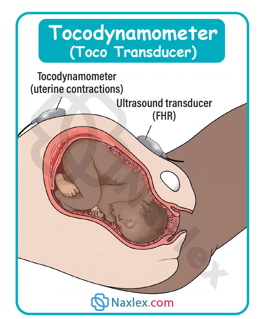
- Measures uterine activity indirectly by detecting changes in abdominal wall tension.
- Placed over the uterine fundus, at the area of greatest contractility.
- Provides data on:
- Frequency of contractions, measured peak to peak.
- Duration of contractions, from onset to relaxation.
- Relative intensity, described qualitatively as mild, moderate, or strong.
- Does NOT measure:
- Actual intrauterine pressure in mmHg.
- True contraction strength objectively.
- Requires periodic repositioning as the fundal height changes throughout labor.
4.2 Ultrasound Transducer (Doppler)

- Utilizes high-frequency sound waves reflected off the fetal heart to detect FHR.
- Placed over the area of maximum fetal heart tone intensity, typically over the fetal back.
- Connected to a cardiotachometer that converts Doppler signals into a continuous FHR tracing.
- Requires ultrasound transmission gel to reduce signal interference.
- Susceptible to artifact from:
- Maternal movement.
- Fetal movement.
- Obesity or excessive abdominal adipose tissue.
4.3 Nursing Care and Client Positioning for External Monitoring
- Position the client in a semi-Fowler or lateral tilt position to prevent supine hypotension syndrome.
- Apply ultrasound gel liberally to the abdominal transducer prior to placement.
- Palpate Leopold maneuvers first to identify fetal back for optimal Doppler placement.
- Secure both transducers using an elastic abdominal belt without over-tightening.
- Reassess transducer placement with every maternal position change.
- Document baseline tracing quality immediately upon application.
4.4 Advantages and Limitations of External Monitoring
|
Aspect |
Advantages |
Limitations |
|
Invasiveness |
Non-invasive, safe for intact membranes |
Cannot be used for precise contraction strength |
|
Application |
Can apply regardless of cervical dilation |
Signal quality affected by maternal obesity |
|
Risk |
No risk of fetal scalp trauma or infection |
Prone to artifact with fetal or maternal movement |
|
Use Case |
Ideal for early labor and antepartum testing |
Less accurate baseline variability detection |
Nursing Insights
- A nurse must reposition the tocodynamometer whenever the client changes position because uterine fundal contour shifts with movement.
- The nurse should recognize that obesity is a leading cause of poor quality external tracings, sometimes necessitating internal monitoring.
- When external Doppler tracing is intermittent or unclear, the nurse should first attempt repositioning before assuming fetal compromise.
Internal Fetal Monitoring
6.1 Fetal Scalp Electrode (FSE)
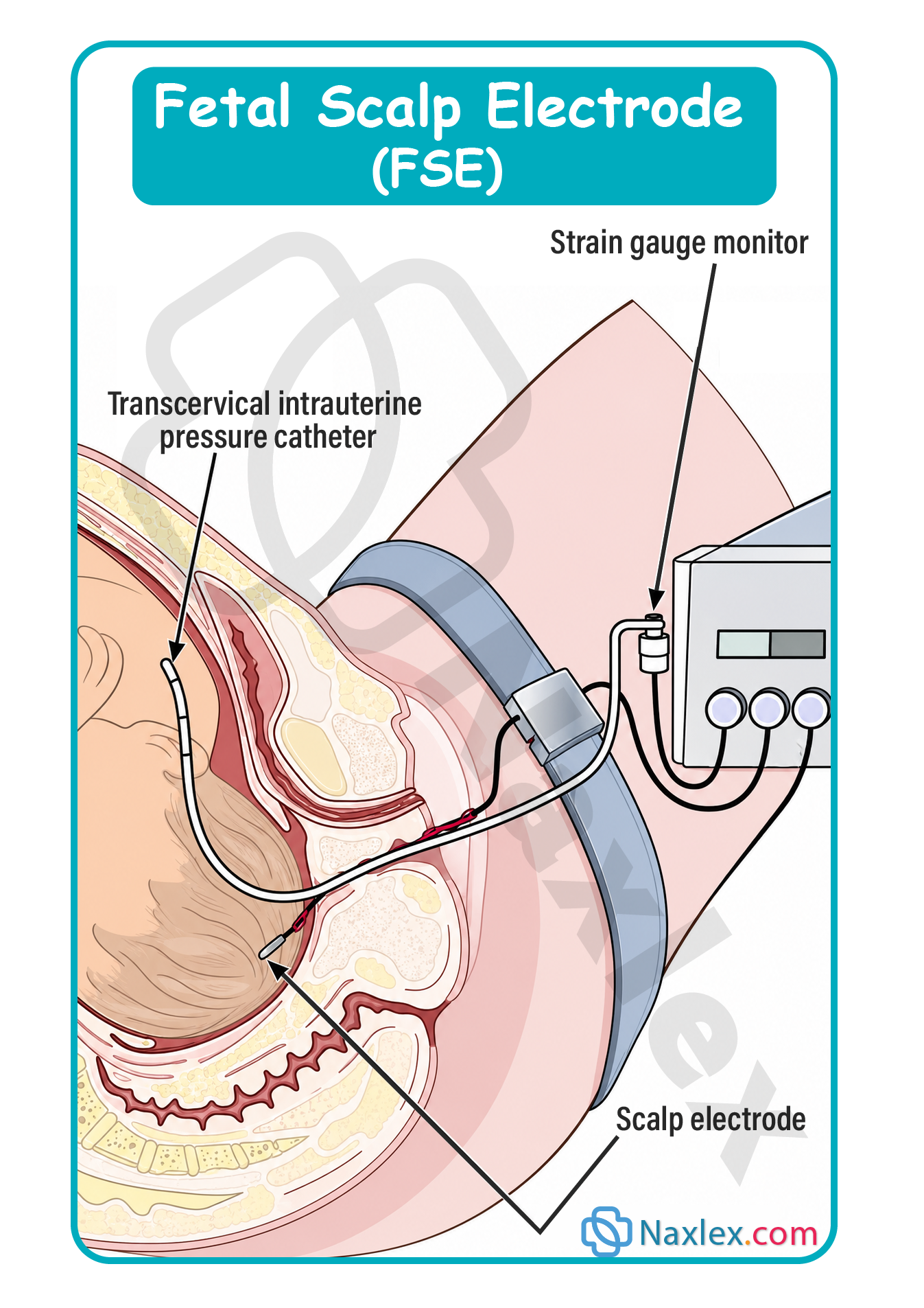
- A spiral wire electrode inserted directly into the fetal presenting part, usually the scalp.
- Provides a direct measurement of the fetal electrocardiogram (ECG) signal.
- Advantages over external ultrasound monitoring:
- Produces a more accurate and continuous baseline FHR.
- Detects beat-to-beat variability with greater precision.
- Unaffected by maternal obesity, movement, or fetal position changes.
- Requirements for insertion:
- Ruptured amniotic membranes.
- Cervical dilation of at least 1-2 cm.
- Known fetal presenting part, most commonly vertex.
- Absence of active maternal infection such as HIV, hepatitis B, or herpes simplex virus.
- Nursing responsibilities:
- Assist the primary health care provider with insertion under sterile technique.
- Confirm correct attachment by observing a clear, continuous FHR tracing.
- Monitor scalp insertion site for signs of infection or bleeding postpartum.
6.2 I ntrauterine Pressure Catheter (IUPC)
A soft, flexible catheter inserted into the uterine cavity alongside the fetus.
- Measures actual intrauterine pressure in mmHg during contractions and at rest.
- Provides objective data on:
- Frequency of contractions.
- Duration of contractions.
- True intensity of contractions, expressed in mmHg.
- Resting tone between contractions, normally 5-15 mmHg.
- Some catheters allow for amnioinfusion, the instillation of warmed sterile normal saline into the uterine cavity.
- Used to relieve variable decelerations caused by umbilical cord compression.
- Used to dilute thick meconium-stained amniotic fluid.
- Montevideo units (MVUs) may be calculated from IUPC data to assess labor adequacy.
- Calculated by summing the pressure generated by each contraction above baseline resting tone within a 10-minute window.
- A value of 200-250 MVUs is generally considered adequate for labor progress.
6.3 Criteria and Prerequisites for Internal Monitoring
- Ruptured amniotic membranes, either spontaneously or artificially.
- Cervical dilation sufficient to allow safe device insertion, typically 1-2 cm or more.
- Engaged fetal presenting part to minimize risk of cord prolapse during insertion.
- Skilled practitioner available for sterile insertion technique.
- Informed consent obtained prior to invasive procedure.
Nursing Insights
- A nurse should remember the mnemonic "ROM" before internal monitoring: Ruptured membranes, Open cervix (dilation), Mobile presenting part must be engaged and identifiable.
- The nurse must verify membrane rupture status and cervical dilation before assisting with FSE or IUPC insertion.
- The nurse should never assist with internal monitoring insertion if the fetal presenting part is unengaged, due to increased cord prolapse risk.
6.4 Risks and Contraindications of Internal Monitoring
- Contraindications include:
- Active maternal genital herpes lesions.
- Maternal human immunodeficiency virus (HIV) infection.
- Maternal hepatitis B or hepatitis C infection.
- Unknown or unengaged fetal presenting part.
- Placenta previa.
- Risks associated with fetal scalp electrode:
- Fetal scalp trauma, abscess, or infection.
- Rare risk of fetal hemorrhage, especially with bleeding disorders.
- Risks associated with intrauterine pressure catheter:
- Uterine perforation during insertion.
- Intrauterine infection, chorioamnionitis.
- Placental laceration if malpositioned.
Nursing Insights
- A nurse caring for a client with active genital herpes lesions must advocate for external monitoring only, since FSE insertion may inoculate the fetus with the virus.
- The nurse should monitor maternal temperature closely after prolonged internal monitoring, as chorioamnionitis risk increases with duration of membrane rupture.
- The mnemonic "TICS" helps recall internal monitoring contraindications: Transmissible infections (HIV, hepatitis, herpes), Incomplete engagement, Cervix undilated, Suspected placenta previa.
Periodic And Episodic Patterns In Fetal Heart Rate
8.1 Baseline Fetal Heart Rate
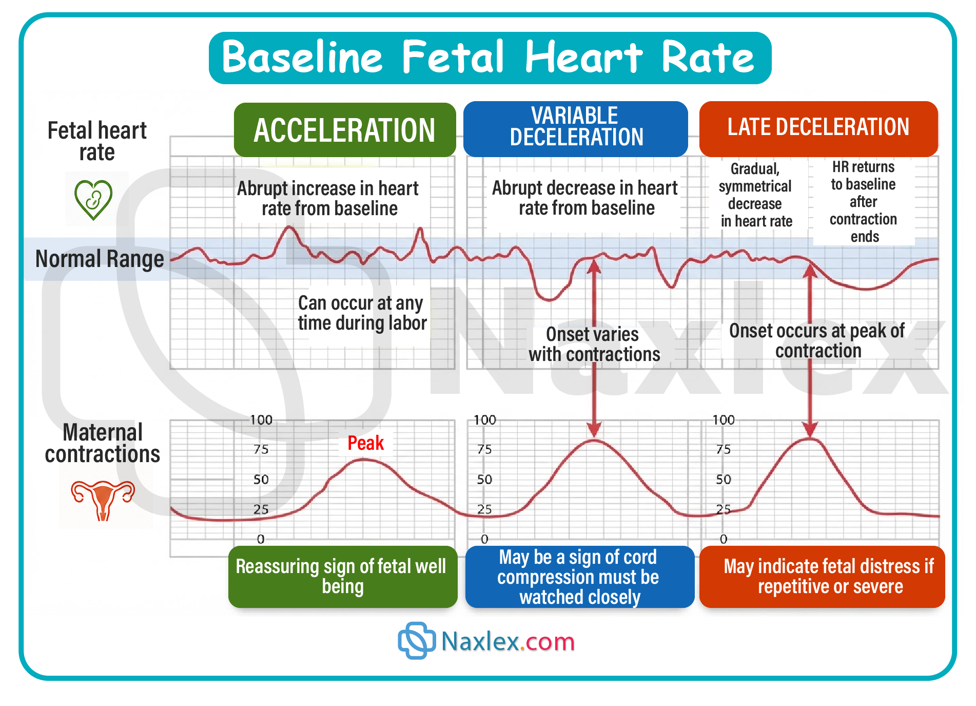
- Defined as the mean FHR rounded to the nearest 5 beats per minute (bpm) during a 10-minute segment.
- Excludes periods of marked variability, periodic changes, and segments differing by more than 25 bpm.
- Normal baseline range is 110-160 bpm.
- Classifications of abnormal baseline:
- Bradycardia: baseline below 110 bpm sustained for 10 minutes or longer.
- Tachycardia: baseline above 160 bpm sustained for 10 minutes or longer.
- Common causes of fetal bradycardia:
- Fetal hypoxia or acidemia.
- Maternal hypotension, often from supine positioning or epidural anesthesia.
- Umbilical cord compression or prolapse.
- Fetal heart block, congenital.
- Common causes of fetal tachycardia:
- Maternal fever or chorioamnionitis.
- Fetal hypoxia, early compensatory stage.
- Maternal or fetal anemia.
- Maternal use of beta-sympathomimetic medications.
- Fetal tachyarrhythmia.
Nursing Insights
- A nurse noting a sustained fetal bradycardia of 90 bpm must first assess maternal blood pressure and reposition the client onto her side.
- The nurse should recognize maternal fever, temperature above 38°C (100.4°F), as a leading cause of fetal tachycardia, warranting temperature reassessment.
8.2 Baseline Variability
- Refers to fluctuations in the baseline FHR, reflecting fetal autonomic nervous system integrity.
- Considered the single most important indicator of adequate fetal oxygenation.
- Classified into 4 categories based on amplitude range:
|
Category |
Amplitude Range |
Clinical Significance |
|
Absent |
Undetectable |
Non-reassuring, requires immediate action |
|
Minimal |
≤5 bpm |
Non-reassuring, warrants close evaluation |
|
Moderate |
6-25 bpm |
Reassuring, indicates adequate oxygenation |
|
Marked |
>25 bpm |
Uncertain significance, requires evaluation |
- Causes of decreased variability:
- Fetal hypoxia or acidemia.
- Fetal sleep cycle, typically lasting 20-40 minutes.
- Maternal narcotic or sedative administration.
- Fetal prematurity, less than 32 weeks gestation.
- Fetal congenital anomalies.
- Causes of increased (marked) variability:
- Early mild fetal hypoxemia, compensatory stage.
- Fetal stimulation or movement.
- Cord compression, transient.
Nursing Insights
- A nurse should differentiate fetal sleep cycles from pathologic minimal variability by attempting vibroacoustic stimulation to elicit an acceleration.
- The nurse must consider recent maternal opioid administration before interpreting minimal variability as fetal compromise.
8.3 Accelerations

- Defined as an abrupt increase in FHR above baseline, onset to peak in less than 30 seconds.
- Criteria for a qualifying acceleration:
- At 32 weeks gestation or greater: peak ≥15 bpm above baseline, lasting ≥15 seconds but <2 minutes.
- Before 32 weeks gestation: peak ≥10 bpm above baseline, lasting ≥10 seconds but <2 minutes.
- Prolonged acceleration: lasts 2 minutes or longer but less than 10 minutes.
- If an acceleration lasts 10 minutes or longer, it is redefined as a baseline change.
- Considered a reassuring sign, indicating fetal well-being and adequate central nervous system oxygenation.
- Often associated with fetal movement, vaginal examination, or vibroacoustic stimulation.
Nursing Insights
- A nurse should document accelerations as a reassuring finding requiring no intervention, reflecting fetal neurological integrity.
- The nurse can use vibroacoustic stimulation to elicit an acceleration and confirm fetal well-being when variability appears minimal.
8.4 Early Decelerations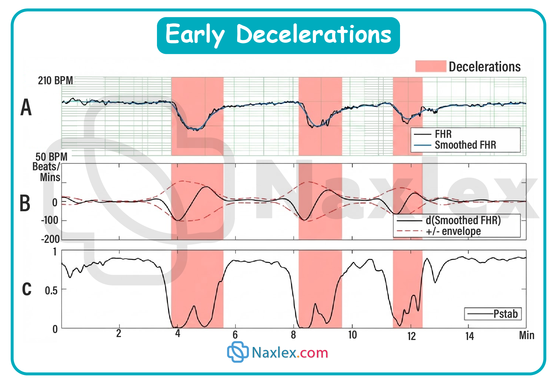
- A gradual decrease and return to baseline FHR, onset to nadir 30 seconds or more.
- Mirror image of the corresponding uterine contraction, with the nadir occurring at the peak of the contraction.
- Caused by fetal head compression during descent, stimulating a vagal response.
- Considered a benign, physiologic pattern requiring no intervention.
- Commonly seen during the active and transition phases of labor.
Nursing Insights
- A nurse should recognize early decelerations as reassuring and continue routine monitoring without intervention.
- The nurse must correctly distinguish early decelerations from late decelerations, since both are gradual but differ in timing relative to the contraction.
8.5 Late Decelerations

- A gradual decrease and return to baseline FHR, onset to nadir 30 seconds or more.
- Onset, nadir, and recovery occur after the corresponding contraction begins, peaks, and ends respectively.
- Caused by uteroplacental insufficiency, resulting in decreased fetal oxygenation.
- Common causes include:
- Maternal hypotension.
- Uterine tachysystole.
- Placental abruption or previa.
- Post-term placental aging.
- Maternal diabetes or hypertension affecting placental perfusion.
- Considered a non-reassuring pattern requiring immediate nursing intervention.
Nursing Insights
- A nurse identifying recurrent late decelerations must immediately reposition the client to the lateral position, administer oxygen, and discontinue oxytocin if infusing.
- The nurse should notify the primary health care provider promptly, as recurrent late decelerations reflect uteroplacental insufficiency and possible fetal acidemia.
8.6 Variable Decelerations
- An abrupt decrease in FHR, onset to nadir less than 30 seconds.
- Decrease is ≥15 bpm below baseline, lasting ≥15 seconds but less than 2 minutes.
- Variable in onset, duration, and depth relative to contractions, hence the name.
- Caused by umbilical cord compression, transient or recurrent.
- Often described as having a U, V, or W shape on the tracing.
- May be preceded and followed by brief accelerations, termed "shoulders."
Nursing Insights
- A nurse detecting variable decelerations should first reposition the client to relieve cord compression, such as a lateral or knee-chest position.
- The nurse should consider amnioinfusion as an intervention for recurrent variable decelerations caused by oligohydramnios.
8.7 Prolonged Decelerations and Sinusoidal Pattern
- Prolonged deceleration:
- A decrease in FHR of ≥15 bpm below baseline, lasting 2 minutes or longer but less than 10 minutes.
- If lasting 10 minutes or longer, reclassified as a baseline change.
- Causes include cord prolapse, uterine rupture, maternal seizure, or prolonged maternal hypotension.
- Sinusoidal pattern:
- A smooth, regular, sine wave-like undulating pattern in baseline FHR.
- Cycle frequency of 3-5 per minute, persisting for 20 minutes or longer.
- Associated with severe fetal anemia, such as from Rh isoimmunization or fetal-maternal hemorrhage.
- Considered an ominous pattern requiring immediate intervention.
Nursing Insights
- A nurse recognizing a sinusoidal pattern must notify the primary health care provider immediately, as this pattern is associated with severe fetal anemia and impending fetal demise.
- The nurse should distinguish a true prolonged deceleration from a sinusoidal pattern, since management priorities differ significantly.
Mnemonic: VEAL CHOP
- A widely used mnemonic connecting periodic FHR patterns to their causes and appropriate nursing response:
- Variable decelerations correspond to Cord compression.
- Early decelerations correspond to Head compression.
- Accelerations correspond to Oxygenation, indicating fetal well-being.
- Late decelerations correspond to Placental insufficiency.
- Explanation for nursing application:
- Variable and Cord: nurse intervenes with maternal repositioning to relieve cord compression.
- Early and Head: nurse recognizes this as benign, requiring no intervention.
- Acceleration and Oxygenation: nurse documents as reassuring, reflecting fetal well-being.
- Late and Placental insufficiency: nurse intervenes urgently to improve uteroplacental blood flow.
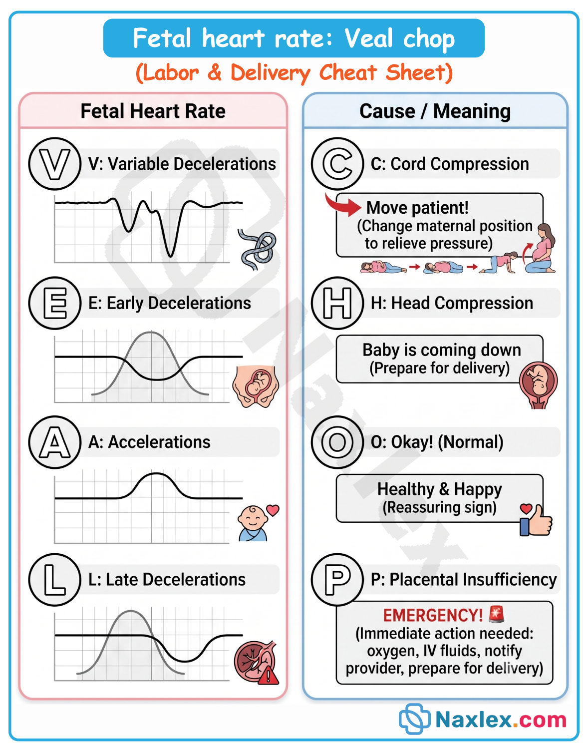
Nursing Insights
- A nurse should use the VEAL CHOP mnemonic at the bedside to rapidly classify a deceleration pattern and determine the corresponding intervention.
- The nurse must remember that while early decelerations require no action, late and variable decelerations demand a structured intrauterine resuscitation response.
Fetal Heart Rate Tracing Classification And Nursing Interventions
10.1 Three-Tier Fetal Heart Rate Interpretation System
- Developed by the National Institute of Child Health and Human Development (NICHD).
- Classifies FHR tracings into 3 categories based on overall risk of fetal acidemia.
- Category I (Normal):
- Baseline 110-160 bpm.
- Moderate baseline variability.
- Absent late or variable decelerations.
- Early decelerations may be present or absent.
- Accelerations may be present or absent.
- Strongly predictive of normal fetal acid-base status at the time of observation.
- Nursing action: continue routine monitoring, no intervention required.
- Category II (Indeterminate):
- Includes all tracings not meeting Category I or Category III criteria.
- Examples include:
- Tachycardia or bradycardia without absent variability.
- Minimal or marked baseline variability.
- Absent variability without recurrent decelerations.
- Recurrent variable decelerations with minimal or moderate variability.
- Recurrent late decelerations with moderate variability.
- Prolonged decelerations lasting 2-10 minutes.
- Not predictive of abnormal fetal acid-base status but requires evaluation, continued surveillance, and possible intrauterine resuscitation.
- Nursing action: implement corrective measures and reassess frequently.
- Category III (Abnormal):
- Absent baseline variability with any of the following:
- Recurrent late decelerations.
- Recurrent variable decelerations.
- Bradycardia.
- Sinusoidal pattern.
- Predictive of abnormal fetal acid-base status at the time of observation.
- Requires prompt evaluation and immediate intervention.
- Nursing action: initiate intrauterine resuscitation immediately and notify the primary health care provider.
- Absent baseline variability with any of the following:
Nursing Insights
- A nurse identifying a Category III tracing must intervene immediately rather than waiting for provider arrival, as this reflects probable fetal acidemia.
- The nurse should reassess Category II tracings frequently, since this category encompasses a wide clinical spectrum requiring clinical judgment.
10.2 Intrauterine Resuscitation Measures
- Goal is to improve fetal oxygenation and uteroplacental blood flow rapidly.
- Standard nursing interventions include:
- Repositioning the client to a lateral position, either left or right side-lying.
- Administering an intravenous fluid bolus of isotonic solution to improve maternal circulating volume.
- Discontinuing oxytocin infusion if uterine tachysystole is present.
- Administering supplemental oxygen via face mask, typically 8-10 liters per minute.
- Correcting maternal hypotension, particularly after epidural anesthesia.
- Preparing for amnioinfusion if variable decelerations persist due to cord compression.
- Administering a tocolytic agent, such as terbutaline, if ordered, to reduce uterine activity.
- Reassessment after interventions:
- Reevaluate the FHR tracing for resolution of the non-reassuring pattern.
- Reassess maternal vital signs, particularly blood pressure and pulse.
- Document response to interventions and notify the primary health care provider of ongoing concerns.
Nursing Insights
- A nurse implementing intrauterine resuscitation should perform interventions simultaneously rather than sequentially when a Category III tracing is identified.
- The nurse must recognize that repositioning and oxygen administration are first-line, low-risk interventions appropriate for nearly all non-reassuring patterns.
10.3 Documentation and Communication (SBAR)
- SBAR is a widely used structured communication framework: Situation, Background, Assessment, Recommendation.
- Situation: concise statement of the current FHR concern, such as recurrent late decelerations.
- Background: relevant maternal-fetal history, gestational age, labor progress, and risk factors.
- Assessment: nurse's clinical interpretation, including FHR category and interventions already performed.
- Recommendation: specific request, such as provider bedside evaluation or order clarification.
- Documentation should include:
- Time of pattern onset and resolution.
- Specific interventions performed and client response.
- Time of provider notification and orders received.
- Category classification of the tracing at the time of the event.
Nursing Insights
- A nurse using SBAR to report a Category III tracing should lead with the situation clearly, stating the specific abnormal finding before background details.
- The nurse must document interventions in real time, since delayed documentation of a non-reassuring tracing carries significant legal and clinical risk.
Summary
- Fetal heart rate monitoring is essential for evaluating fetal oxygenation status during labor.
- External monitoring uses a tocodynamometer for contraction timing and an ultrasound transducer for FHR detection, both non-invasive but limited by maternal body habitus and movement artifact.
- Internal monitoring, using a fetal scalp electrode and intrauterine pressure catheter, provides direct and objective data but requires ruptured membranes, adequate cervical dilation, and absence of contraindications such as active maternal infection.
- Baseline FHR normally ranges from 110-160 bpm, with bradycardia and tachycardia reflecting distinct underlying etiologies.
- Baseline variability is the most reliable indicator of fetal central nervous system oxygenation, classified as absent, minimal, moderate, or marked.
- Periodic patterns include accelerations, early decelerations, late decelerations, and variable decelerations, each with distinct causes summarized by the VEAL CHOP mnemonic.
- The NICHD three-tier system classifies tracings into Category I, II, or III, guiding the urgency of nursing intervention.
- Intrauterine resuscitation measures, including repositioning, oxygen administration, intravenous fluid bolus, and discontinuing oxytocin, are foundational nursing responses to non-reassuring patterns.
- Clear, timely documentation and SBAR communication with the primary health care provider are essential to ensure prompt escalation of care and optimal maternal-fetal outcomes.
Naxlex
Videos
Login to View Video
Click here to loginTake Notes on Fetal Monitoring
This filled cannot be empty
Join Naxlex Nursing for nursing questions & guides! Sign Up Now