





 Video Library


Electrocardiography and Dysrhythmia Monitoring
Study Questions
Practice Exercise 1
A nurse is conducting staff education about ECG monitoring. Which statement accurately describes the process.
Explanation
The cardiac conduction system initiates electrical impulses through a specialized sequence to ensure coordinated myocardial contraction. The sinoatrial node acts as the primary pacemaker, while leads provide specific electrophysiological monitoring views. Accurate lead placement is vital for capturing precise waveforms, preventing signal distortion, and ensuring correct interpretation of cardiac rhythm disturbances.
Rationale for correct answer
3. Standard precordial lead placement requires V1 to be positioned at the fourth intercostal space to the right of the sternum. This anatomical landmark ensures accurate diagnostic visualization of septal cardiac activity. Proper placement remains essential for capturing clear electrical signals. This placement protocol is mandatory for obtaining valid data.
Rationale for incorrect answers
1. Ventricular depolarization is represented by the QRS complex, not the T wave. The T wave signifies ventricular repolarization, which is the electrical recovery phase of the myocardium. The QRS complex captures the active contraction of the ventricles. This choice misidentifies the waveform component.
2. An artifact is an unwanted signal caused by motion or electrical interference. A premature beat originating in the atria or ventricles is an ectopic rhythm, not a technical error. Confusing technical noise with clinical rhythm disturbances leads to diagnostic inaccuracies. This description incorrectly defines clinical events.
4. The atrioventricular junction functions as a latent pacemaker with an intrinsic rate of 40 to 60 beats per minute. A rate of 30 to 40 beats per minute characterizes a ventricular escape rhythm. The junctional rate does not fall into the lower ventricular range. This choice provides inaccurate physiological firing rates.
Test-taking strategy
- Analyze the Scenario and Question: The question requires identifying the accurate statement regarding ECG monitoring and lead placement principles.
- The goal is to distinguish between correct physiological nomenclature and common clinical misconceptions.
- Apply Cardiac Electrophysiology Principles: Evaluate the definition of waves and pacemaker rates to Rule out incorrect clinical statements.
- Rule out 1: Depolarization refers to the QRS complex rather than the T wave.
- Rule out 2: Artifacts are external interference signals, not cardiac impulses.
- Rule out 4: Junctional pacemakers fire at 40 to 60 beats per minute.
- Identify Correct Diagnostic Protocol: Determine the anatomical accuracy of electrode placement.
- Confirm 3: Standard precordial placement for V1 is correctly identified as the fourth intercostal space at the right sternal border.
Take home points
- The QRS complex represents ventricular depolarization while the T wave represents ventricular repolarization.
- Artifacts are technical errors caused by external factors rather than abnormal electrical cardiac impulses.
- The sinoatrial node functions at 60 to 100 bpm, the AV junction at 40 to 60 bpm, and ventricles at 20 to 40 bpm.
- Proper precordial lead placement is critical for obtaining diagnostic quality tracings and identifying specific wall ischemia.
What accurately describes the PR interval? Select all that apply
Explanation
The PR interval is a fundamental measurement in electrocardiography, representing the time required for an electrical impulse to travel from the sinoatrial node through the atria, the AV node, and the bundle of His to reach the ventricular myocardium. Assessing this interval is critical for diagnosing conduction delays or blocks within the atrio-ventricular junction.
Rationale for correct answer
4. The normal clinical range for the PR interval is 0.12 to 0.20 seconds. This timeframe accounts for the necessary physiological delay at the AV node, which allows the atria to contract fully and fill the ventricles before ventricular systole begins.
6. The PR interval is defined by the measurement from the beginning of the P wave (atrial depolarization) to the beginning of the QRS complex (ventricular depolarization). This covers the entire duration of atrial conduction and the nodal delay.
Rationale for incorrect answers
1. While 0.16 seconds is a normal value, it is a specific example rather than a description of the interval itself, and including it as a primary descriptor is redundant given the specified range.
2. An interval less than 0.12 seconds is not the standard range and typically indicates a pre-excitation syndrome, such as Wolff-Parkinson-White, where impulses bypass the AV node delay.
3. The range 0.06 to 0.12 seconds is incorrect for the PR interval. This timeframe is actually characteristic of the normal QRS complex duration, which represents ventricular depolarization.
5. The PR interval does not measure repolarization. Ventricular repolarization is represented by the ST segment and the T wave. Misidentifying this can lead to incorrect assessments of cardiac repolarization phases.
Test-taking strategy
- Analyze the Question: The focus is on identifying the definition and normal values of the PR interval.
- The goal is to differentiate between the PR interval, the QRS complex, and other cardiac intervals.
- Apply ECG Interpretation Principles:
- Rule out 2: Less than 0.12 indicates pre-excitation, not a normal range.
- Rule out 3: 0.06 to 0.12 describes the QRS width, not the PR interval.
- Rule out 5: PR does not involve ventricular repolarization (T wave).
- Confirm 4, 6: Recognize the standard 0.12 to 0.20 range and the definition of the start-to-start measurement.
Take home points
- The PR interval reflects the time for atrial depolarization and the AV node delay.
- The normal PR interval duration is strictly 0.12 to 0.20 seconds.
- Intervals shorter than 0.12 seconds suggest bypass tracts, while intervals longer than 0.20 seconds indicate AV blocks.
- Distinguishing between the PR interval and QRS duration is essential for accurate rhythm analysis and identifying conduction abnormalities.
What describes the SA node’s ability to discharge an electrical impulse spontaneously?
Explanation
The sinoatrial (SA) node acts as the heart's primary pacemaker, responsible for initiating each cardiac cycle. This unique function relies on the specialized property of cardiac cells to spontaneously generate an electrical action potential without external neural or hormonal stimulation. This intrinsic rhythmicity ensures a continuous, self-sustaining heartbeat, which is the foundational requirement for maintaining effective systemic perfusion.
Rationale for correct answer
4. Automaticity is the specific property of cardiac cells—most notably those in the SA node—to spontaneously depolarize and generate an electrical impulse. This capability allows the heart to initiate its own electrical activity, independent of the central nervous system, establishing the baseline heart rate under normal physiological conditions.
Rationale for incorrect answers
1. Excitability refers to the ability of cardiac cells to respond to an electrical stimulus once it is received. While necessary for the propagation of an impulse, it does not describe the spontaneous generation of that impulse itself.
2. Contractility describes the ability of the myocardial muscle fibers to shorten and generate force in response to an electrical stimulus. This is a mechanical property related to the physical pumping action of the heart, not the electrical triggering of a heartbeat.
3. Conductivity is the ability of cardiac cells to transmit an electrical impulse from one cell to the next. This facilitates the rapid spread of electricity throughout the heart's conduction system, but like excitability, it is a response mechanism rather than the act of initiation.
Test-taking strategy
- Analyze the Question: The focus is on identifying the specific electrical property that allows the SA node to start an impulse on its own.
- The goal is to distinguish between the four core electrophysiological properties of cardiac tissue.
- Apply Electrophysiology Principles:
- Rule out 1: Excitability is about reacting to a stimulus.
- Rule out 2: Contractility is about the mechanical pumping action.
- Rule out 3: Conductivity is about transmitting an existing signal.
- Confirm 4: Automaticity is the "automatic" generation of an impulse.
- Identify Definitions:
- Focus on the key term: "Spontaneously" is synonymous with "automatic."
Take home points
- Automaticity is the unique ability of pacemaker cells (like those in the SA node) to discharge impulses spontaneously.
- Excitability, conductivity, and contractility are essential supportive functions but do not initiate the rhythm.
- The SA node has the highest rate of automaticity, which is why it serves as the dominant pacemaker for a healthy heart.
- Understanding these electrophysiological terms is critical for diagnosing conduction disorders and rhythm disturbances.
What describes refractoriness?
Explanation
Refractoriness is a fundamental electrophysiological property of cardiac tissue that prevents the heart from becoming tetanized or contracting prematurely. During this period, the cardiac cells are physically unable to respond to a new stimulus because their internal electrical state is undergoing restoration. This safety mechanism ensures a coordinated, rhythmic, and effective pumping action by the heart muscle.
Rationale for correct answer
2. Refractoriness is defined as the period during which heart tissue is unresponsive to electrical stimulation. This phase is divided into absolute (where no stimulus can trigger a contraction) and relative (where a very strong stimulus might trigger a premature, abnormal response), safeguarding the integrity of the cardiac cycle.
Rationale for incorrect answers
1. Abnormal electrical impulses are referred to as ectopic foci or dysrhythmias. These represent a failure of the heart's normal electrical conduction, whereas refractoriness is a normal, healthy, and essential physiological feature of cardiac cell function.
3. Areas of the heart that do not repolarize at the same rate due to depressed conduction are associated with dispersion of repolarization. This state creates electrical heterogeneity in the myocardium, which is a major mechanism for re-entrant tachyarrhythmias rather than being a definition of refractoriness.
4. Sodium migrating rapidly into the cell to make it positive compared with the outside is the description of depolarization. This phase is when the heart muscle actually receives the stimulus and triggers a contraction, which is the opposite of the refractory state.
Test-taking strategy
- Analyze the Question: The focus is on defining the term "refractoriness" in the context of cardiac physiology.
- The goal is to differentiate between normal electrical properties and pathological rhythm states or depolarization events.
- Apply Electrophysiology Principles:
- Rule out 1: Abnormal impulses are pathology, not a structural property of tissue.
- Rule out 3: Non-uniform repolarization describes re-entry mechanics.
- Rule out 4: Sodium influx describes depolarization.
- Confirm 2: The definition of refractoriness is the "unresponsive" or "recovery" period.
- Identify Definitions:
- Focus on the key term: "Refractory" means resistant or unresponsive to stimuli.
Take home points
- Refractoriness is a mandatory recovery period that allows cardiac cells to reset after a contraction.
- The absolute refractory period prevents the heart from accepting any new stimulus, protecting against dangerously high heart rates.
- Depolarization and repolarization are the electrical processes of excitation and recovery, while refractoriness describes the tissue's responsiveness throughout these phases.
- Understanding these cycles is essential to interpreting how dysrhythmias emerge from abnormalities in the heart's recovery phase.
A client’s PR interval comprises six small boxes on the ECG graph. What should the nurse conclude about this finding?
Explanation
Cardiac conduction assessment relies on precisely measuring intervals to ensure electrical impulses travel through the heart within established timeframes. Each small box on a standard ECG graph represents 0.04 seconds. An interval measuring six small boxes totals 0.24 seconds, which exceeds the normal limit of 0.20 seconds, signaling a significant conduction delay.
Rationale for correct answer
4. A PR interval of 0.24 seconds indicates a first-degree AV block. This means the electrical impulse originating in the SA node is being delayed excessively as it travels through the AV node, the Bundle of His, or the bundle branches before reaching the ventricular myocardium.
Rationale for incorrect answers
1. A normal PR interval is defined as 0.12 to 0.20 seconds (three to five small boxes). Since six boxes equal 0.24 seconds, this finding is clearly outside the range of normal conduction time and represents a clinical abnormality.
2. Ventricular depolarization is measured by the duration of the QRS complex, not the PR interval. Problems with ventricular depolarization would appear as a widened QRS complex (greater than 0.12 seconds), not as an abnormality in the PR interval.
3. The PR interval reflects conduction through the atria and the AV node; it does not specifically measure the repolarization of the atria. Atrial repolarization actually occurs simultaneously with ventricular depolarization and is buried within the QRS complex, making it invisible on a standard surface ECG.
Test-taking strategy
- Analyze the Scenario/Question: Calculate the duration (6 boxes $\times$ 0.04 seconds = 0.24 seconds) and identify the clinical implication.
- The goal is to link a prolonged PR interval to a specific site of conduction disturbance.
- Apply ECG Principles:
- Rule out 1: The math (0.24s) exceeds the normal range (0.20s).
- Rule out 2: Ventricular issues relate to the QRS complex.
- Rule out 3: Atrial repolarization is not measured by the PR interval.
- Confirm 4: The PR interval specifically measures the time from the SA node to the ventricles; a delay here means a conduction problem.
Take home points
- Each small ECG box equals 0.04 seconds.
- A PR interval exceeding 0.20 seconds signifies a conduction delay, commonly identified as a first-degree AV block.
- The PR interval is the diagnostic window for assessing conduction velocity from the atria to the ventricles.
- Clinical assessment must differentiate between atrial/nodal delays (PR interval) and ventricular conduction delays (QRS width).
A client is undergoing electrophysiologic testing and requires close cardiac monitoring. Which reason best explains why the nurse should be prepared to respond rapidly to changes in the client’s cardiac rhythm?
Explanation
Electrophysiologic testing (EPS) is an invasive diagnostic procedure where electrodes are placed within the heart to map its electrical pathways and intentionally trigger potential rhythm disturbances. The primary goal of the study is to identify the precise mechanism of a patient's tachycardia or conduction abnormality by reproducing the rhythm in a controlled environment. Because these induced rhythms can be hemodynamically unstable, the nursing team must be prepared to intervene immediately to restore a sinus rhythm.
Rationale for correct answer
4. The nature of EPS involves electrical stimulation designed to provoke the patient's symptomatic dysrhythmia. If the induced rhythm results in significant hypotension or loss of consciousness, the provider must perform cardioversion or defibrillation to terminate the rhythm and return the heart to a stable, perfusing state.
Rationale for incorrect answers
1. While contrast dye is sometimes used during cardiac catheterization or angiography, it is not the primary component of an electrophysiologic study. The inherent risks of EPS are related to the electrical induction of rhythms, not the chemical irritation of the myocardium by dyes.
2. Although any invasive procedure carries risks, the EPS procedure is not specifically designed to cause myocardial ischemia. While the induction of certain tachycardias can increase oxygen demand, the clinical focus is on the electrical trigger itself rather than a primary ischemic insult.
3. Anticoagulation is a standard safety protocol during many invasive cardiac procedures to prevent clot formation on catheters. While managing anticoagulation is part of the overall care, it is not the reason the nurse must be prepared for emergency rhythm correction; the risk of induced, unstable rhythms is the direct rationale for vigilance.
Test-taking strategy
- Analyze the Scenario/Question: The focus is on the specific purpose and risk profile of an electrophysiologic study (EPS).
- The goal is to link the procedure's diagnostic mechanism (rhythm induction) to the necessary clinical preparation (rhythm correction).
- Apply Procedure-Specific Knowledge:
- Rule out 1: Dye is not the primary diagnostic agent for EPS.
- Rule out 2: Ischemia is not the target mechanism; electrical induction is.
- Rule out 3: Anticoagulation is a general safety measure, not the reason for rapid rhythm response.
- Confirm 4: The procedure's defining characteristic is the intentional creation of dysrhythmias, which may need to be stopped if they become dangerous.
Take home points
- Electrophysiologic studies are diagnostic procedures that map cardiac electrical activity by inducing specific rhythms.
- Induced rhythms during an EPS can become hemodynamically unstable, necessitating immediate conversion.
- Nurses must be prepared for rapid intervention, including defibrillation, cardioversion, or administration of antiarrhythmic medications.
- Close monitoring is non-negotiable throughout the duration of the test due to the unpredictability of the provoked rhythms.
The nurse is preparing to obtain a standard 12-lead ECG on a client experiencing acute chest discomfort. Which action by the nurse is appropriate?
Explanation
The 12-lead ECG is a critical diagnostic tool for identifying acute myocardial infarction and dysrhythmias. Precise electrode placement is essential to ensure that the electrical vectors are recorded from the standard anatomical views of the heart. Even minor deviations in lead positioning can create artifacts or misleading tracings, potentially delaying the recognition of life-threatening ischemic changes.
Rationale for correct answer
3. The V4 electrode is correctly placed in the fifth intercostal space at the left midclavicular line. This position is a standard landmark for recording the electrical activity of the heart's apex and is essential for obtaining an accurate view of the left ventricular wall.
Rationale for incorrect answers
1. V1 is correctly placed in the fourth intercostal space at the right sternal border, not the left. Placing V1 at the left sternal border is a common error that compromises the accuracy of the septal view on the ECG.
2. V2 is correctly placed in the fourth intercostal space at the left sternal border, not the right. Similar to V1, swapping the side of the sternal border results in inaccurate recordings of the electrical potential across the cardiac septum.
4. V6 is placed in the fifth intercostal space, not the sixth, at the left midaxillary line. Placing the electrode in the sixth intercostal space is too low and results in an inaccurate recording of the lateral wall of the left ventricle.
Test-taking strategy
- Analyze the Question: The focus is on the correct anatomical landmarks for precordial lead placement on a 12-lead ECG.
- The goal is to identify the one correct landmark from the list of provided options.
- Apply Clinical Knowledge:
- Review the standard landmarks:
- V1: 4th ICS, Right Sternal Border.
- V2: 4th ICS, Left Sternal Border.
- V3: Midpoint between V2 and V4.
- V4: 5th ICS, Midclavicular Line.
- V5: 5th ICS, Anterior Axillary Line.
- V6: 5th ICS, Midaxillary Line.
- Rule out 1: Incorrect side (Left vs Right).
- Rule out 2: Incorrect side (Right vs Left).
- Rule out 4: Incorrect ICS (6th vs 5th).
- Confirm 3: Correct ICS and correct landmark (Midclavicular).
- Review the standard landmarks:
Take home points
- Accurate electrode placement is mandatory for the valid interpretation of cardiac electrical activity.
- The precordial leads (V1 to V6) must follow the standard 4th and 5th intercostal space landmarks relative to the sternum and axillary lines.
- Nurses must master these landmarks to ensure that the diagnostic results accurately reflect the patient's myocardial status during an emergency.
The nurse reviews a 12-lead ECG and observes that the tracing demonstrates unusual abnormalities in the inferior leads. The client is asymptomatic, and the tracing does not correlate with the clinical assessment. Which action should the nurse take first?
Explanation
When clinical findings do not correlate with diagnostic test results, the nurse must first verify the integrity of the data. Inaccurate ECG tracings are frequently caused by technical errors rather than underlying cardiac pathology. Assessing the quality of the technical setup ensures that the healthcare team does not misdiagnose the client or initiate unnecessary, high-risk cardiac interventions based on false data.
Rationale for correct answer
3. Inspecting limb lead placement is the priority action. Reversing leads (such as swapping the arm leads) is a common technical error that can create "abnormalities" mimicking ischemia in specific leads, such as the inferior leads. Repeating the ECG after correcting the placement confirms whether the findings were an artifact or a genuine clinical event.
Rationale for incorrect answers
1. Notifying the provider of an acute MI based on an unverified, clinically inconsistent tracing is premature. This could lead to inappropriate administration of emergency cardiac drugs or unnecessary activation of the catheterization lab, which carries significant risks for the client.
2. Administering oxygen and starting continuous monitoring are appropriate only after confirming the presence of a real cardiac event. If the ECG abnormality is merely a technical artifact, these interventions are unnecessary and do not address the primary issue of data validation.
4. Removing the electrodes without verifying the result is negligent. If the client were truly experiencing an MI, delaying the correct diagnosis could be life-threatening. The nurse must troubleshoot the technical issue or escalate the concern, rather than discarding the test and documenting it as inconclusive.
Test-taking strategy
- Analyze the Scenario/Question: The client is asymptomatic but the ECG shows an inferior MI.
- The goal is to resolve the discrepancy between the clinical picture (stable) and the diagnostic result (abnormal).
- Apply Clinical Logic:
- "If the clinical picture and the test don't match, verify the test."
- Rule out 1: Premature diagnosis leads to harmful errors.
- Rule out 2: Symptom-driven treatment is not yet indicated for an unconfirmed artifact.
- Rule out 4: Documentation must be accurate; ignoring a potential issue is dangerous.
- Confirm 3: Technical troubleshooting is the first, safest step in validating diagnostic results.
Take home points
- Always treat the patient, not just the monitor. An asymptomatic patient with a "dramatic" ECG should trigger a check for technical error.
- Lead reversal (e.g., limb lead swaps) is a frequent cause of false inferior or lateral wall changes on a 12-lead ECG.
- Nursing judgment involves validating data through systematic troubleshooting before escalating a clinical alert.
- Promptly repeating an ECG or correcting lead placement saves time, reduces medical errors, and prevents unnecessary alarm.
A nurse is evaluating an ECG tracing for evidence of normal ventricular depolarization. Which finding is expected?
Explanation
The QRS complex represents the electrical activation of the ventricles. In a healthy heart, the conduction system efficiently delivers the impulse through the Bundle of His, the bundle branches, and the Purkinje fibers, resulting in a rapid, synchronized contraction. A QRS duration of less than 0.12 seconds is the standard for normal, efficient ventricular depolarization; anything wider suggests an electrical delay or a non-sinus origin of the beat.
Rationale for correct answer
1. A QRS duration of less than 0.12 seconds (less than three small boxes) indicates that the electrical impulse is traveling through the ventricles via the normal, rapid conduction pathways. This confirms that the ventricles are depolarizing in a coordinated and efficient manner.
Rationale for incorrect answers
2. A QRS duration greater than 0.20 seconds is highly abnormal and indicates a significant delay or block in ventricular conduction, such as a complete bundle branch block or a ventricular escape rhythm. This is not a finding of normal depolarization.
3. The absence of a QRS complex after every P wave describes a heart block, where atrial impulses fail to conduct to the ventricles. Normal conduction requires that every P wave is followed by a QRS complex, indicating successful AV node transmission.
4. A QRS complex occurring before the P wave is characteristic of a junctional or ventricular rhythm. In a normal sinus rhythm, the P wave (atrial depolarization) must precede the QRS complex (ventricular depolarization) to maintain the correct electrical sequence.
Test-taking strategy
- Analyze the Question: The goal is to identify the technical definition of normal ventricular depolarization on an ECG.
- Focus on the two main criteria for a normal QRS: width (duration) and morphology/timing.
- Apply Electrophysiology Principles:
- Rule out 2: A wide complex (greater than 0.12s) is pathological.
- Rule out 3: Every P wave must be followed by a QRS for normal conduction.
- Rule out 4: The P wave must precede the QRS in a normal heartbeat.
- Confirm 1: The duration must be narrow (less than 0.12s) to represent normal, rapid conduction.
Take home points
- Normal ventricular depolarization is represented by a narrow QRS complex.
- A QRS width of 0.12 seconds or greater signifies a ventricular conduction delay (e.g., bundle branch block).
- In a healthy conduction sequence, every QRS complex should be preceded by a P wave and represent a successful atrial-to-ventricular impulse.
- Understanding these baseline values is essential for quickly identifying rhythm disturbances that threaten cardiac output.
The nurse is preparing to obtain an ECG from a client who is diaphoretic and has significant chest hair. Which interventions should the nurse implement? Select all that apply
Explanation
Diagnostic accuracy during electrocardiography depends on minimizing electrical impedance and motion artifacts. Proper skin preparation and patient stabilization are essential to ensure the conductive gel interface remains intact. High quality tracings require reducing electrical interference from diaphoresis or body hair. Failure to optimize these factors yields poor diagnostic data.
Rationale for correct answers
1. Ensuring the client is relaxed prevents somatic muscle tremors. These involuntary muscle contractions create excessive electrical noise on the tracing. Static positioning is essential for obtaining a clean, diagnostic quality rhythm strip during the procedure.
3. Cleaning and drying the skin is a vital step for adherence. Sweat acts as a conductive medium that can cause short circuits between electrodes. Proper skin preparation reduces signal impedance and ensures the electrode remains securely attached to the skin.
4. Removing hair at the electrode site reduces physical barriers. Hair follicles create air pockets that prevent the conductive gel from reaching the epidermal layer. Trimming improves electrical conductivity and ensures the sensor maintains a consistent connection throughout the recording process.
Rationale for incorrect answers
2. Applying electrodes over hair prevents necessary contact with the skin surface. Hair creates an insulating gap that significantly degrades the signal quality of the ECG. This oversight leads to excessive baseline wander and makes accurate interpretation of the cardiac rhythm impossible.
5. Asking the client to perform continuous deep breathing induces significant chest wall movement. This mechanical motion generates large artifacts that distort the electrocardiographic waveform. Respiratory variations cause inconsistent baseline shifts, which frequently mimic pathological changes or conceal underlying cardiac dysrhythmias.
6. Placing electrodes over clothing is physically inappropriate for electrical signal detection. Fabric acts as a dielectric barrier that blocks the transmission of cardiac electrical potentials to the sensors. This action results in a completely unreadable or blank output on the monitor during testing.
Test-taking strategy
- Analyze the Scenario/Question: The client presents with diaphoresis and dense chest hair, both of which are common technical barriers to recording a clear electrocardiogram. The goal is to maximize signal clarity by eliminating artifact sources.
- Apply technical preparation principles:
- Rule out 2: Excessive hair creates a physical barrier to the electrode, which must be addressed by trimming to obtain a signal.
- Rule out 5: Continuous deep breathing introduces motion, which generates baseline wander and distorts the wave morphology on the recording.
- Rule out 6: Clothing prevents the electrode from achieving direct contact with the skin, rendering the test results invalid.
- Confirm 1: Stillness is required to eliminate skeletal muscle artifacts, which create high frequency noise on the ECG.
- Confirm 3: Drying the skin removes sweat, which minimizes resistance and prevents electrode displacement.
- Confirm 4: Trimming hair facilitates direct contact between the electrode gel and the epidermis, ensuring signal integrity.
Take home points
- Quality ECGs depend on a stable, low resistance interface between the skin and the electrode.
- Diaphoresis requires drying the skin, and excessive hair requires trimming to allow the electrode to adhere.
- Nurses must address technical barriers before recording to avoid misleading or unusable diagnostic data.
- Minimizing artifact through patient positioning and proper skin preparation is a core competency for accurate cardiac monitoring.
Practice Exercise 2
What should the nurse reading the monitor strip call a rhythm with a regular PR interval but a blocked QRS complex?
Explanation
Second-degree atrioventricular block, type II, involves intermittent interruption of conduction through the bundle of His or Purkinje system. This rhythm is characterized by a constant PR interval on conducted beats, followed by a non-conducted atrial impulse resulting in a dropped QRS complex. This condition indicates significant conduction disease and poses a risk for progression to complete heart block.
Rationale for correct answer
4. Type II second-degree AV block is defined by the sudden absence of a QRS complex despite a constant, non-prolonged PR interval. This reflects a fixed block at the level of the bundle branches, necessitating urgent hemodynamic monitoring.
Rationale for incorrect answers
1. Asystole signifies a total absence of cardiac electrical activity, resulting in a flatline on the monitor. This is a lethal rhythm requiring immediate advanced cardiac life support initiation and emergency resuscitation measures without delay.
2. Atrial fibrillation is characterized by an irregularly irregular rhythm with no discernible P waves. The electrical activity in the atria is chaotic, leading to the absence of a coordinated atrial contraction before the ventricles.
3. First-degree AV block presents with a constant PR interval that is consistently prolonged beyond 0.20 seconds. In this rhythm, every atrial impulse is conducted to the ventricles, so there are no missing QRS complexes or blocked beats.
Test-taking strategy
Analyze the Scenario/Question: The question asks to identify a rhythm characterized by a regular PR interval and a blocked QRS complex, requiring the identification of specific AV conduction patterns on an ECG.
Apply Cardiac Conduction Principles:
- Rule out 1: Asystole has no QRS or P waves, it is not a block.
- Rule out 2: Atrial fibrillation lacks P waves and has no fixed PR interval.
- Rule out 3: First-degree AV block has a consistent 1:1 conduction ratio, so no beats are blocked.
- Rule in 4: Type II second-degree AV block specifically features a fixed, non-prolonged PR interval and the characteristic intermittent dropped QRS complexes.
Take home points
- Type II second-degree AV block is identified by a consistent PR interval followed by a non-conducted P wave.
- This rhythm often progresses to third-degree AV block, requiring close observation and potential pacemaker intervention.
- First-degree AV block is a conduction delay, not a block that results in missing QRS complexes.
- Asystole is defined by the complete cessation of electrical activity and represents a cardiac arrest state.
A client has undergone defibrillation, and the advanced cardiac life support (ACLS) nurse identifies pulseless electrical activity (PEA). Which characteristic should the nurse understand about this rhythm?
Explanation
Pulseless electrical activity describes a clinical state characterized by the presence of organized electrical rhythm on a cardiac monitor despite the absence of a palpable peripheral pulse or effective cardiac output. This condition represents a profound disconnect between electrical depolarization and the heart's ability to generate myocardial contraction, necessitating immediate initiation of high-quality cardiopulmonary resuscitation.
Rationale for correct answer
4. This rhythm demonstrates organized cardiac electrical activity on the ECG tracing, yet the patient remains pulseless. This occurs because the myocardium is either too weak to contract or the ventricular filling is inadequate to support effective systemic hemodynamic perfusion.
Rationale for incorrect answers
1. The heart rate in pulseless electrical activity is not restricted to 40 to 60 beats/min and can present as any organized rhythm. A slow rate may be present, but the diagnostic criteria rely on the lack of a mechanical pulse rather than rate.
2. Hypoxemia is a primary reversible cause, but hypervolemia is not; rather, hypovolemia is a classic etiological factor. Addressing the underlying H's and T's, such as volume resuscitation for severe hypovolemia, is critical during the management of this arrest.
3. Dissociated activity between the atria and the ventricles is the diagnostic hallmark of third-degree atrioventricular block, not pulseless electrical activity. PEA can present with sinus rhythm or other organized electrical patterns that simply fail to produce a mechanical response.
Test-taking strategy
Analyze the Scenario/Question: The nurse must recognize the defining pathophysiological characteristic of pulseless electrical activity (PEA) within the context of ACLS protocols.
Apply Cardiac Arrest Principles:
- Rule out 1: PEA is defined by the absence of a pulse, not a specific heart rate range.
- Rule out 2: Hypovolemia is a reversible cause, while hypervolemia is not typically a trigger for PEA arrest.
- Rule out 3: Atrioventricular dissociation is characteristic of heart block pathologies, not PEA.
- Rule in 4: The fundamental definition of PEA is the presence of an organized electrical rhythm that produces no detectable mechanical contraction or pulse.
Take home points
- Pulseless electrical activity is defined by organized electrical complexes without a corresponding palpable pulse.
- Management must prioritize identifying and treating reversible causes such as hypovolemia, hypoxia, and tension pneumothorax.
- Defibrillation is not indicated for PEA because there is no organized ventricular fibrillation present to terminate.
- Continuous high-quality chest compressions are required immediately, as the heart is failing to pump effectively.
A client with no history of heart disease has a rhythm strip showing an occasional distorted P wave followed by normal atrioventricular and ventricular conduction. Which question should the nurse ask the client?
Explanation
Premature atrial contractions are characterized by an ectopic focus within the atria that fires before the next expected sinus impulse. The resulting premature beat features a distorted or abnormal P wave morphology, while the subsequent atrioventricular and ventricular conduction usually remain normal. These complexes are common in healthy individuals and are often triggered by cardiac irritants or emotional stress.
Rationale for correct answer
1. Caffeine is a potent central nervous system and cardiac stimulant that lowers the threshold for ectopic atrial firing. Frequent consumption of caffeinated beverages is a classic, modifiable trigger for the development of premature atrial contractions in patients without structural disease.
Rationale for incorrect answers
2. Sedatives typically decrease sympathetic nervous system activity and induce myocardial depression rather than triggering premature electrical impulses. These agents are more likely to cause bradycardia or conduction delays rather than the atrial irritability associated with premature contractions.
3. Aerobic training generally improves myocardial efficiency and autonomic tone, reducing the resting heart rate and overall cardiac workload. Regular physical exercise is associated with a reduced incidence of ectopic activity rather than the induction of atrial arrhythmias.
4. Holding the breath, or the Valsalva maneuver, primarily affects venous return and baroreceptor-mediated vagal tone. While this maneuver can alter heart rate and terminate certain tachycardias, it is not a primary cause of isolated atrial ectopy in the absence of other factors.
Test-taking strategy
Analyze the Scenario/Question: The client presents with premature atrial contractions (distorted P wave followed by normal conduction) and no structural heart disease, requiring the identification of an exogenous trigger.
Apply Cardiac Assessment Principles:
- Rule in 1: Caffeine is a well-documented atrial stimulant known to provoke ectopic beats in otherwise healthy individuals.
- Rule out 2: Sedatives act as nervous system depressants and do not stimulate atrial irritability.
- Rule out 3: Aerobic exercise promotes cardiovascular health and is unlikely to be the cause of isolated premature beats.
- Rule out 4: Valsalva maneuvers influence vagal tone and are not standard causes of ectopic rhythm initiation.
Take home points
- Premature atrial contractions present with a premature, abnormal P wave followed by a normal QRS complex.
- Caffeine, alcohol, tobacco, and stress are common triggers for atrial ectopy in patients without heart disease.
- Management primarily involves identifying and eliminating the underlying stimulant or triggering lifestyle factor.
- Isolated premature atrial contractions are generally benign and rarely require clinical intervention unless symptoms are severe.
A client’s rhythm strip shows a normal heart rate and rhythm with normal P waves and QRS complexes, but the PR interval measures 0.26 second. What action should the nurse take?
Explanation
First-degree atrioventricular block is a conduction disorder where every atrial impulse is conducted to the ventricles, but the conduction time through the atrioventricular node is delayed. This results in a prolonged PR interval that exceeds the normal limit of 0.20 seconds. In the absence of other symptoms or co-existing pathologies, this rhythm is typically benign and requires no acute clinical intervention.
Rationale for correct answer
1. First-degree AV block is usually asymptomatic and does not compromise hemodynamic stability. Since the heart rate and rhythm are normal, the nurse should continue routine monitoring to observe for any progression to higher-degree blocks.
Rationale for incorrect answers
2. Atropine is a vagolytic medication used to increase the heart rate in symptomatic bradycardia. Because the patient’s heart rate is within normal limits and there is no evidence of cardiac compromise, this medication is not indicated for conduction delays.
3. Synchronized cardioversion is a treatment for unstable tachyarrhythmias, such as atrial fibrillation or ventricular tachycardia. It is contraindicated in this scenario because there is no tachyarrhythmia, and the patient is currently in a stable rhythm with a normal ventricular rate.
4. A temporary pacemaker is indicated for symptomatic bradycardia or high-degree AV blocks that cause hypoperfusion. Since this patient has a normal heart rate and no evidence of symptomatic instability, invasive intervention is not required to maintain cardiac output.
Test-taking strategy
Analyze the Scenario/Question: The nurse must identify the correct clinical action for a patient presenting with a first-degree AV block (prolonged PR interval) while maintaining a normal heart rate.
Apply Cardiac Management Principles:
- Rule in 1: First-degree AV block is typically a stable, benign finding; continuous assessment is the standard care.
- Rule out 2: Atropine is reserved for symptomatic bradycardia, not isolated PR prolongation.
- Rule out 3: Cardioversion is for unstable tachycardias, which this patient does not demonstrate, making it clinically unnecessary.
- Rule out 4: Pacemaker therapy is for advanced heart blocks or unstable bradycardia, neither of which is present in this patient.
Take home points
- First-degree AV block is defined by a PR interval greater than 0.20 seconds with a 1:1 atrial-to-ventricular conduction ratio.
- This conduction delay is often asymptomatic and rarely requires treatment unless the patient becomes symptomatic.
- The nurse should prioritize assessment and observation to detect any potential progression to second- or third-degree heart blocks.
- Interventions like atropine or pacing are inappropriate for stable rhythms that do not impact hemodynamic status.
Which rhythm pattern finding is indicative of PVCs?
Explanation
Premature ventricular contractions originate from an ectopic focus within the ventricles, bypassing the normal rapid conduction pathway of the His-Purkinje system. This slower, cell-to-cell myocardial depolarization results in a wide, distorted QRS complex measuring greater than or equal to 0.12 second. This abnormal ventricular depolarization is typically followed by a compensatory pause, with a retrograde P wave occasionally occurring afterward if the impulse conducts backward into the atria.
Rationale for correct answer
1. A premature ventricular contraction is characterized by a premature, widened QRS complex because the stimulus initiates in the ventricles. The slow, sequential spread of electrical activity through the myocardium causes delayed depolarization, which is frequently followed by a non-conducted or retrograde P wave.
Rationale for incorrect answers
2. Continuous wide QRS complexes at a rate of 160 beats/min describe ventricular tachycardia, which is a run of consecutive premature ventricular contractions. While it originates from the ventricles, this continuous, rapid sequence is classified as a tachyarrhythmia rather than isolated, indicative rhythm patterns of PVCs.
3. P waves buried within QRS complexes during a regular rhythm of 120 beats/min are characteristic of supraventricular tachycardia or junctional tachycardia. These rhythms involve rapid, upper-chamber pacing where the atrial activation is masked by the simultaneous and fast ventricular depolarization, indicating a supraventricular origin.
4. Saw-toothed waves with an unmeasurable PR interval and an irregular ventricular response describe atrial flutter with variable conduction. This rhythm represents a rapid, organized atrial reentry circuit that is entirely different from the isolated, wide ventricular complexes seen with atrial-independent PVCs.
Test-taking strategy
Analyze the Scenario/Question: The question asks to identify the specific ECG rhythm pattern finding that is diagnostic and indicative of premature ventricular contractions (PVCs).
Apply Cardiac Conduction Principles:
- Rule out 2: Continuous wide complexes at a high rate define ventricular tachycardia, not isolated, indicative PVC beats.
- Rule out 3: Hidden P waves with a regular tachycardia indicate a junctional or supraventricular origin, not ventricular ectopy.
- Rule out 4: Saw-toothed waves represent atrial flutter, which is an atrial conduction pathology.
- Rule in 1: PVCs are fundamentally characterized by a widened QRS complex (equal to or greater than 0.12 second) because ventricular conduction is slow and abnormal.
Take home points
- Premature ventricular contractions are characterized by early, wide, and distorted QRS complexes wider than 0.12 second.
- These ectopic beats are not preceded by a premature P wave because the impulse originates below the AV node.
- Frequent PVCs can decrease cardiac output and may progress to more dangerous ventricular arrhythmias like ventricular tachycardia.
- The presence of a compensatory pause following the wide QRS complex is a classic diagnostic feature of a PVC.
The nurse is reviewing an ECG that demonstrates regular “sawtooth” atrial waves occurring at a rapid rate. Which dysrhythmia is most consistent with this finding?
Explanation
Atrial flutter is a tachydysrhythmia arising from a macro-reentrant circuit within the right atrium, typically rotating around the tricuspid valve annulus. This localized electrical activity produces a characteristic pattern of rapid, uniform atrial oscillations known as flutter waves. These waves create a distinct sawtooth appearance on the electrocardiogram, reflecting continuous, organized atrial depolarization that occurs at a rate of approximately 250 to 350 beats per minute.
Rationale for correct answer
1. The sawtooth morphology is the pathognomonic feature of atrial flutter. This pattern occurs because the atria are depolarizing in a rapid, rhythmic fashion, resulting in a consistent flutter wave baseline that is easily identified on the ECG tracing.
Rationale for incorrect answers
2. Atrial fibrillation is characterized by a chaotic, irregular baseline with no identifiable P waves. This dysrhythmia reflects disorganized atrial electrical activity rather than the organized, repetitive circuit seen in atrial flutter.
3. Ventricular tachycardia involves rapid, wide QRS complexes originating from an ectopic ventricular focus. This rhythm is unrelated to atrial activity and does not feature the characteristic atrial flutter waves or the specific sawtooth baseline.
4. Sinus bradycardia is defined by a regular rhythm originating from the sinoatrial node with a rate below 60 beats/min. It maintains normal P wave morphology and 1:1 conduction, displaying none of the rapid oscillations or atrial dysfunction associated with flutter.
Test-taking strategy
Analyze the Scenario/Question: The question asks to identify a dysrhythmia based on the specific ECG pattern of regular, rapid, sawtooth atrial waves.
Apply ECG Interpretation Principles:
- Rule out 2: Atrial fibrillation displays a chaotic, fibrillatory baseline, not organized sawtooth waves.
- Rule out 3: Ventricular tachycardia is a ventricular rhythm and lacks the P-wave patterns associated with atrial dysrhythmias.
- Rule out 4: Sinus bradycardia is a slow, organized SA node rhythm with normal morphology.
- Rule in 1: The sawtooth pattern is the definitive, diagnostic hallmark of atrial flutter, representing organized atrial reentry.
Take home points
- Atrial flutter is identified by rapid, regular, sawtooth-shaped waves on the ECG baseline.
- The atrial rate in flutter is typically very high, usually requiring AV node filtering to determine the ventricular response.
- Unlike atrial fibrillation, which is irregularly irregular, atrial flutter often maintains a more consistent rhythm.
- Proper identification of the sawtooth pattern allows the nurse to distinguish flutter from other atrial dysrhythmias and initiate appropriate care.
The nurse in the telemetry unit observes the following pattern on the client’s monitor. The client is unresponsive and has no palpable pulse. Which action is the priority?
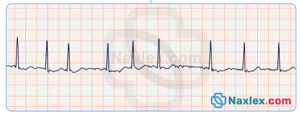
Explanation
Ventricular fibrillation is a life-threatening dysrhythmia characterized by chaotic, disorganized electrical activity that renders the heart incapable of effective mechanical contraction. This condition results in an immediate cessation of cardiac output and tissue perfusion. Management is governed by Advanced Cardiac Life Support (ACLS) protocols, which prioritize the restoration of organized electrical activity through rapid electrical intervention and sustained chest compressions to maintain cerebral perfusion.
Rationale for correct answer
3. Immediate high-quality cardiopulmonary resuscitation must be started to facilitate blood flow to the heart and brain. Defibrillation is the definitive treatment for ventricular fibrillation, as it delivers an electrical shock to depolarize the entire myocardium, allowing the sinoatrial node to resume control.
Rationale for incorrect answers
1. Atropine is indicated for the treatment of symptomatic bradycardia to increase the firing rate of the sinoatrial node. It is entirely ineffective in the treatment of ventricular fibrillation and would delay the delivery of life-saving defibrillation therapy.
2. Synchronized cardioversion is used to treat tachyarrhythmias where an organized QRS complex is present, such as supraventricular tachycardia. Using synchronization during ventricular fibrillation is inappropriate because the machine cannot identify a R wave to deliver the shock.
4. Obtaining a 12-lead ECG is a diagnostic measure for stable or stable-adjacent cardiac events. This action is contraindicated in an unresponsive patient who lacks a pulse, as it wastes critical time and delays essential resuscitation efforts.
Test-taking strategy
Analyze the Scenario/Question: The client is in ventricular fibrillation and is pulseless/unresponsive, representing a state of clinical death requiring immediate emergency intervention.
Apply ACLS Prioritization:
- Rule out 1: Atropine is for slow rhythms, not cardiac arrest.
- Rule out 2: Synchronization is for organized rhythms, not the chaotic activity of fibrillation.
- Rule out 4: A 12-lead ECG is a diagnostic delay and provides no therapeutic benefit in cardiac arrest.
- Rule in 3: Basic and advanced life support, specifically chest compressions and defibrillation, are the only evidence-based actions for pulseless ventricular fibrillation.
Take home points
- Ventricular fibrillation requires immediate defibrillation and the initiation of high-quality cardiopulmonary resuscitation.
- Synchronized cardioversion is reserved for unstable tachycardias with a pulse, whereas defibrillation is for pulseless rhythms.
- Time to defibrillation is the most critical factor in improving survival outcomes for patients in ventricular fibrillation.
- Never delay chest compressions for diagnostic testing or rhythm analysis in an unresponsive, pulseless patient.
The nurse is caring for a client whose ECG tracing is as shown below. The nurse should recognize the tracing as?
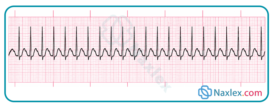
Explanation
Supraventricular tachycardia originates from an ectopic focus above the bifurcation of the bundle of His, causing a rapid, sustained heart rate typically ranging between 150 and 220 beats per minute. This dysrhythmia is characterized by a narrow QRS complex and a regular rhythm where the P waves are often buried within the preceding T wave or QRS complex. The rapid rate compromises diastolic filling time, which may lead to clinical symptoms of decreased cardiac output.
Rationale for correct answer
3. The tracing shows a narrow, regular, and rapid rhythm without clearly identifiable P waves, which is diagnostic of supraventricular tachycardia. This pattern reflects the rapid depolarization of the ventricles driven by an ectopic pacemaker located within the atria or AV junction.
Rationale for incorrect answers
1. Second-degree AV block, Mobitz type II, is characterized by intermittent, non-conducted P waves and a fixed PR interval in the conducted beats. This rhythm is generally slower and exhibits an atrial-ventricular dissociation of rhythm, which does not match the rapid, narrow-complex tachycardia presented.
2. Ventricular tachycardia is defined by a wide QRS complex (greater than or equal to 0.12 second) and an ectopic focus originating in the ventricular myocardium. It lacks the narrow QRS configuration and organized, fast conduction characteristic of the tachycardia originating from supraventricular sites.
4. A right bundle branch block presents as a widened QRS complex with a characteristic rsR' pattern in lead V1. This is a conduction delay within the ventricular system rather than a tachycardia rhythm, and it does not demonstrate the sustained elevation in heart rate seen here.
Test-taking strategy
Analyze the Scenario/Question: The nurse must identify a narrow-complex tachycardia characterized by a regular, rapid heart rate and absent P waves, which is indicative of atrial ectopy.
Apply ECG Interpretation Principles:
- Rule out 1: Mobitz II block has dropped beats, whereas this rhythm is a rapid, sustained tachycardia.
- Rule out 2: Ventricular tachycardia displays wide QRS complexes, while the prompt refers to narrow-complex SVT.
- Rule out 4: Right bundle branch block is a conduction delay, not a tachycardia rhythm.
- Rule in 3: Supraventricular tachycardia is confirmed by the narrow complex morphology, rapid rate, and the classic absence of visible P waves due to high-speed atrial firing.
Take home points
- Supraventricular tachycardia is identified by a regular, narrow-complex rhythm with a rate typically exceeding 150 beats per minute.
- The rapid heart rate results in decreased coronary artery perfusion and reduced diastolic filling, which can lead to hemodynamic instability.
- First-line interventions include vagal maneuvers to increase parasympathetic tone and the administration of adenosine.
- Distinguishing between SVT and ventricular tachycardia is critical, as treatment pathways for these rhythms are fundamentally different.
The nurse reviews an ECG and observes that every P wave is followed by a QRS complex. The PR interval is consistently prolonged at 0.24 seconds. Which conduction abnormality does this finding indicate?
Explanation
First-degree atrioventricular block is a conduction disorder where every atrial impulse is conducted to the ventricles, but the electrical signal experiences a delay as it passes through the atrioventricular node. This results in a prolonged PR interval that measures greater than 0.20 seconds. This rhythm maintains a 1:1 atrial-to-ventricular conduction ratio, meaning no QRS complexes are dropped or missing from the tracing.
Rationale for correct answer
2. The defining criterion for first-degree AV block is a fixed, prolonged PR interval (greater than 0.20 seconds) with every P wave successfully conducting to a QRS complex. This indicates a consistent conduction delay rather than a true blockade of the electrical impulse.
Rationale for incorrect answers
1. Second-degree AV block, Mobitz type II, is characterized by intermittent, non-conducted P waves, resulting in dropped QRS complexes. The PR interval remains constant in the conducted beats, but the presence of blocked beats excludes this diagnosis.
3. Complete heart block, or third-degree AV block, involves a total failure of conduction between the atria and ventricles. This rhythm shows complete atrioventricular dissociation, where P waves and QRS complexes march out independently at their own intrinsic rates.
4. Right bundle branch block is a conduction abnormality within the ventricles, manifesting as a widened QRS complex (equal to or greater than 0.12 seconds). It does not affect the PR interval duration and is not considered a block of the atrioventricular junction.
Test-taking strategy
Analyze the Scenario/Question: The nurse must classify an ECG rhythm that features 1:1 conduction and a PR interval of 0.24 seconds, identifying the specific conduction pathology.
Apply ECG Interpretation Principles:
- Rule out 1: Mobitz II requires missing QRS complexes, which contradicts the prompt's finding of 1:1 conduction.
- Rule out 3: Third-degree block displays no relationship between atrial and ventricular activity, which is not present here.
- Rule out 4: Right bundle branch block involves ventricular morphology changes, not PR interval prolongation.
- Rule in 2: A PR interval greater than 0.20 seconds with consistent AV conduction is the classic diagnostic finding for first-degree AV block.
Take home points
- First-degree AV block is defined by a prolonged PR interval exceeding 0.20 seconds with 1:1 conduction.
- The delay in conduction typically occurs within the AV node and does not result in dropped beats.
- This rhythm is usually benign and asymptomatic, often requiring no acute clinical intervention.
- Distinguishing between AV blocks depends on assessing the relationship between P waves and QRS complexes and the consistency of the PR interval.
The nurse is evaluating a client with acute chest pressure. The ECG demonstrates ST-segment elevation in contiguous leads. Which interpretation is accurate?
Explanation
ST-segment elevation indicates significant myocardial injury caused by an acute disruption of coronary blood flow, often due to a total occlusion of a coronary artery. This finding reflects the repolarization abnormality resulting from transmural ischemia, where the damaged tissue fails to maintain its resting membrane potential. Prompt identification is critical to minimize myocardial necrosis and preserve ventricular function through urgent reperfusion therapies.
Rationale for correct answer
1. ST-segment elevation in contiguous leads is the diagnostic hallmark of an ST-elevation myocardial infarction (STEMI). This finding represents active cardiac tissue damage and requires immediate medical intervention to restore coronary perfusion and limit permanent injury.
Rationale for incorrect answers
2. Normal ventricular repolarization is represented by a flat, isoelectric ST segment at the same baseline as the PR interval. ST-segment elevation is a pathological marker of severe ischemia and never occurs during the process of healthy, physiological repolarization.
3. First-degree AV block is identified solely by a prolonged PR interval exceeding 0.20 seconds with 1:1 conduction. It is a conduction delay and does not produce changes in the ST segment or indicate acute injury.
4. Sinus bradycardia is a rhythm with a normal P-QRS-T configuration originating from the sinoatrial node at a rate below 60 beats per minute. This rhythm is unrelated to ST-segment changes or the presence of an acute infarction.
Test-taking strategy
Analyze the Scenario/Question: The nurse is evaluating a patient with acute chest pressure and ST-segment elevation, which is a critical sign of myocardial ischemia.
Apply Cardiac Interpretation Principles:
- Rule out 2: ST elevation is never normal; it is a sign of pathological injury.
- Rule out 3: First-degree AV block refers to PR interval timing, not ventricular repolarization.
- Rule out 4: Sinus bradycardia is a heart rate finding and does not account for ST-segment elevation.
- Rule in 1: ST-segment elevation in contiguous leads is the clinical standard for identifying an acute myocardial infarction.
Take home points
- ST-segment elevation in contiguous leads is a medical emergency indicating an acute myocardial infarction.
- Normal ventricular repolarization should show an isoelectric ST segment, not elevation.
- Conductive delays like first-degree AV block are distinct from the ischemic patterns seen in injury.
- The nurse must prioritize rapid assessment and activation of protocols for cardiac catheterization or fibrinolysis.
Practice Exercise 3
A client is experiencing ventricular fibrillation (VF), and the nurse prepares for cardiac defibrillation. What is the rationale for using defibrillation in this situation?
Explanation
Ventricular fibrillation is a life-threatening, chaotic dysrhythmia characterized by rapid, disorganized electrical activity that prevents effective ventricular contraction. During this state, the heart lacks a coordinated electrical impulse, rendering it unable to maintain cardiac output. Defibrillation works by delivering a high-energy, non-synchronized electrical shock to the entire myocardium simultaneously, creating a state of temporary electrical silence.
Rationale for correct answer
3. The electrical shock depolarizes all myocardial cells at once, effectively stopping the chaotic electrical activity. This momentary cessation provides the heart's natural pacemaker, the sinoatrial node, with the opportunity to recover and resume its role as the dominant rhythm generator.
Rationale for incorrect answers
1. Defibrillation does not enhance repolarization or relaxation; rather, it forces depolarization. By overriding the disorganized ventricular firing, the device resets the myocardium, which is fundamentally different from merely assisting in the cell's resting phase.
2. Defibrillation does not provide a stimulus for normal contraction; it does not pace the heart. Instead, the shock clears the abnormal rhythm so that the heart’s own internal conduction system can spontaneously restart and maintain a perfusing rhythm.
4. Synchronized delivery is inappropriate for ventricular fibrillation because there is no organized QRS or R wave to detect. Defibrillation must be delivered asynchronously to ensure the myocardial depolarization is successful in terminating the fibrillatory activity.
Test-taking strategy
Analyze the Scenario/Question: The patient is in ventricular fibrillation and requires defibrillation, necessitating an understanding of the electrophysiological mechanism of the intervention.
Apply Cardiac Resuscitation Principles:
- Rule out 1: Defibrillation induces depolarization, not repolarization.
- Rule out 2: Defibrillation is not a pacemaker; it does not stimulate contractions.
- Rule out 4: Synchronization is contraindicated in fibrillation because the heart lacks an identifiable rhythm.
- Rule in 3: The goal of defibrillation is the global depolarization of the heart, allowing the sinoatrial node to regain control.
Take home points
- Defibrillation is the definitive treatment for pulseless ventricular fibrillation and pulseless ventricular tachycardia.
- The mechanism of action is the simultaneous depolarization of the myocardium to stop chaotic electrical firing.
- Asynchronous delivery is required for defibrillation because no organized ventricular contraction exists to trigger a sync pulse.
- Successful defibrillation enables the sinoatrial node to reset and restore a normal, perfusing sinus rhythm.
What action is included in the nurse’s responsibilities in preparing to administer defibrillation?
Explanation
Defibrillation requires meticulous preparation to ensure safe and effective energy delivery to the myocardium. The process involves establishing proper contact between the electrode pads and the skin to decrease transthoracic impedance. This reduction in resistance facilitates the successful passage of the high-energy electrical shock through the heart, which is necessary to achieve simultaneous myocardial depolarization and terminate the lethal fibrillatory rhythm.
Rationale for correct answer
1. Applying conductive gel or using pre-gelled pads is essential to minimize skin resistance. This ensures the maximum amount of electrical current is delivered directly to the heart rather than dissipating through the epidermal layers of the chest wall.
Rationale for incorrect answers
2. A setting of 50 joules is insufficient for adult defibrillation, which typically requires much higher energy levels to overcome the myocardial resistance. Standard biphasic protocols recommend an initial dose of 120 to 200 joules to effectively terminate ventricular fibrillation.
3. Setting the defibrillator to synchronized mode is strictly contraindicated in the treatment of ventricular fibrillation. Synchronization requires a detectable R wave to trigger the shock, and because this rhythm is chaotic and lacks organized complexes, the device will fail to deliver the emergency energy.
4. Sedation is indicated for elective synchronized cardioversion in a stable patient experiencing a tachyarrhythmia. In a life-threatening, pulseless arrest like ventricular fibrillation, the client is unresponsive; therefore, providing pharmacological sedation is unnecessary and would dangerously delay the delivery of life-saving intervention.
Test-taking strategy
Analyze the Scenario/Question: The nurse is preparing for an emergency defibrillation procedure for a patient in ventricular fibrillation, requiring the selection of a critical safety step.
Apply Resuscitation Principles:
- Rule out 2: Low-energy shocks are ineffective for the massive myocardial depolarization needed in fibrillation.
- Rule out 3: Synchronization is only for organized rhythms with identifiable R waves.
- Rule out 4: Sedation is for stable patients, whereas this client is pulseless and unresponsive, making time-to-shock the only priority.
- Rule in 1: Maximizing electrical conductivity through proper pad placement and gel application is a mandatory preparation step.
Take home points
- Defibrillation requires conductive media (gel pads) to lower transthoracic impedance and ensure energy delivery.
- Energy levels for adult defibrillation are significantly higher than those used for synchronized cardioversion.
- The defibrillator must be in asynchronous mode for ventricular fibrillation because no organized cardiac cycle exists to synchronize with.
- Emergency defibrillation in a cardiac arrest setting never involves sedation, as the client is already unconscious and requires immediate electrical therapy.
A client is being discharged after insertion of an implantable cardioverter-defibrillator (ICD). What should the nurse instruct the client to do if the ICD delivers a shock?
Explanation
An implantable cardioverter-defibrillator is designed to monitor cardiac rhythm continuously and deliver electrical therapy if life-threatening arrhythmias such as ventricular tachycardia or ventricular fibrillation occur. When the device discharges, it signifies that a significant cardiac event has been detected. Patients must understand that device activation requires prompt medical evaluation to assess the rhythm status, potential underlying causes, and the integrity of the generator function.
Rationale for correct answer
2. Contacting the cardiologist or emergency services is mandatory after an ICD shock. This ensures an immediate analysis of the device data to determine if the shock was appropriate for a lethal rhythm or inappropriate due to a technical malfunction.
Rationale for incorrect answers
1. Lying down is not the primary action required after a device discharge. While the patient should minimize activity, the priority is notifying medical personnel for urgent assessment of their hemodynamic status and the potential need for further intervention.
3. Implantable cardioverter-defibrillator devices are programmed with internal safety parameters and do not feature a manual reset button for patient use. Attempting to manipulate the site or the device itself is dangerous and ineffective for managing dysrhythmias.
4. Taking an extra dose of antidysrhythmic medication after a shock is not the recommended first response. Such action can lead to adverse effects or toxicity and does not address the cause of the electrical discharge, which requires clinical evaluation by a specialist.
Test-taking strategy
Analyze the Scenario/Question: The patient has an ICD and experiences a shock, requiring an action that ensures medical safety and appropriate device follow-up.
Apply Patient Safety Principles:
- Rule out 1: While resting is helpful, contacting the physician is the critical priority for evaluation.
- Rule out 3: Manual resetting is impossible and shows a lack of understanding regarding implanted technology.
- Rule out 4: Adjusting medication dosages independently is unsafe and ineffective for rhythm management.
- Rule in 2: Expert clinical assessment is the gold standard after a device intervention to ensure patient stability.
Take home points
- Patients must notify their cardiologist immediately if the implantable cardioverter-defibrillator delivers a shock.
- The device serves as a life-saving monitor and shock generator, not a device requiring manual manipulation or resets.
- Any shock delivered by the ICD is considered a medical event requiring professional data interrogation.
- Patients should not alter their medication regimen independently in response to device activity.
A client on the cardiac telemetry unit develops ventricular fibrillation and becomes unresponsive. After initiating the emergency call system, what is the nurse’s next priority action?
Explanation
Ventricular fibrillation is a fatal dysrhythmia characterized by the total loss of coordinated ventricular contraction, resulting in immediate circulatory collapse. Because the brain and myocardium cannot tolerate prolonged hypoxia, the fundamental goal of resuscitative care is to re-establish oxygenated blood flow through external myocardial compression. High-quality chest compressions act as a mechanical substitute for the heart's pumping action, maintaining minimal cerebral perfusion while the team prepares for definitive electrical therapy.
Rationale for correct answer
1. The initiation of cardiopulmonary resuscitation is the most urgent priority to prevent irreversible damage. Establishing circulation via chest compressions takes precedence over all other interventions, as it provides the necessary substrate to sustain vital organs until the defibrillator is ready for shock delivery.
Rationale for incorrect answers
2. While retrieving the crash cart is a necessary step in the resuscitation sequence, it is not the immediate priority. Compressions must start first, and the cart should be brought to the bedside by other personnel or after the nurse has ensured circulatory support is established.
3. Intravenous amiodarone is an antiarrhythmic agent used to treat persistent or refractory ventricular fibrillation. It is administered only after initial attempts at defibrillation and CPR have failed to convert the rhythm; therefore, it is not the first action in the resuscitation sequence.
4. Although defibrillation is the definitive treatment for ventricular fibrillation, it cannot be performed instantaneously. Compressions must continue until the defibrillator pads are applied and the machine is charged, ensuring that there is no interruption in cardiac support during the transition to shock.
Test-taking strategy
Analyze the Scenario/Question: The client is in cardiac arrest due to ventricular fibrillation, requiring the application of ACLS priority interventions to maximize survival outcomes.
Apply Resuscitation Prioritization:
- Rule out 2: Equipment retrieval is secondary to the physiological need for blood flow.
- Rule out 3: Pharmacological therapy is for refractory rhythms, not the initial management of arrest.
- Rule out 4: Immediate defibrillation is the goal, but chest compressions are the immediate and mandatory starting action.
- Rule in 1: CPR is the cornerstone of resuscitation science and must be started immediately when a patient is found pulseless and unresponsive.
Take home points
- Immediate initiation of high-quality cardiopulmonary resuscitation is the primary action in any unwitnessed or witnessed cardiac arrest.
- Compressions must continue without interruption while the defibrillator is being prepared and charged.
- Pharmacological interventions like amiodarone are secondary to the delivery of electrical therapy and effective mechanical compressions.
- The focus of the initial response is to restore oxygen delivery to the brain and heart as quickly as possible.
A client with chest pain unrelieved by nitroglycerin is admitted to the coronary care unit for observation and diagnostic evaluation. Which ECG finding during continuous monitoring should concern the nurse most?
Explanation
ST-segment elevation indicates significant myocardial injury caused by an acute disruption of coronary blood flow, often due to a total occlusion of a coronary artery. This finding reflects the repolarization abnormality resulting from transmural ischemia, where the damaged tissue fails to maintain its resting membrane potential. Prompt identification is critical to minimize myocardial necrosis and preserve ventricular function through urgent reperfusion therapies.
Rationale for correct answer
3. ST-segment elevation in contiguous leads is the diagnostic hallmark of an ST-elevation myocardial infarction (STEMI). This finding represents active cardiac tissue damage and requires immediate medical intervention to restore coronary perfusion and limit permanent injury.
Rationale for incorrect answers
1. Occasional premature ventricular contractions are common in patients with cardiac disease and are often triggered by myocardial ischemia or electrolyte imbalances. While they warrant monitoring, they are not as immediately diagnostic of a transmural infarction as ST-segment elevation.
2. An inverted T wave indicates myocardial ischemia or strain, representing a lack of oxygen to the myocardial tissue. While clinically significant, it is a marker of ischemia rather than the acute, ongoing myocardial cell death indicated by ST-segment elevation.
4. A PR interval of 0.18 second is within the normal range of 0.12 to 0.20 seconds. This finding represents normal atrial-to-ventricular conduction and does not indicate any acute cardiac pathology or injury in this patient.
Test-taking strategy
Analyze the Scenario/Question: The patient has persistent chest pain, and the nurse must identify the most life-threatening ECG finding associated with acute myocardial infarction.
Apply Cardiac Interpretation Principles:
- Rule out 1: PVCs are common ectopy and generally do not signify total vessel occlusion.
- Rule out 2: Inverted T waves indicate ischemia, which is less urgent than the acute injury marked by ST elevation.
- Rule out 4: This is a normal conduction finding and has no relation to acute ischemia.
- Rule in 3: ST-segment elevation is the definitive sign of a STEMI, requiring immediate reperfusion protocols.
Take home points
- ST-segment elevation is the most concerning finding, as it signifies transmural myocardial injury and an acute infarction.
- Inverted T waves indicate ischemia, which is significant but distinct from the acute injury pattern of ST elevation.
- PVCs are common in ischemic heart disease but are less diagnostic of an acute STEMI than ST-segment changes.
- A normal PR interval indicates appropriate conduction and is not an indicator of acute myocardial damage.
A client with acute chest pain has new ST-segment elevation on the ECG. Which action is the priority?
Explanation
ST-segment elevation indicates significant myocardial injury caused by an acute disruption of coronary blood flow, often due to a total occlusion of a coronary artery. This finding reflects the repolarization abnormality resulting from transmural ischemia, where the damaged tissue fails to maintain its resting membrane potential. Prompt identification is critical to minimize myocardial necrosis and preserve ventricular function through urgent reperfusion therapies.
Rationale for correct answer
1. Activating the acute coronary syndrome response is the immediate priority to expedite reperfusion therapy, such as fibrinolytic administration or primary percutaneous coronary intervention. Continuous cardiac monitoring ensures the nurse can detect further arrhythmias or hemodynamic deterioration in real-time.
Rationale for incorrect answers
2. Waiting for cardiac biomarkers, such as troponin, is inappropriate and dangerously delays critical reperfusion treatment. Clinical decisions in the setting of persistent chest pain and ST-segment elevation must be made based on ECG findings to prevent irreversible damage.
3. Delaying further assessment by 24 hours is negligent and ignores the time-sensitive nature of myocardial infarction. Immediate intervention is required to open the occluded artery and restore blood flow to the threatened myocardial muscle.
4. Ambulation is strictly contraindicated in patients with acute myocardial injury, as physical exertion increases the myocardial oxygen demand. This can exacerbate ischemia, provoke life-threatening ventricular dysrhythmias, and lead to rapid clinical deterioration.
Test-taking strategy
Analyze the Scenario/Question: The patient shows ST-segment elevation, which is a medical emergency requiring rapid intervention to limit myocardial infarction size.
Apply Cardiac Management Principles:
- Rule out 2: Biomarkers are secondary; the ECG change is the primary driver for immediate action.
- Rule out 3: A 24-hour delay would likely lead to massive, irreversible injury or patient death.
- Rule out 4: Exercise is fatal during an acute myocardial infarction due to excessive oxygen demand.
- Rule in 1: Immediate activation of the ACS protocol and continuous monitoring are the only evidence-based priorities for ST-elevation.
Take home points
- ST-segment elevation in the setting of chest pain requires immediate activation of emergency cardiac protocols.
- Reperfusion therapy must be administered without delay, regardless of initial biomarker results.
- Physical exertion is contraindicated during the acute phase of myocardial infarction due to the risk of hemodynamic collapse.
- Continuous ECG monitoring is essential to identify lethal arrhythmias that often complicate acute myocardial injury.
The nurse is teaching a client about a Holter monitor prescribed for intermittent palpitations. Which statement by the client indicates correct understanding?
Explanation
A Holter monitor is a portable device used for continuous ambulatory electrocardiographic monitoring, typically for 24 to 48 hours, to detect transient cardiac arrhythmias. The diagnostic efficacy of this tool relies on the correlation between the patient's reported symptoms and the electrical activity recorded by the device. Proper documentation allows clinicians to identify whether reported palpitations correspond to atrial or ventricular ectopy, conduction abnormalities, or benign sinus rhythm.
Rationale for correct answer
2. The client must keep a detailed diary of their daily activities, including the exact time, nature of symptoms, and triggers. This information is vital for the provider to correlate symptoms with the recorded ECG findings during the monitoring period.
Rationale for incorrect answers
1. The monitor must remain attached to the skin throughout the entire duration of the study to capture data. Removing the device during episodes of palpitations would cause a data gap, preventing the diagnosis of the underlying cardiac rhythm.
3. The primary purpose of a Holter monitor is to record the heart's activity during normal daily life, including movement and stressors. Bed rest is not required and would fail to capture physiological changes or palpitations triggered by routine physical activity.
4. The event button should be pressed whenever the patient experiences a symptom, such as palpitations, dizziness, or chest pain, regardless of whether the monitor alarms. The event recording serves as a clinical marker for the provider to analyze that specific rhythmic segment.
Test-taking strategy
Analyze the Scenario/Question: The nurse is assessing the client's understanding of Holter monitor use, requiring identification of correct ambulatory monitoring procedures.
Apply Cardiac Monitoring Principles:
- Rule out 1: The device must stay on to capture continuous ECG data.
- Rule out 3: Normal activity is preferred to document symptom-rhythm correlation.
- Rule out 4: The event button is for documenting patient-reported symptoms, not device alarms.
- Rule in 2: Accurate documentation of events is the cornerstone of diagnostics for Holter monitoring.
Take home points
- Holter monitoring requires the device to remain in place for the entire prescribed duration, including during sleep.
- Patients must maintain a diary recording the time and type of symptoms experienced during the monitoring period.
- The event button is used to mark the time of symptoms, not to respond to device alerts.
- Normal activities should be maintained to ensure the ECG recording captures real-world heart rhythm patterns.
A client with symptomatic atrial fibrillation is scheduled for elective synchronized cardioversion. Which nursing action is essential before the procedure?
Explanation
Synchronized cardioversion is an elective procedure performed to terminate persistent tachyarrhythmias, such as atrial fibrillation, by delivering an electrical shock timed precisely to the R wave. Because atrial fibrillation promotes the formation of mural thrombi, particularly in the left atrial appendage, the restoration of sinus rhythm carries a significant risk of thromboembolic events, including stroke. Consequently, establishing an adequate anticoagulation status or performing transesophageal echocardiography is mandatory before the procedure to ensure patient safety.
Rationale for correct answer
1. Evaluation for thromboembolism is the most critical safety intervention. If the patient has been in atrial fibrillation for more than 48 hours without adequate anticoagulation therapy, they must be bridged with anticoagulants or screened for atrial thrombi to prevent a post-procedure stroke.
Rationale for incorrect answers
2. Patients must remain NPO for several hours prior to the procedure to prevent aspiration pneumonia during sedation. Consuming a large meal immediately before the procedure is a serious safety violation that would mandate postponement of the cardioversion attempt.
3. Synchronization is the defining requirement for elective cardioversion. The machine must be set to the synchronized mode to ensure the electrical discharge occurs during ventricular repolarization, which avoids the vulnerable period of the T wave and prevents the induction of ventricular fibrillation.
4. The patient should remove all metal jewelry, but ECG electrodes must remain in place for continuous rhythm monitoring. Removing the leads would prevent the device from sensing the R wave, making it impossible to perform the synchronized delivery required for this specific procedure.
Test-taking strategy
Analyze the Scenario/Question: The patient is undergoing elective synchronized cardioversion for atrial fibrillation, which requires careful risk mitigation regarding stroke prevention.
Apply Procedure Safety Principles:
- Rule out 2: Aspiration is a life-threatening complication; NPO status is strictly required.
- Rule out 3: Unsynchronized shock is for fibrillation/arrest; synchronized mode is required to prevent ventricular irritability.
- Rule out 4: Monitoring leads must remain to ensure the machine can sense electrical activity and time the shock appropriately.
- Rule in 1: Assessing for thromboembolic risk is the primary safety prerequisite for converting long-standing atrial fibrillation.
Take home points
- Synchronized cardioversion is only performed if the machine is set to detect the R wave, preventing the delivery of a shock during the T wave.
- The risk of stroke is high during rhythm conversion if the patient has not been therapeutically anticoagulated.
- Pre-procedure fasting (NPO status) is essential to minimize the risk of aspiration during moderate sedation.
- Continuous ECG monitoring via chest leads is required throughout the procedure to facilitate R-wave sensing.
The nurse is preparing a client for synchronized cardioversion. Which statement accurately describes synchronized cardioversion?
Explanation
Synchronized cardioversion is an elective procedure performed to terminate supraventricular tachycardias or atrial fibrillation that are unresponsive to medical management. The synchronization mechanism is a critical safety feature that ensures the electrical shock is delivered at a specific point in the cardiac cycle. This precise timing allows the device to avoid the vulnerable period of the cardiac cycle, thereby preventing the transition of an organized rhythm into a lethal ventricular dysrhythmia.
Rationale for correct answer
2. The defibrillator is programmed to detect the R wave of the QRS complex, ensuring the electrical discharge is timed to occur during ventricular depolarization. Delivering energy during this period prevents the shock from falling on the T wave, which corresponds to the vulnerable period of ventricular repolarization.
Rationale for incorrect answers
1. Delivering an electrical shock randomly during the cardiac cycle is the definition of unsynchronized defibrillation. This approach is reserved for emergency situations where no organized rhythm is present, as it carries a high risk of inducing ventricular fibrillation in a patient with an existing rhythm.
3. Synchronized cardioversion is contraindicated in pulseless ventricular fibrillation because the machine requires an identifiable R wave to function. In the absence of an organized rhythm, unsynchronized defibrillation is the only safe and effective treatment to terminate the chaotic electrical activity.
4. Continuous ECG monitoring is an absolute requirement for synchronized cardioversion. The defibrillator must continuously sense the electrical activity of the heart to trigger the shock at the exact moment of the R wave, a process that is impossible without reliable rhythm surveillance.
Test-taking strategy
Analyze the Scenario/Question: The nurse must correctly define the mechanism of synchronized cardioversion, distinguishing it from emergency defibrillation protocols.
Apply Procedural Safety Principles:
- Rule out 1: Random delivery characterizes asynchronous shocks, not synchronized cardioversion.
- Rule out 3: Synchronization is only for organized tachycardias, not for pulseless cardiac arrest.
- Rule out 4: Monitoring is essential for the device to perform its sensing and timing function.
- Rule in 2: The goal of synchronization is to avoid the vulnerable T-wave period, which significantly enhances the procedure's safety profile.
Take home points
- Synchronized cardioversion uses a controlled electrical discharge timed to the R wave to treat unstable or symptomatic tachycardias.
- The synchronization process prevents the induction of dangerous rhythms by avoiding the vulnerable repolarization phase (T wave).
- This procedure requires continuous ECG monitoring to ensure the device detects the QRS complex accurately.
- Never use synchronized mode for pulseless rhythms; always use unsynchronized defibrillation for ventricular fibrillation or pulseless ventricular tachycardia.
The nurse is assisting with synchronized cardioversion for a client with an unstable tachydysrhythmia who has a pulse. Which intervention is appropriate?
Explanation
Synchronized cardioversion for an unstable patient requires a rapid, systematic approach to ensure both electrical and physiological safety. Because the patient is unstable, the nurse must balance the urgency of restoring a perfusing rhythm with the necessity of safe sedation and continuous hemodynamic monitoring. Preparing the equipment, securing venous access, and providing supplemental oxygen are standard components of the peri-procedural care, ensuring the patient is supported during the transition from a pathologic tachycardia to a normal sinus rhythm.
Rationale for correct answer
1. These actions establish the necessary clinical environment for a safe procedure. Continuous monitoring allows the device to sense the R wave for synchronization, IV access is required for the administration of sedatives or analgesics, and supplemental oxygen mitigates the hypoxemia often exacerbated by rapid heart rates and increased myocardial oxygen consumption.
Rationale for incorrect answers
2. Patients undergoing elective or semi-urgent synchronized cardioversion must remain NPO for several hours to prevent aspiration pneumonia due to the administration of sedative agents. Oral fluids are strictly prohibited immediately prior to the procedure.
3. Removing monitoring equipment is contraindicated. The defibrillator requires the signal from ECG leads to detect the R wave for proper synchronization. If the rhythm is not sensed, the shock cannot be delivered in the synchronized mode, which would be unsafe for a patient who has a pulse.
4. Patients must never handle electrical equipment during a procedure. It is the responsibility of the nurse or physician to ensure proper pad placement and safe electrical discharge. The client should be positioned correctly, and all personnel must stand clear of the bed to prevent accidental electrical injury during the shock delivery.
Test-taking strategy
Analyze the Scenario/Question: The patient requires urgent cardioversion for an unstable tachycardia, necessitating a combination of procedural safety and supportive care.
Apply Procedure Safety Principles:
- Rule out 2: Always maintain NPO status to prevent aspiration.
- Rule out 3: Monitoring is the primary means of rhythm sensing for the device.
- Rule out 4: Only staff should handle equipment; the patient must remain passive for electrical safety.
- Rule in 1: Preparation for sedation, oxygenation, and continuous monitoring are the pillars of safe synchronized delivery.
Take home points
- Synchronized cardioversion for an unstable patient necessitates continuous cardiac monitoring to maintain R-wave sensing.
- Patient safety requires that the client be sedated yet monitored for airway patency throughout the procedure.
- Always maintain NPO status prior to the procedure, regardless of the urgency, unless the situation is a life-threatening arrest.
- Electrical safety protocols, including standing clear of the patient during the shock, are essential for all personnel.
Comprehensive Questions
A student nurse is observing a cardioversion procedure and hears the team leader call out, “Stand clear.” The student should recognize the purpose of this action is to alert personnel that?
Explanation
Cardioversion is a medical procedure used to terminate tachyarrhythmias by delivering a synchronized electrical shock to the heart. During the delivery phase, the electrical current is discharged across the thoracic cavity to reset the rhythm. Ensuring safety for all personnel is a critical component of preventing accidental myocardial electrical injury.
Rationale for correct answer
3. The instruction to stand clear ensures that no personnel are touching the patient or connected equipment during the electrical discharge. This prevents the transfer of high-voltage energy, which could cause significant myocardial trauma or injury to the medical staff present.
Rationale for incorrect answers
1. Charging the cardioverter is a preparatory step that occurs prior to the synchronization and discharge phase. The alert is specifically timed to the moment of delivery, not the charging process, to manage the risk of electrical conduction throughout the environment.
2. Cardioversion is indicated for patients with a pulse, such as atrial fibrillation or flutter. Initiating cardiopulmonary resuscitation would be inappropriate, as the patient is hemodynamically stable enough to require rhythm correction rather than immediate life support or advanced resuscitation.
4. A time out is a standardized protocol performed before a procedure to verify patient identity and site, not during the delivery of a shock. The urgent command to stand clear is intended for immediate safety during the high-energy discharge, not for procedural verification.
Test-taking strategy
- Analyze the Scenario and Question: The question requires understanding the safety protocols during a synchronized cardioversion.
- The goal is to determine the specific safety objective behind the command to stand clear during high-voltage electrical delivery.
- Apply Safety and Risk Reduction Principles: Use the principle of electrical safety to evaluate why personnel must move away from the patient.
- Rule out 1: The charge state does not require personnel to stand clear; only the discharge phase carries this risk.
- Rule out 2: Cardioversion requires a pulse; resuscitation is for pulseless states.
- Rule out 4: A time out is a pre-procedural safety check, not a mid-procedure safety command.
- Identify Critical Safety Logic:
- Confirm 3: Preventing conduction of current to staff is the primary safety rationale for the command.
Take home points
- Synchronized cardioversion is used for patients with tachyarrhythmias who remain hemodynamically stable with a pulse.
- Personnel must avoid all contact with the patient, bed, or connected cables during electrical discharge to prevent injury.
- The command to stand clear is an essential safety requirement for both defibrillation and cardioversion procedures.
- Pre-procedure time-outs verify patient identity and consent, while safety alerts during discharge prevent electrical hazards to staff.
A client on a cardiac telemetry unit has a heart rate of 46/min, and the nurse notifies the provider. Which management strategy should the nurse anticipate for this client?
Explanation
Symptomatic bradycardia occurs when the sinoatrial node fails to maintain an adequate heart rate to support cardiac output. Management focuses on restoring hemodynamic stability using pharmacologic agents or electrical intervention. The nurse must identify signs of hypoperfusion and anticipate definitive therapy, such as long term rhythm regulation for persistent conduction failure.
Rationale for correct answer
2. Pacemaker insertion is the definitive treatment for persistent symptomatic bradycardia that does not respond to atropine or transcutaneous pacing. This device provides consistent electrical stimulation to maintain an adequate ventricular rate, ensuring sustained perfusion and preventing further hemodynamic compromise.
Rationale for incorrect answers
1. Defibrillation is used to treat life threatening arrhythmias like pulseless ventricular tachycardia or ventricular fibrillation. It delivers a high energy asynchronous shock to depolarize the entire myocardium. Using this for bradycardia is ineffective and physically damaging to the heart tissue.
3. Synchronized cardioversion is utilized for tachyarrhythmias such as atrial fibrillation or supraventricular tachycardia to restore a normal rhythm. It times the discharge to the R wave to avoid inducing ventricular fibrillation. It is contraindicated in patients with slow heart rates.
4. Lidocaine is an antiarrhythmic medication used to suppress ventricular irritability and treat ventricular tachycardia or ventricular ectopy. Administering this to a bradycardic patient would further suppress cardiac conduction, potentially leading to asystole or a complete cessation of electrical activity.
Test-taking strategy
- Analyze the Scenario and Question: The client presents with a heart rate of 46/min, which is defined as bradycardia.
- The nurse must determine the appropriate management strategy for a slow rhythm using cardiac electrophysiology knowledge.
- Apply Pathophysiological Principles: Evaluate the mechanism of each intervention to determine which is indicated for slow heart rates versus fast or irritable rhythms.
- Rule out 1: Defibrillation is for pulseless, chaotic ventricular rhythms, not for pacing slow heart rates.
- Rule out 3: Synchronized cardioversion is for rapid rhythms that require a timed shock, not for bradycardia.
- Rule out 4: Lidocaine is a cardiac depressant used for fast or irritable rhythms and would worsen bradycardia.
- Identify Correct Treatment:
- Confirm 2: Pacemaker insertion is the standard intervention to provide reliable electrical stimulation in cases of persistent symptomatic bradycardia.
Take home points
- Bradycardia is defined as a heart rate below 60 beats per minute, which requires intervention if symptomatic.
- Pacemakers are the definitive treatment for persistent bradycardia caused by sinus node dysfunction or heart block.
- Antiarrhythmic drugs like lidocaine are contraindicated in bradycardia because they further slow the heart rate.
- Defibrillation and cardioversion are electrical therapies reserved for tachyarrhythmias and are not used for bradycardia.
A client has undergone defibrillation, and the nurse is completing documentation of the procedure. Which findings should the nurse include in the documentation? Select all that apply
Explanation
Defibrillation is a critical, life-saving procedure involving the delivery of an asynchronous shock to terminate lethal ventricular rhythms like ventricular fibrillation. Detailed documentation ensures clinical continuity and evaluates the myocardial response to electrical intervention. Accurate recording of procedural data, including electrical energy and patient tissue integrity, is vital for clinical outcomes and ongoing monitoring.
Rationale for correct answer
1. A follow-up ECG strip is required to document the return of a spontaneous rhythm or the persistence of the arrhythmia. This provides scientific evidence of the electrical response to the energy delivered during the procedure.
2. Energy settings must be recorded to monitor the total joules delivered during the resuscitation event. Precise documentation of the joules utilized is necessary for evaluating the effectiveness of the therapy and for providing a complete record of care.
5. Skin condition assessment is essential after high-energy shocks to identify potential electrical burns. Documenting the integrity of the tissue confirms the patient's dermatologic safety following the application of conductive pads and ensures appropriate post-procedural wound monitoring and care.
Rationale for incorrect answers
3. Intravenous fluid intake is part of general resuscitation and circulatory management. It is not a direct component of the immediate post-defibrillation documentation for the procedure itself, as fluids address systemic perfusion rather than the electrical activity of the heart.
4. Urinary output is a measure of end-organ perfusion and systemic hemodynamic status over time. While it is important for the overall care of a stable patient, it does not pertain to the specific technical documentation required for the defibrillation procedure.
Test-taking strategy
- Analyze the Scenario and Question: The nurse must identify which data points are specific to the documentation of a defibrillation procedure.
- The goal is to differentiate between general patient monitoring and specific procedure-related documentation requirements.
- Apply Procedure-Specific Logic: Evaluate whether each choice relates directly to the electrical delivery and its immediate effects on the heart and skin.
- Rule out 3: Fluid intake is for systemic management, not procedural recording.
- Rule out 4: Urinary output measures organ function, not the procedure results.
- Identify Procedural Requirements:
- Confirm 1: The ECG confirms the rhythm outcome.
- Confirm 2: Energy records the dose of electrical therapy.
- Confirm 5: Skin condition records safety and potential injury.
Take home points
- Documentation for defibrillation must include rhythm strips, energy levels, and patient tissue assessments.
- Assessing for electrical burns under electrode pads is a standard safety measure following high-energy shock delivery.
- Post-procedure rhythm strips serve as proof of effectiveness and guide the next steps in advanced cardiac life support.
- Systemic monitoring like fluid balance is important for recovery but is separate from the documentation of the shock procedure.
A client is being admitted to the cardiac unit for cardioversion, and the nurse reviews the health record before the procedure. Which information should cause the nurse to notify the provider because the procedure may need to be canceled?
|
MAR |
VITAL SIGNS |
HISTORY AND PHYSICAL |
|
Ferrous Sulfate (Feosol) 200 mg PO 0800 and 2000 Diazepam (Valium) 2 mg PO 0800 and 2000 Isosorbide (Isordil) 2.5 mg PO 4 times a day AC and HS |
0800 T 99° F (37.2° C) Blood pressure 142/86 mm Hg Heart rate 88/min and irregular Respirations 20/min |
Bariatric surgery 10 years ago Dyspnea with exertion for 3 years Atrial fibrillation began 3 years ago Client reports taking the following medications for the past 6 weeks: iron supplement, multivitamin, antilipemic, and nitroglycerin |
Explanation
Cardioversion requires precise management of medications to ensure clinical safety and procedure efficacy. A major concern before elective synchronization is the potential for subtherapeutic anticoagulation in patients with atrial fibrillation. The nurse must meticulously review the patient's medication history to identify any omitted pharmacotherapy that could increase the risk of systemic thromboembolism.
Rationale for correct answer
3. The client reports taking only an iron supplement, multivitamin, antilipemic, and nitroglycerin. The health record fails to show anticoagulant therapy, which is mandatory for patients with atrial fibrillation before elective cardioversion. Absence of this medication increases the risk of thromboembolic stroke during rhythm conversion.
Rationale for incorrect answers
1. Respiratory history and associated conditions like bariatric surgery are significant for overall care but do not directly contraindicate elective cardioversion. Stable airway status is typically maintained throughout the procedure. These findings do not necessitate the immediate cancellation of the scheduled procedure.
2. Vital signs reflect a hemodynamic state compatible with elective cardioversion. The blood pressure and heart rate are within acceptable ranges for a patient in atrial fibrillation. These findings do not indicate an unstable condition that would require the immediate cancellation of the scheduled procedure.
4. The medications to be administered include iron and diazepam, which are not contraindicated for cardioversion. The dose of diazepam may be used for sedation during the procedure. None of these medications pose an immediate risk or require the cancellation of the planned electrical therapy.
Test-taking strategy
- Analyze the Scenario/Question: The goal is to identify a clinical contraindication that requires canceling elective cardioversion.
- Focus on safety risks, specifically the need for anticoagulation in atrial fibrillation.
- Apply Clinical Safety Principles:
- Rule out 1: Respiratory history does not contraindicate the procedure.
- Rule out 2: Current vital signs are hemodynamically stable.
- Rule out 4: The medications scheduled are safe for the procedure.
- Confirm 3: Medication history is incomplete because the patient lacks necessary anticoagulation therapy to prevent strokes.
- Identify Critical Missing Data:
- Atrial fibrillation for 3 years requires chronic anticoagulation before restoring normal sinus rhythm.
- Failure to document this therapy suggests the patient is at high embolic risk.
Take home points
- Elective cardioversion for atrial fibrillation requires therapeutic anticoagulation to prevent post-procedural stroke.
- Nursing assessment must include a thorough medication reconciliation to ensure essential prophylactic therapy is present.
- Hemodynamic stability and airway management are essential considerations for procedural safety and sedation.
- Always verify the duration of atrial fibrillation to determine if the patient requires a transesophageal echocardiogram before cardioversion.
A nurse on a cardiac unit is caring for a group of clients. The nurse should recognize which of the following clients as being at risk for the development of a dysrhythmia? Select all that apply
Explanation
Electrolyte imbalances and structural cardiac disease significantly alter myocardial excitability, predisposing clients to life threatening dysrhythmias. Maintaining acid base homeostasis and optimal oxygenation is essential for stabilizing membrane potentials in cardiac cells. Nurses must identify patients with predisposing comorbidities or metabolic disturbances to prevent acute rhythm disturbances and hemodynamic instability.
Rationale for correct answer
1. Metabolic alkalosis frequently coincides with hypokalemia, which alters the resting membrane potential of cardiac myocytes. This shift in electrolyte concentration increases the risk of ventricular ectopy and serious dysrhythmias by delaying repolarization and increasing myocardial irritability.
4. COPD creates a chronic state of hypoxia and hypercapnia, which triggers compensatory sympathetic nervous system activation. This constant hypoxemia stresses the cardiac conduction system and increases the myocardial oxygen demand, significantly elevating the risk for atrial and ventricular dysrhythmias.
5. Stent placement indicates significant underlying coronary artery disease, which involves structural changes to the myocardium. Chronic ischemia or the presence of previous infarctions provides a substrate for re-entry circuits, which are a primary cause of lethal cardiac dysrhythmias.
Rationale for incorrect answers
2. A serum potassium level of 4.3 mEq/L is within the normal reference range of 3.5 to 5.0 mEq/L. This level provides adequate electrophysiological stability for cardiac conduction and does not place the client at an increased risk for dysrhythmias.
3. An SaO2 of 96% is within the normal range of 95% to 100%. This level indicates effective pulmonary gas exchange and tissue oxygenation. Adequate arterial saturation supports normal myocardial metabolic function and does not trigger electrical rhythm disturbances.
Test-taking strategy
- Analyze the Scenario/Question: The goal is to identify clinical conditions that disrupt cardiac electrical stability, leading to dysrhythmias.
- Focus on physiology affecting electrolyte balance, myocardial oxygenation, and structural heart health.
- Apply Pathophysiological Principles: Evaluate each choice based on its potential to alter cardiac conduction.
- Rule out 2: Potassium of 4.3 is normal; it is not a risk factor.
- Rule out 3: SaO2 of 96% indicates adequate oxygenation; it is not a risk factor.
- Confirm 1: Alkalosis impacts ion channels, specifically potassium movement.
- Confirm 4: COPD causes chronic hypoxia and cardiac strain.
- Confirm 5: Stents represent significant structural disease.
- Identify High Risk Triggers:
- Focus on conditions that stress the heart, disrupt ion gradients, or create re-entry pathways.
Take home points
- Electrolyte disturbances, particularly potassium and magnesium imbalances, are primary causes of cardiac dysrhythmias.
- Chronic respiratory conditions increase the risk of arrhythmias due to persistent hypoxemia and acid-base disturbances.
- Structural heart disease, such as coronary artery disease, provides the anatomical substrate for lethal re-entry rhythms.
- Normal diagnostic values for oxygen saturation and serum electrolytes indicate baseline stability and lower arrhythmia risk.
A client is scheduled for a radiofrequency catheter ablation procedure to treat a cardiac dysrhythmia. Which information should the nurse include when explaining the purpose of the procedure?
Explanation
Radiofrequency catheter ablation is an invasive electrophysiological procedure designed to terminate tachyarrhythmias by targeting aberrant electrical pathways. By delivering controlled thermal energy, the provider performs selective destruction of the ectopic focus. This intervention successfully disrupts re-entry circuits, restoring normal sinus rhythm and improving the patient's long-term cardiac hemodynamic stability.
Rationale for correct answer
3. The primary purpose of ablation is to identify and destroy the specific myocardial tissue responsible for generating or propagating rapid heart rhythms. This eliminates the source of the dysrhythmia, preventing the heart from reverting to its previous rapid, inefficient, and symptomatic electrical pattern.
Rationale for incorrect answers
1. Ablation is not used to induce and treat ventricular bradycardia. The procedure is specifically indicated for treating tachyarrhythmias, such as atrial fibrillation or supraventricular tachycardia, by eliminating pathways that cause rapid rates, rather than correcting slow heart rhythms.
2. Standard catheter access for this procedure typically involves the femoral vein, not the arteries. Utilizing the venous system allows for direct access to the cardiac chambers for mapping electrical pathways, while arterial access is generally reserved for procedures requiring left-sided myocardial intervention.
4. A general anesthetic is not routinely required for this procedure. Most patients receive conscious sedation to remain comfortable while staying alert enough to communicate with the medical team. The purpose of sedation is patient comfort, not the prevention of awareness regarding sudden cardiac death.
Test-taking strategy
- Analyze the Scenario and Question: The question requires identifying the primary therapeutic purpose of radiofrequency catheter ablation for dysrhythmias.
- The goal is to distinguish the procedure's actual mechanism from common misconceptions regarding access, anesthesia, or indications.
- Apply Procedure-Specific Knowledge:
- Rule out 1: Ablation treats tachycardia, not bradycardia.
- Rule out 2: Access is typically venous, not arterial, to reach cardiac chambers.
- Rule out 4: Conscious sedation is standard; general anesthesia is rarely necessary.
- Confirm 3: Ablation functions by destroying the specific tissue causing the rapid electrical pathways.
- Identify Correct Therapeutic Goal:
- Focus on the definition of ablation: the removal or destruction of abnormal electrical tissue to stop rapid heart rates.
Take home points
- Radiofrequency ablation is a definitive treatment for refractory tachyarrhythmias caused by ectopic foci or re-entry pathways.
- The procedure utilizes thermal energy to create small scars, which block abnormal electrical impulses from propagating.
- Vascular access is usually obtained through the femoral vein to navigate the catheters directly into the heart.
- Patients remain under conscious sedation, allowing them to cooperate with clinicians while maintaining comfort throughout the procedure.
A client has received a new permanent pacemaker and is preparing for discharge with a caregiver. Which teaching points should the nurse include? Select all that apply
Explanation
A permanent pacemaker is a sophisticated device providing electrical stimulation to regulate heart rate in clients with chronic conduction failure. Postoperative education emphasizes device safety and monitoring for signs of complications. Instructing the client on activity restrictions and routine pulse assessment ensures the integrity of the surgical pocket and pacing function.
Rationale for correct answer
2. Recording a daily pulse rate allows the client to detect deviations from the programmed pacemaker rate. Monitoring confirms that the device is successfully capturing and maintaining an adequate heart rate, serving as an early indicator of potential battery failure or lead malfunction.
3. Wearing a Medic Alert device is critical for alerting emergency personnel to the presence of an implanted electronic device. This is vital during emergencies where healthcare providers must avoid procedures like MRI or diathermy, which can damage the pacemaker or interfere with its programmed settings.
4. Avoiding lifting the affected arm above the shoulder level prevents tension on the lead wires and the surgical incision site. This restriction is crucial during the immediate post-operative period to ensure proper tissue anchoring and prevent lead displacement before the incision has fully healed.
Rationale for incorrect answers
1. Air travel is safe for clients with pacemakers. Modern devices are shielded, and airport security metal detectors will not cause reprogramming, though they may trigger an alarm. Clients should carry their device identification card and simply inform security personnel of their implant.
5. Microwave ovens are safe to use and do not interfere with the function of modern pacemakers. The shielding in present-day technology prevents electromagnetic interference from common household appliances. This outdated concern about household electronics does not restrict the client's normal daily activities.
Test-taking strategy
- Analyze the Scenario and Question: The focus is on discharge education for a client with a newly implanted permanent pacemaker.
- The goal is to differentiate between essential safety and monitoring requirements and outdated or incorrect myths.
- Apply Post-Procedural Care Principles:
- Rule out 1: Air travel is safe; no need to avoid or limit it.
- Rule out 5: Microwaves do not cause interference; this is a myth.
- Confirm 2: A daily pulse check provides baseline monitoring data.
- Confirm 3: Identification is essential for emergency safety.
- Confirm 4: Protecting the incision and leads prevents displacement.
- Identify Correct Teaching Points:
- Focus on activity restrictions for surgical healing and identification for medical emergencies.
Take home points
- Daily pulse checks are essential for verifying the pacemaker is capturing at the programmed rate.
- Medical identification bracelets notify clinicians of implanted devices to prevent hazardous medical procedures like MRI.
- Lifting the arm above the shoulder is restricted initially to prevent dislodging the lead wires from the myocardium.
- Most modern household appliances and airport security scanners are safe for clients with current pacemaker technology.
The nurse prepares a client for synchronized cardioversion knowing that cardioversion differs from defibrillation in that:
Explanation
Synchronized cardioversion utilizes low energy electrical delivery timed to the cardiac cycle to terminate tachyarrhythmias. Unlike asynchronous defibrillation, which is used for lethal, unstable rhythms, elective cardioversion requires careful procedural planning. This includes patient sedation to manage discomfort during the delivery of an electrical impulse to the myocardium.
Rationale for correct answer
4. Elective cardioversion is an uncomfortable, painful procedure due to the delivery of electrical energy. Therefore, the client should receive conscious sedation to provide analgesia and amnesia. This ensures patient comfort and cooperation during the delivery of the electrical discharge in a non-emergent clinical setting.
Rationale for incorrect answers
1. Defibrillation typically requires a higher dose of electrical energy compared to cardioversion. Defibrillation is used for chaotic, pulseless rhythms and delivers a high-energy, asynchronous shock to depolarize the entire cardiac mass, whereas cardioversion uses lower energy delivered in a synchronized manner.
2. Cardioversion is indicated to treat symptomatic tachyarrhythmias, not bradydysrhythmias. Treating a slow heart rate with electrical shock is contraindicated, as it may cause asystole. Bradycardia is managed through pacing or pharmacologic agents designed to increase the heart rate and improve cardiac output.
3. Defibrillation is an asynchronous shock, meaning it does not sync with the cardiac cycle. It is the synchronized feature that defines cardioversion, which specifically times the delivery of energy to the R wave of the QRS complex to avoid the vulnerable period of the T wave.
Test-taking strategy
- Analyze the Scenario/Question: The question requires identifying the fundamental procedural differences between synchronized cardioversion and asynchronous defibrillation.
- The goal is to differentiate these procedures based on electrical timing, indications, energy levels, and patient management.
- Apply Electrophysiology Principles:
- Rule out 1: Defibrillation uses higher energy levels, not lower.
- Rule out 2: Cardioversion treats tachycardia, not bradycardia.
- Rule out 3: Defibrillation is asynchronous; cardioversion is synchronized.
- Confirm 4: Sedation is standard and necessary for nonemergency, conscious procedures.
- Identify Critical Procedural Distinctions:
- Focus on the timing mechanism (synchronization) and the hemodynamic state of the patient, which dictates the need for sedation.
Take home points
- Synchronized cardioversion delivers energy during the R wave of the QRS complex to prevent R-on-T phenomenon.
- Defibrillation is an asynchronous, high-energy procedure reserved for pulseless rhythms like ventricular fibrillation.
- Conscious sedation is required for elective cardioversion to ensure patient comfort during the painful electrical discharge.
- Cardioversion is strictly indicated for hemodynamically unstable tachyarrhythmias and never for bradydysrhythmias.
A client in the cardiac care unit has ventricular bigeminy at a rate of 50 beats/min following a myocardial infarction (MI). What should the nurse do first?
Explanation
Ventricular bigeminy represents a pattern of premature ventricular contractions where every normal beat is followed by an ectopic beat, often indicating myocardial irritability after an acute infarction. The nurse must prioritize clinical assessment to determine if the patient is symptomatic. Establishing patient stability guides the necessity for immediate intervention versus continuous hemodynamic monitoring and electrolyte correction.
Rationale for correct answer
4. The initial nursing action must be to assess the client's clinical status. Determining if the patient is stable or experiencing symptoms of hypoperfusion, such as hypotension or chest pain, is the vital first step that dictates the urgency and type of subsequent interventions required.
Rationale for incorrect answers
1. Defibrillation is reserved for pulseless rhythms like ventricular fibrillation. The client has a heart rate of 50 beats/min, indicating a palpable pulse and perfusion. Applying an asynchronous shock to a patient with an organized cardiac rhythm is dangerous and clinically inappropriate practice.
2. Amiodarone is an antiarrhythmic used to suppress ventricular ectopy. However, before initiating drug therapy, the nurse must assess the client's tolerance to the rhythm. Administering medication without a clinical assessment could mask progression or ignore other treatable causes like electrolyte imbalances.
3. A temporary pacemaker is utilized for symptomatic bradycardia or high grade heart blocks. While the heart rate is 50 beats/min, the nurse must first evaluate the patient's hemodynamic response. Pacemaker insertion is a complex intervention not indicated until hemodynamic compromise is confirmed by clinical assessment.
Test-taking strategy
- Analyze the Scenario and Question: The client is exhibiting ventricular bigeminy at 50 beats/min after an MI.
- The goal is to determine the highest priority nursing action using the nursing process.
- Apply Prioritization and Assessment Principles:
- Rule out 1: Defibrillation is for pulseless patients only.
- Rule out 2: Do not initiate medications before assessing clinical stability.
- Rule out 3: Pacemakers are for specific conduction blocks, not primary assessment.
- Confirm 4: Assessment is the first step in the nursing process to determine the need for action.
- Identify Clinical Priorities:
- Nursing practice dictates that you must assess the patient before initiating treatments, especially in a cardiac patient where symptoms change rapidly.
Take home points
- Assessment is always the first step in the nursing process to determine hemodynamic stability and symptom burden.
- Ventricular bigeminy is an ectopic rhythm that requires evaluation of underlying causes, such as electrolyte status or ischemia.
- Defibrillation is an emergency intervention reserved solely for pulseless, lethal cardiac rhythms like ventricular fibrillation.
- Interventions like antiarrhythmics or pacing are only initiated after identifying that the client is symptomatic or unstable.
A client admitted with acute coronary syndrome (ACS) is undergoing continuous ECG monitoring. Which ECG finding would most strongly suggest myocardial ischemia?
Explanation
Myocardial ischemia occurs when coronary perfusion fails to meet the metabolic demands of the ventricular myocardium. The ECG serves as a critical diagnostic tool to detect these electrical disturbances. Clinicians must recognize specific waveform changes, particularly those involving the repolarization phase, to identify ongoing subendocardial injury and prevent infarction progression.
Rationale for correct answer
3. ST segment depression is a classic hallmark of myocardial ischemia. It reflects inadequate oxygen supply to the subendocardial layers of the heart, resulting in abnormal repolarization. This finding is highly specific for ischemic distress in patients presenting with clinical symptoms of acute coronary syndrome.
Rationale for incorrect answers
1. A pathologic Q wave indicates an area of previous myocardial infarction or permanent scar tissue. While it confirms significant past damage, it does not specifically reflect ongoing acute ischemia or current myocardial cell distress requiring immediate intervention in the same way as dynamic ST changes.
2. An elevated ST segment represents a transmural injury and is the hallmark of an ST-elevation myocardial infarction (STEMI). While this indicates a more severe form of coronary occlusion, ST depression is the classic manifestation of primary ischemia compared to full-thickness injury.
4. Premature atrial contractions are electrical ectopy originating in the atria. While they may indicate cardiac irritability, they do not specifically indicate myocardial ischemia. These beats can occur due to stress, caffeine, or electrolyte shifts rather than primary coronary artery supply-demand imbalances.
Test-taking strategy
- Analyze the Scenario/Question: The goal is to identify the ECG change that specifically characterizes myocardial ischemia during ACS.
- Focus on differentiating between signs of past damage, active ischemia, acute infarction, and general cardiac irritability.
- Apply ECG Interpretation Principles:
- Rule out 1: Pathologic Q waves signify necrosis from old damage.
- Rule out 2: ST elevation indicates acute transmural injury (STEMI).
- Rule out 4: PACs represent electrical irritability, not ischemia.
- Confirm 3: ST depression represents active subendocardial ischemia.
- Identify Diagnostic Hallmarks:
- Ischemia typically presents with ST depression or T wave inversion, whereas injury presents with ST elevation.
Take home points
- ST segment depression is the most definitive electrical indicator of myocardial ischemia in patients with ACS.
- Pathologic Q waves suggest established myocardial scarring from previous infarcts rather than acute ischemia.
- ST elevation characterizes acute transmural myocardial injury and requires immediate reperfusion therapy.
- Atrial ectopy is a sign of myocardial irritability but is not specific to coronary perfusion deficits.
A client admitted with acute coronary syndrome (ACS) has continuous ECG monitoring that shows an atrial rate of 74 beats/min and a regular rhythm, a ventricular rate of 62 beats/min and an irregular rhythm, normal P waves, progressively lengthening PR intervals, and an intermittently nonconducted P wave. Which nursing intervention is the priority?
Explanation
The cardiac rhythm described is a second-degree AV block, Mobitz type I, also known as Wenckebach phenomenon. This rhythm is characterized by a progressive PR interval lengthening until a P wave is blocked. The nurse must prioritize monitoring the patient's hemodynamic tolerance to this conduction delay, as symptoms dictate the urgency of treatment.
Rationale for correct answer
3. The priority intervention for Mobitz type I is to observe for signs of decreased cardiac output. This AV block is often transient and benign in ACS. Assessing for hypotension or angina helps determine if the client remains stable or requires urgent pharmacological or electrical pacing intervention.
Rationale for incorrect answers
1. Synchronized cardioversion is utilized for rapid tachyarrhythmias to restore normal sinus rhythm. Applying this to a client with a conduction block is incorrect and potentially dangerous, as it does not address the AV node delay and may further suppress the already impaired electrical conduction system.
2. Epinephrine is indicated for pulseless electrical activity or as a secondary intervention for severe, symptomatic bradycardia. The client currently has an organized rhythm with an adequate ventricular rate. Using a powerful stimulant is inappropriate and may increase myocardial oxygen demand, worsening the acute coronary syndrome.
4. Transcutaneous pacing is reserved for symptomatic bradycardia or high grade AV blocks, such as Mobitz type II or third degree blocks, that fail to respond to atropine. Since the client is currently hemodynamically stable with an adequate rate, prophylactic pacing is not the required first-line nursing intervention.
Test-taking strategy
- Analyze the Scenario and Question: The client demonstrates a Mobitz type I (Wenckebach) AV block.
- The goal is to determine the priority nursing action based on the patient's current hemodynamic status and the stability of the rhythm.
- Apply Cardiac Assessment Principles:
- Rule out 1: Cardioversion is for rapid rhythms, not heart blocks.
- Rule out 2: Epinephrine is for severe bradycardia or cardiac arrest, not stable blocks.
- Rule out 4: Pacing is for symptomatic or unstable blocks, not asymptomatic ones.
- Confirm 3: Observe first, as assessment determines if escalation is necessary.
- Identify Priority of Care:
- Nursing practice mandates assessment of symptoms (angina, hypotension) before intervening in stable conduction delays.
Take home points
- Mobitz type I AV block is characterized by a progressive PR interval lengthening and an intermittent nonconducted P wave.
- Hemodynamic assessment is the primary nursing responsibility to differentiate stable conduction delays from symptomatic high grade blocks.
- Pharmacological agents like epinephrine are reserved for advanced life support or severe symptomatic bradycardia.
- Electrical pacing is a secondary intervention required only when the client becomes symptomatic or hemodynamically unstable.
A client is brought to the emergency department with acute coronary syndrome (ACS) and undergoes continuous ECG monitoring. Which ECG change should the nurse expect if myocardial injury has occurred?
Explanation
Myocardial injury represents a critical stage of ischemia where sustained lack of oxygen causes permanent damage to the cardiac muscle. The ST segment reflects the electrical state of the ventricles during the period between depolarization and repolarization. Detecting these specific electrocardiographic changes is essential for immediate diagnosis and the initiation of rapid reperfusion therapies.
Rationale for correct answer
4. ST-segment elevation is the diagnostic hallmark of acute transmural myocardial injury. It indicates that the damage has extended through the full thickness of the ventricular wall. This finding is the primary indicator for activating cardiac catheterization protocols and immediate coronary intervention.
Rationale for incorrect answers
1. An absent P wave typically indicates a junctional rhythm or atrial fibrillation rather than myocardial injury. While it signifies a loss of atrial conduction or electrical activity, it does not specifically reflect the extent of damage to the ventricular muscle tissue.
2. A wide Q wave is a sign of an old or completed myocardial infarction, representing a zone of necrotic, non-conductive scar tissue. While it documents significant past damage, it does not indicate ongoing or acute myocardial injury that requires emergency reperfusion treatment.
3. An inverted T wave is often associated with myocardial ischemia or chronic injury. However, it is less specific for acute, severe myocardial injury than ST-segment elevation. T wave inversion typically represents the recovery phase of the heart and does not signal a transmural transmural emergency.
Test-taking strategy
- Analyze the Scenario/Question: The goal is to identify the ECG feature that specifically indicates acute myocardial injury.
- Focus on differentiating between signs of necrosis, ischemia, and acute transmural injury.
- Apply ECG Interpretation Principles:
- Rule out 1: Absent P waves relate to atrial rhythm changes.
- Rule out 2: Wide Q waves indicate old, healed infarcts (necrosis).
- Rule out 3: Inverted T waves are common in ischemia but not acute injury.
- Confirm 4: ST-segment elevation is the classic indicator of acute transmural myocardial injury.
- Identify Critical Diagnostic Findings:
- Recognize that ST elevation is the gold standard ECG indicator for a STEMI, necessitating urgent medical attention.
Take home points
- ST-segment elevation is the most reliable ECG indicator of acute transmural myocardial injury.
- Pathologic Q waves are signs of established necrosis from previous myocardial infarctions.
- Myocardial ischemia often presents with T wave inversion or ST-segment depression rather than elevation.
- Acute injury requires immediate assessment and potential surgical or pharmacological reperfusion to preserve cardiac function.
A client with no structural heart disease experiences an episode of syncope and undergoes an upright tilt-table test to evaluate for neurocardiogenic syncope. If neurocardiogenic syncope is present, which response should the nurse expect?
Explanation
Neurocardiogenic syncope is a reflex condition triggered by autonomic nervous system dysfunction, leading to sudden vasovagal episodes. During tilt-table testing, the cardiovascular response involves an exaggerated response where compensatory mechanisms fail, resulting in profound hemodynamic collapse. The nurse must monitor for these classic signs of acute circulatory failure to confirm the diagnosis of this syndrome.
Rationale for correct answer
4. A positive response to the tilt-table test is defined by a significant drop in blood pressure and a marked decrease in heart rate, known as bradycardia. This paradoxical reflex demonstrates the patient's autonomic inability to maintain vascular resistance and heart rate during orthostatic challenge.
Rationale for incorrect answers
1. A lack of change in vitals indicates a negative tilt-table test result. In patients with neurocardiogenic syncope, the physiologic goal of the test is to replicate the patient's syncopal symptoms, which requires observing specific, symptomatic changes in heart rate and blood pressure.
2. Palpitations and dizziness may be reported by the client during the test as symptoms of lightheadedness, but they do not define a positive diagnostic outcome. Clinical confirmation requires the documented hemodynamic changes, specifically the combined presence of significant bradycardia and sudden hypotensive crisis.
3. Tachydysrhythmias are typically associated with primary electrical or structural cardiac disorders, not the reflex-mediated vasovagal response seen in neurocardiogenic syncope. Chest pain is also more specific to cardiac ischemia and does not reliably identify the reflex mechanisms responsible for this form of syncope.
Test-taking strategy
- Analyze the Scenario/Question: The client is undergoing a tilt-table test to diagnose neurocardiogenic syncope (vasovagal syncope).
- The goal is to identify the specific physiological indicators that confirm this diagnosis.
- Apply Pathophysiological Principles:
- Rule out 1: No change is a negative result; not diagnostic.
- Rule out 2: Symptoms like palpitations are subjective; they do not fulfill hemodynamic criteria.
- Rule out 3: Tachydysrhythmias relate to electrical pathology, not autonomic reflex.
- Confirm 4: Bradycardia and hypotension are the hallmark clinical signs of a vasovagal syncopal reflex.
- Identify Diagnostic Criteria:
- Focus on the mechanism: Autonomic overreaction leading to rapid drops in both heart rate and vascular pressure.
Take home points
- Neurocardiogenic syncope is a common, reflex-mediated condition that causes transient loss of consciousness.
- The tilt-table test is a standard diagnostic tool used to provoke and observe these autonomic reflex responses.
- A positive test result is characterized by a paradoxical combination of significant bradycardia and severe hypotension.
- The procedure must be performed in a monitored setting where emergency equipment is readily available for circulatory support.
The use of catheter ablation therapy to “burn” areas of the cardiac conduction system is indicated for the treatment of:
Explanation
Radiofrequency catheter ablation is an invasive electrophysiological procedure designed to terminate tachyarrhythmias by targeting aberrant electrical pathways. By delivering controlled thermal energy, the provider performs selective destruction of the ectopic focus. This intervention successfully disrupts re-entry circuits, restoring normal sinus rhythm and improving the patient's long-term cardiac hemodynamic stability.
Rationale for correct answer
3. The primary purpose of ablation is to identify and destroy the specific myocardial tissue responsible for generating or propagating rapid heart rhythms. This eliminates the source of the dysrhythmia, preventing the heart from reverting to its previous rapid, inefficient, and symptomatic electrical pattern.
Rationale for incorrect answers
1. Sinus arrest occurs when the sinoatrial node fails to discharge an impulse. Treatment involves pacing to maintain an adequate heart rate, not destroying tissue. Ablating tissue would further suppress electrical activity and exacerbate the underlying failure of the primary pacemaker to initiate cardiac cycles.
2. Heart blocks represent a disruption or delay in electrical conduction through the AV node or bundle branches. Destruction of conduction tissue via ablation would worsen the block rather than restore electrical continuity. These conditions are managed with pacemakers to ensure reliable ventricular stimulation and output.
4. Premature ventricular complexes or beats are often symptomatic manifestations of underlying irritability. While catheter ablation can be used to treat frequent, symptomatic PVCs if they originate from a focal source, it is not used for ventricular tachycardia specifically defined by a "premature" descriptor.
Test-taking strategy
- Analyze the Scenario/Question: The question asks to identify the clinical indication for radiofrequency catheter ablation therapy.
- The goal is to link the procedure's mechanism (destruction of tissue) to the correct electrical indication (rapid, unwanted rhythms).
- Apply Pathophysiological Principles:
- Rule out 1: Sinus arrest is a failure of conduction; destroying tissue worsens the problem.
- Rule out 2: Heart blocks are conduction delays; ablation would aggravate the existing block.
- Rule out 4: Premature ventricular tachycardia is not a standard clinical term; ablation targets focal tachycardias.
- Confirm 3: Tachydysrhythmias are caused by excessive electrical foci, which ablation successfully removes.
- Identify Clinical Indications:
- Focus on the mechanism: Ablation targets abnormal tissue creating rapid rhythms (tachycardia) rather than fixing slow rhythms or conduction blocks.
Take home points
- Radiofrequency ablation is a definitive treatment for refractory tachyarrhythmias caused by ectopic foci or re-entry pathways.
- The procedure utilizes thermal energy to create small scars, which block abnormal electrical impulses from propagating.
- Catheter ablation is contraindicated for bradyarrhythmias and heart blocks, as it further impairs the electrical conduction system.
- The procedure is the standard of care for restoring sinus rhythm in patients with symptomatic, medication-resistant tachycardia.
A client with sinus node dysfunction has had a permanent pacemaker inserted. Which instruction should the nurse include in the client’s discharge teaching?
Explanation
A permanent pacemaker is a sophisticated device providing electrical stimulation to regulate heart rate in clients with chronic conduction failure. Postoperative education emphasizes device safety and monitoring for signs of complications. Instructing the client on environmental hazards and routine follow up ensures the integrity of the surgical pocket and pacing function.
Rationale for correct answer
2. Antitheft devices in doorways can create electromagnetic interference that may temporarily disrupt the function of the pacemaker. The client should be advised to pass through these areas at a normal pace and avoid lingering or leaning against the equipment to ensure the device maintains consistent electrical output.
Rationale for incorrect answers
1. Modern microwave ovens are safely shielded and do not pose a risk of interference with pacemaker function. Clients can use these appliances without any restrictions, as current manufacturing standards have effectively eliminated concerns regarding household electrical devices and their impact on the internal pacing mechanism.
3. Spasms caused by the pacing current are not a normal or expected finding after implantation. Pacing stimuli should be imperceptible to the client. The presence of chest spasms suggests a potential lead malfunction or inappropriate electrical conduction, which requires immediate medical evaluation rather than management with mild analgesics.
4. Lifting the arm above the shoulder level is restricted initially to prevent tension on the lead wires and the surgical incision site. Premature movement of the arm can cause lead displacement, which could lead to loss of capture or sensing, requiring secondary surgical intervention to reposition the wires.
Test-taking strategy
- Analyze the Scenario/Question: The focus is on discharge education for a client with a newly implanted permanent pacemaker.
- The goal is to differentiate between essential safety measures, incorrect myths, and potentially dangerous postoperative behaviors.
- Apply Post-Procedural Care Principles:
- Rule out 1: Microwave ovens are safe for use; this is a common myth.
- Rule out 3: Pacing should be imperceptible; any spasm indicates a complication.
- Rule out 4: Lifting the arm is restricted initially to prevent lead displacement.
- Confirm 2: Antitheft devices represent electromagnetic interference that should be avoided by not lingering.
- Identify Correct Teaching Points:
- Focus on environmental hazards and maintaining postoperative stability for the internal device.
Take home points
- Pacemakers are shielded against most common household items like microwaves and cell phones.
- Electromagnetic interference from sources like antitheft gates should be avoided by moving through them quickly without loitering.
- Pacing stimuli are programmed to be painless; any sensation of chest spasms or pain requires prompt medical investigation.
- Arm movement restrictions are vital in the immediate postoperative period to allow the lead wires to anchor firmly in the myocardium.
A client with an acute myocardial infarction (MI) is experiencing multifocal premature ventricular contractions (PVCs) and ventricular couplets. The client is alert, has a blood pressure of 118/78 mm Hg, and has an irregular pulse of 86 beats/min. What is the priority nursing action?
Explanation
Myocardial infarction frequently precipitates electrical instability due to localized tissue injury, leading to ventricular ectopy like multifocal PVCs and couplets. These rhythms signify heightened myocardial irritability, which may progress to lethal arrhythmias. Rapid intervention is essential to stabilize the conduction system and restore a stable, organized cardiac rhythm.
Rationale for correct answer
3. The presence of multifocal PVCs and couplets in the post-MI period indicates a high risk for malignant arrhythmias like ventricular tachycardia. Initiating antidysrhythmic drugs, such as amiodarone or lidocaine, is the standard protocol to suppress irritable foci and prevent the progression to hemodynamic collapse.
Rationale for incorrect answers
1. Continuing only to assess the client is inadequate given the documented rhythm progression. Multifocal PVCs and couplets are indicators of serious myocardial instability. Relying solely on observation increases the risk of the client suddenly developing a life-threatening rhythm without immediate pharmacological protection.
2. The Valsalva maneuver is used to terminate supraventricular tachycardias by increasing vagal tone. It is ineffective and contraindicated for ventricular arrhythmias, as it does not address the underlying irritability of the ventricular myocardium and may potentially trigger further ectopy in an already compromised ischemic heart.
4. CPR is indicated only for patients who are pulseless and apneic. The client currently remains alert with a palpable blood pressure and pulse. Initiating resuscitation procedures for a stable client is premature and inappropriate, as the focus is on proactive management to prevent the need for emergency resuscitation.
Test-taking strategy
- Analyze the Scenario/Question: The client shows multifocal PVCs and couplets after an MI but remains hemodynamically stable.
- The goal is to provide the correct prophylactic intervention to prevent progression to lethal rhythms.
- Apply Prioritization and Management Principles:
- Rule out 1: Observation alone is dangerous for this specific high-risk rhythm.
- Rule out 2: The Valsalva maneuver is for supraventricular rhythms, not ventricular ectopy.
- Rule out 4: CPR is strictly for pulseless arrest, not stable rhythm management.
- Confirm 3: Antidysrhythmic medication is the gold standard for suppressing symptomatic or dangerous ventricular irritability.
- Identify Priority of Care:
- Proactive pharmacological intervention is required to manage the electrical irritability caused by acute myocardial infarction.
Take home points
- Multifocal PVCs and couplets are warning signs of serious electrical instability and potential progression to ventricular tachycardia.
- Antidysrhythmic drugs are the priority medical management for stabilizing irritable ventricular myocardium post-MI.
- Valsalva maneuvers are indicated only for narrow-complex supraventricular tachycardias and should not be used for ventricular rhythms.
- Cardiopulmonary resuscitation is reserved solely for patients who have progressed to cardiac arrest and are pulseless.
A client is experiencing a dysrhythmia and requires assessment for signs of decreased cardiac output. Which finding should the nurse recognize as an indication of decreased cardiac output?
Explanation
Cardiac output represents the volume of blood pumped by the heart per minute, essential for systemic perfusion. When an electrical dysrhythmia impairs this function, the heart cannot meet the body’s metabolic demands. Recognizing signs of hypoperfusion is the priority in clinical assessment to identify acute hemodynamic compromise and determine the need for immediate, life-saving interventions.
Rationale for correct answer
2. Chest pain and decreased mentation are classic clinical indicators of inadequate oxygen delivery to critical organs. Myocardial ischemia presents as chest pain due to an imbalance in oxygen supply and demand, while altered mentation (such as confusion or lethargy) suggests that the brain is not receiving sufficient cerebral perfusion, a hallmark of low cardiac output.
Rationale for incorrect answers
1. Hypertension indicates increased systemic vascular resistance, not low output. While bradycardia can reduce output, the combination of high pressure and slow rate is not the primary indicator of hemodynamic collapse. Low cardiac output typically presents with hypotension, as the heart fails to generate adequate pressure to maintain perfusion.
3. Abdominal distention and hepatomegaly are signs of chronic venous congestion, typically associated with right-sided heart failure. These are long-term manifestations of fluid overload and venous backup rather than the acute, systemic hypoperfusion symptoms that indicate a sudden drop in cardiac output due to a dysrhythmia.
4. Bounding pulses and a ventricular heave are findings associated with hyperdynamic states or structural cardiac enlargement. Bounding pulses suggest high stroke volume, and a heave indicates ventricular hypertrophy. These are inconsistent with low cardiac output, which is characterized by weak, thready pulses and decreased systemic perfusion pressure.
Test-taking strategy
- Analyze the Scenario and Question: The focus is on identifying acute physical findings of reduced systemic perfusion caused by a dysrhythmia.
- The goal is to link poor cardiac output to clinical symptoms of oxygen deprivation in major organ systems (heart and brain).
- Apply Hemodynamic Assessment Principles:
- Rule out 1: Hypertension is inconsistent with acute low cardiac output.
- Rule out 3: Hepatomegaly is a chronic finding of venous congestion, not acute hypoperfusion.
- Rule out 4: Bounding pulses reflect high output, not low output.
- Confirm 2: Chest pain (myocardial) and decreased mentation (cerebral) are key organs signaling oxygen deficit.
- Identify Critical Symptoms:
- Look for symptoms related to the "brain and heart" rule, as these are the most sensitive indicators of acute hypoperfusion.
Take home points
- Decreased cardiac output manifests primarily as signs of systemic and end-organ hypoperfusion.
- Altered mental status and chest pain are the most urgent clinical signs of inadequate perfusion to the brain and heart.
- Hypotension is a typical finding in acute low-output states, contrasting with hypertensive responses.
- Assessment findings such as bounding pulses or chronic organ congestion do not reflect the acute failure of cardiac output characteristic of a primary dysrhythmia.
A client with an acute myocardial infarction (MI) has sinus tachycardia at a rate of 126 beats/min. If this dysrhythmia remains untreated, which complication should the nurse anticipate?
Explanation
Sinus tachycardia is a common response to the stress of an acute myocardial infarction, but it poses a severe threat to the compromised myocardium. The persistent elevation in heart rate drastically reduces diastolic filling time and coronary artery perfusion, while simultaneously increasing myocardial oxygen demand. This imbalance forces an ischemic heart to work harder with less fuel, directly promoting the extension of the necrotic zone.
Rationale for correct answer
4. An increase in infarct size is the most anticipated complication. By raising the heart rate, the oxygen demand of the myocardium exceeds the limited oxygen supply provided by obstructed coronary arteries. This supply-demand mismatch causes further tissue death, extending the area of the myocardial infarction.
Rationale for incorrect answers
1. Hypertension is unlikely to persist in a patient experiencing the hemodynamic stress of a large MI and rapid tachycardia. In fact, the tachycardia often represents a compensatory response to reduced stroke volume and falling blood pressure as the heart fails to maintain adequate perfusion at higher rates.
2. Escape rhythms are protective mechanisms that emerge when the heart’s primary pacemaker fails or conduction is blocked, leading to bradycardia. Tachycardia involves an overly active primary pacemaker, not the failure of the conduction system that would necessitate an escape mechanism.
3. While ventricular tachycardia is a risk after an MI due to myocardial irritability, it is not the immediate complication caused by the physiological burden of sinus tachycardia. The primary, direct consequence of the sustained tachycardia itself is the metabolic ischemia that exacerbates the infarct size, which in turn increases the later risk for ventricular arrhythmias.
Test-taking strategy
- Analyze the Scenario/Question: A client has an acute MI and sinus tachycardia (126 beats/min).
- The goal is to connect sustained rapid heart rate in an ischemic heart to the most severe long-term prognostic complication.
- Apply Pathophysiological Principles:
- Rule out 1: Hypertension does not follow MI-related tachycardia; perfusion failure is more common.
- Rule out 2: Escape rhythms are for bradycardia, not tachycardia.
- Rule out 3: Ventricular tachycardia is an electrical outcome, but infarct extension is the physiological cause.
- Confirm 4: Infarct size increases when the heart works too hard (high rate) with limited oxygen.
- Identify Priority Outcome:
- Focus on how physiological demands (tachycardia) directly worsen anatomical injury (infarct size).
Take home points
- Sinus tachycardia is detrimental post-MI because it limits coronary perfusion time and increases myocardial oxygen consumption.
- The primary goal in managing post-MI tachycardia is to lower the heart rate to improve perfusion and limit the extension of tissue death.
- Infarct size extension leads to decreased left ventricular function, increasing the risk of cardiogenic shock and mortality.
- Nurses must proactively address tachycardia through clinical interventions like beta-blockers or pain management to stabilize the heart rate.
Which rhythm abnormality has an increased risk of ventricular tachycardia and ventricular fibrillation?
Explanation
Ventricular vulnerability describes the period during ventricular repolarization (represented by the T wave) when the myocardium is susceptible to the induction of lethal arrhythmias. Electrical impulses occurring during this vulnerable phase can trigger re-entry circuits, rapidly degrading into ventricular tachycardia or ventricular fibrillation. Understanding this concept of "R-on-T" is fundamental for identifying high risk electrical patterns in acute cardiac care.
Rationale for correct answer
2. A PVC on the T wave (R-on-T phenomenon) occurs during the relative refractory period of the ventricular cycle. Because some myocardial cells are repolarizing while others are not, an electrical stimulus can initiate a disorganized, re-entrant electrical wave, immediately precipitating life threatening ventricular dysrhythmias.
Rationale for incorrect answers
1. A PAC originates in the atria and typically does not pose a direct threat to ventricular stability. While frequent PACs may indicate underlying atrial irritability or stress, they do not possess the capacity to directly trigger ventricular electrical instability.
3. An accelerated idioventricular rhythm (AIVR) is often a benign, reperfusion rhythm seen after myocardial infarction or successful thrombolytic therapy. It typically occurs at a rate slightly faster than a standard escape rhythm and is generally considered stable, not a precursor to lethal ventricular dysrhythmias.
4. A PVC couplet (two consecutive PVCs) represents increased myocardial irritability and warrants close monitoring. However, a single PVC occurring directly on the T wave of a preceding beat presents a much more immediate and severe risk for the development of ventricular fibrillation than a simple couplet.
Test-taking strategy
- Analyze the Scenario/Question: The question asks which specific rhythm abnormality carries the highest risk for lethal ventricular dysrhythmias.
- The goal is to distinguish between general markers of irritability and the specific, dangerous "R-on-T" electrical phenomenon.
- Apply Electrophysiology Principles:
- Rule out 1: PACs are atrial and low risk.
- Rule out 3: AIVR is a stable, often benign, reperfusion rhythm.
- Rule out 4: Couplets signify irritability, but not the same acute vulnerability as an R-on-T beat.
- Confirm 2: The R-on-T phenomenon is the most critical trigger for acute ventricular decompensation.
- Identify Critical Risks:
- Focus on the timing of the stimulus relative to the electrical cycle of the heart.
Take home points
- The "vulnerable period" of the cardiac cycle coincides with the T wave, where R-on-T stimuli can trigger lethal rhythms.
- Ventricular fibrillation is most likely to be induced when an ectopic beat strikes during the vulnerable repolarization phase.
- While ventricular couplets indicate myocardial instability, they do not carry the same immediate danger as an R-on-T PVC.
- Early identification of R-on-T patterns allows clinicians to intervene promptly with antiarrhythmic therapy to stabilize the myocardium.
A client with an acute myocardial infarction (MI) develops an atrial rate of 82 beats/min and a ventricular rate of 46 beats/min, with normal P waves and QRS complexes but no relationship between them. Which dysrhythmia should the nurse identify, and which treatment should the nurse anticipate?
Explanation
The cardiac rhythm described is a third-degree AV block, also known as complete heart block. This rhythm is characterized by AV dissociation, where the atria and ventricles beat independently because no electrical impulses from the atria are conducted to the ventricles. The nurse must recognize this as a life-threatening complication of an acute myocardial infarction that necessitates immediate stabilization of the ventricular rate to maintain cardiac output.
Rationale for correct answer
2. Complete heart block is confirmed by the lack of relationship between regular P waves and the ventricular rhythm. Because the ventricles are relying on an unreliable escape pacemaker with an inadequate rate, the priority treatment is the insertion of a pacemaker to ensure the heart maintains a stable, sufficient rate to perfuse vital organs.
Rationale for incorrect answers
1. Sinus bradycardia would manifest with a 1:1 relationship between P waves and QRS complexes, where every P wave is followed by a QRS. The presence of AV dissociation excludes sinus bradycardia, making atropine an ineffective treatment for this conduction failure.
3. Atrial fibrillation presents with a chaotic, irregular atrial baseline rather than normal, regular P waves. Treating complete heart block with cardioversion is contraindicated and dangerous, as the issue is a failure of conduction, not an atrial rhythm disorder requiring rhythm conversion.
4. Type I second-degree AV block (Wenckebach) involves progressive lengthening of the PR interval until a beat is dropped. The rhythm described here shows complete dissociation rather than progressive conduction delay, meaning observation is inappropriate for such a severe, symptomatic conduction impairment.
Test-taking strategy
- Analyze the Scenario/Question: The client shows atrial and ventricular rates without any relationship (AV dissociation).
- The goal is to match the rhythm to the correct diagnosis and the necessary intervention.
- Apply Cardiac Assessment Principles:
- Rule out 1: Sinus bradycardia requires a 1:1 P-QRS ratio.
- Rule out 3: Atrial fibrillation lacks organized P waves.
- Rule out 4: Second-degree block requires a relationship between P waves and QRS complexes.
- Confirm 2: Third-degree heart block is defined by AV dissociation and requires pacing.
- Identify Priority of Care:
- Recognize that in complete heart block, the conduction system is fundamentally severed, making electrical pacing the only definitive treatment.
Take home points
- Third-degree AV block is marked by independent atrial and ventricular activity due to total conduction failure at the AV node.
- This rhythm is a medical emergency in the context of an acute MI, as it significantly compromises cardiac output.
- Permanent or temporary pacing is the standard of care to override the conduction block and ensure adequate ventricular perfusion.
- Distinguishing between conduction delays (second-degree) and total blocks (third-degree) is vital for determining the urgency of intervention.
Lessons
 Naxlex
Just Now
Naxlex
Just Now
- Objectives
- Obtaining And Understanding A 12 Lead ECG
- Normal ECG And A Systematic Integration Approach
- Practice Exercise 1
- Recognizing Abnormal ECG Findings
- Common Dysrhythmias
- Practice Exercise 2
- ECG Changes In Important Clinical Conditions
- Ambulatory ECG Monitoring
- The Nurse’s Role In Cardioversion
- Practice Exercise 3
- Summary
- Comprehensive Questions
Notes Highlighting is available once you sign in. Login Here.
Objectives
- Demonstrate proficiency in the accurate placement of 12 lead ECG electrodes to ensure high quality and diagnostic tracings.
- Systematically analyze cardiac rhythms by identifying P waves, PR intervals, QRS complexes, and ST segments against standard grid measurements.
- Distinguish between normal sinus rhythm and clinically significant dysrhythmias including atrial fibrillation, ventricular tachycardia, and heart block.
- Correlate specific ECG waveform changes with underlying clinical conditions such as myocardial infarction, electrolyte imbalances, and pericarditis.
- Articulate the nursing responsibilities required for continuous ambulatory monitoring and the patient education needed for successful data capture.
- Implement safe, standardized protocols for synchronized cardioversion, emphasizing sedation, synchronization mode, and post procedure monitoring.
- Develop a critical thinking framework that integrates ECG data with the patient’s hemodynamic status to facilitate prompt clinical decision making.
Obtaining And Understanding A 12 Lead ECG
Essential Electrode Placement
Successful ECG acquisition relies on precise anatomical positioning of the 10 electrodes to create the 12 standard views of the heart.
- Limb Leads (6 Leads):
- Placed on the fleshy parts of the four limbs (Right Arm, Left Arm, Right Leg, Left Leg).
- The Right Leg electrode serves as a ground to minimize electrical interference.
- Precordial Leads (V1–V6): Placed on the chest wall in specific intercostal spaces (ICS).
- V1: 4th ICS, right sternal border.
- V2: 4th ICS, left sternal border.
- V3: Directly between V2 and V4.
- V4: 5th ICS, midclavicular line.
- V5: 5th ICS, anterior axillary line (horizontal to V4).
- V6: 5th ICS, midaxillary line (horizontal to V4 and V5).
Nursing Insight: Accurate placement is critical. Even a slight deviation (e.g., placing V1/V2 too high) can alter the morphology of the QRS complex and lead to misdiagnosis of septal infarction or bundle branch block.
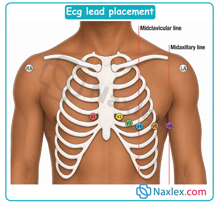
Lead Territories
The 12 leads view the heart from different angles (vectors), allowing clinicians to localize ischemia or infarction.
- Inferior Leads: II, III, aVF (View the bottom surface of the heart; supplied by the Right Coronary Artery).
- Septal Leads: V1, V2 (View the interventricular septum).
- Anterior Leads: V3, V4 (View the front of the left ventricle; supplied by the Left Anterior Descending artery).
- Lateral Leads: I, aVL, V5, V6 (View the lateral wall of the left ventricle; supplied by the Circumflex artery).
ECG Paper Basics
ECG paper is a standardized grid used to measure voltage and time. Understanding this grid is essential for manual interpretation and verifying machine measurements.
- Horizontal Axis (Time):
- 1 Small Box = 1 mm = 0.04 seconds.
- 1 Large Box (5 small boxes) = 5 mm = 0.20 seconds.
- Therefore, 5 Large Boxes = 1.0 second.
- Vertical Axis (Voltage/Amplitude):
- 1 Small Box = 1 mm = 0.1 mV.
- 1 Large Box = 5 mm = 0.5 mV.
- Standard Calibration: The machine should be set to 25 mm/sec speed and 10 mm/mV standardization (a calibration spike should be 2 large boxes high).
Role of the Nurse in Performing an ECG
The nurse is responsible for the entire procedure, ensuring quality control and patient comfort.
- Preparation: Explain the procedure to the patient, ensuring they understand they must remain still and supine. Minimize anxiety, as shivering or movement causes artifact.
- Skin Preparation:
- Clean the skin sites with alcohol wipes to remove oils and lotions.
- If the patient is diaphoretic, use a skin prep solution or gently abrade the superficial skin layer to ensure good electrode adherence.
- Clip excessive chest hair (do not shave) if it prevents good contact.
- Electrode Application: Apply electrodes firmly, ensuring the conductive gel is moist and making full contact with the skin.
- Lead Attachment: Attach the lead wires to the correct electrodes before connecting the cable to the machine to prevent pulling on the electrodes.
- Acquisition and Documentation: Ensure the tracing is labeled with the patient's name, ID number, date, and time. Document any relevant symptoms the patient is experiencing during the recording.
Normal ECG And A Systematic Integration Approach
The Normal Waveforms and Intervals
A single cardiac cycle (one heartbeat) consists of the following waves and measurements:
- P Wave:
- Represents atrial depolarization.
- Normal: Upright in lead II, smooth, rounded, precedes the QRS complex. Duration < 0.12s.
- PR Interval:
- Represents the time from atrial depolarization to the start of ventricular depolarization (includes conduction through the AV node).
- Normal duration: 0.12 to 0.20 seconds (3 to 5 small boxes).
- QRS Complex:
- Represents ventricular depolarization.
- Normal duration: < 0.10 seconds (less than 2.5 small boxes).
- Note: The QRS complex may be composed of multiple waves (Q wave, R wave, S wave).
- ST Segment:
- Represents the early phase of ventricular repolarization. It is normally isoelectric (flat), lying on the baseline (the T-P segment).
- T Wave:
- Represents ventricular repolarization (the resting phase of the ventricles).
- Normal: Typically upright in leads I, II, V3–V6. It should be rounded and asymmetric.
- QT Interval:
- Represents the total time for ventricular depolarization and repolarization.
- This interval varies significantly with heart rate and must be corrected (QTc). Normal QTc is generally < 0.44s in men and < 0.46s in women.
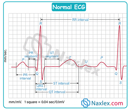
A Practical Step-by-Step Interpretation Method
Always use a consistent method to avoid missing subtle abnormalities.
- Determine Heart Rate:
- Quick Method (300 Method): Count the number of large boxes between two consecutive R waves (R-R interval). Divide 3 by the number of large boxes. (e.g., 4 large boxes = 300 / 4 = 75 bpm).
- 6-Second Method (Best for Irregular Rhythms): Count the number of R waves in a 6-second strip (indicated by markings on the top of the paper) and multiply by 10.
- Determine Heart Rhythm: Assess the regularity of the R-R intervals.
- Regular: R-R intervals vary by less than 0.12 seconds (3 small boxes).
- Irregular: R-R intervals vary significantly.
- Analyze P Waves:
- Are P waves present?
- Are they all identical in shape?
- Is there a P wave before every QRS complex?
- Measure the PR Interval: Count the small boxes from the start of the P to the start of the QRS. Is it consistently within the 0.12–0.20s range?
- Measure the QRS Duration: Count the small boxes across the widest part of the QRS complex. Is it < 0.10s?
- Evaluate ST Segments and T Waves: Are the ST segments at the baseline? Are the T waves upright and normal morphology?
Nursing Tip: Always interpret the ECG in the context of the patient. A rhythm that is technically abnormal may be well-tolerated by one patient (e.g., chronic atrial fibrillation) but require immediate intervention in another (e.g., new-onset atrial fibrillation causing hypotension).
Recognizing Abnormal ECG Findings
P-Wave and Atrial Abnormalities
Changes in the P wave morphology often indicate atrial enlargement or abnormal pacemaker sites.
- Left Atrial Enlargement (P-mitrale): The P wave becomes wide and notched (M-shaped) in lead II, with a deep terminal negative deflection in V1.
- Right Atrial Enlargement (P-pulmonale): The P wave becomes tall, peaked, and > 2.5 mm high in lead II.
- Atrial Premature Beats (APBs/PACs): An early, different-looking P wave interrupts the regular rhythm. The QRS complex that follows is usually normal.
PR Abnormalities and AV Blocks
AV blocks represent a delay or interruption in conduction between the atria and ventricles.
- First-Degree AV Block: The PR interval is prolonged (> 0.20s) but constant. Every P wave is followed by a QRS.
- Second-Degree AV Block, Type I (Mobitz I / Wenckebach): Progressive lengthening of the PR interval until a P wave is blocked (a P wave occurs without a following QRS). The cycle then repeats.
- Second-Degree AV Block, Type II (Mobitz II): The PR interval is constant (often normal) for all conducted beats, but P waves are intermittently blocked (e.g., every 2nd or 3rd P wave has no QRS). This type is dangerous as it often progresses to complete heart block.
- Third-Degree AV Block (Complete Heart Block): There is complete dissociation between P waves and QRS complexes. The atria and ventricles beat independently, controlled by separate pacemakers. P waves march through the tracing unrelated to the QRS complexes.
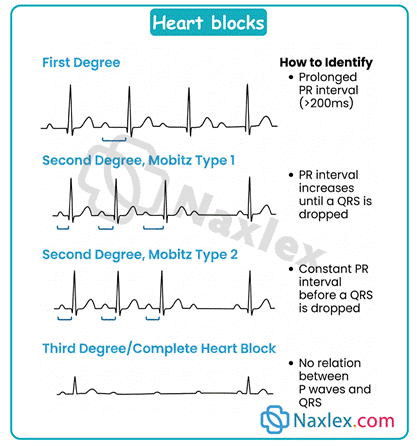
QRS Abnormalities
Changes in the QRS complex usually indicate issues with ventricular depolarization or conduction pathways.
- Pathological Q Waves: A Q wave that is > 0.04s wide or deeper than one-third the height of the R wave. This is a classic sign of a previous myocardial infarction (irreversible myocardial death).
- Low Voltage: All QRS complexes in the limb leads are < 5 mm (0.5 mV) high. This can occur in obesity, pericardial effusion, or emphysema.
Bundle Branch Blocks (BBB)
BBB occurs when there is a delay or block in conduction through the right or left bundle branch, forcing the ventricles to depolarize sequentially rather than simultaneously, resulting in a widened QRS complex (≥ 0.12s).
- Right Bundle Branch Block (RBBB): Characterized by an "RSR'" (rabbit ears) pattern in leads V1 and V2, and a wide, slurred S wave in lead I and V6.
- Left Bundle Branch Block (LBBB): Characterized by a wide, notched, or "M-shaped" R wave in leads I, aVL, V5, and V6, and deep S waves in V1 and V2. LBBB often obscures the diagnosis of a concurrent myocardial infarction.
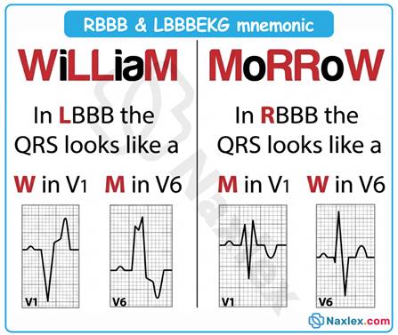
Ventricular Hypertrophy
Increased muscle mass in the ventricles increases the amplitude of the QRS complex.
- Left Ventricular Hypertrophy (LVH): Diagnosed by various voltage criteria. A common one is the Sokolow-Lyon criteria: S in V1 + R in V5 or V6 > 35 mm. Often accompanied by ST depression and T wave inversion in the lateral leads (known as the "strain pattern").
ST-Segment Changes
These changes are critical markers of acute myocardial ischemia or injury.
- ST-Segment Elevation: The ST segment is elevated above the baseline in two or more contiguous leads.
- Significant Elevation: Typically defined as > 1 mm in limb leads or > 2 mm in precordial leads.
- This is the hallmark of acute myocardial injury (e.g., STEMI).
- Note: Concave (smiling) ST elevation can also occur in benign conditions like early repolarization or pericarditis.
- ST-Segment Depression: The ST segment is below the baseline. It can be horizontal, downsloping, or upsloping.
- Significant Depression: > 0.5 mm in two or more contiguous leads.
- This is a primary indicator of subendocardial ischemia (e.g., NSTEMI or angina).
T-Wave Changes
T wave morphology is sensitive to ischemia, electrolyte shifts, and metabolic changes.
- T-Wave Inversion: The T wave points downward. This is a sign of ischemia.
- Hyperacute T Waves: T waves that are very tall, broad, and peaked. This is often an early sign of myocardial infarction, preceding ST elevation.
QT Abnormalities
Prolonged QT interval is a major risk factor for developing life-threatening ventricular dysrhythmias.
- QT Prolongation: The QT interval is extended beyond normal limits. It can be congenital or acquired (e.g., caused by medications like amiodarone, sotalol, or haloperidol; or electrolyte imbalances like hypokalemia or hypomagnesemia).
- U Waves: A small, rounded deflection sometimes seen after the T wave. Prominent U waves are strongly associated with hypokalemia.
Common Dysrhythmias
Atrial Fibrillation (AF)
AF is the most common sustained dysrhythmia, characterized by disorganized electrical activity in the atria.
- ECG Characteristics:
- Rhythm: Irregularly irregular (no pattern to the R-R intervals).
- P Waves: Absent; replaced by chaotic, fine, or coarse fibrillatory waves (f-waves).
- QRS Complex: Usually narrow.
- Nursing Implications: The primary risk is thrombus formation in the atria, leading to stroke. Assess the patient for hemodynamic stability (rate may be too fast or too slow). Treatment goals include rate control (beta-blockers, calcium channel blockers) and anticoagulation.
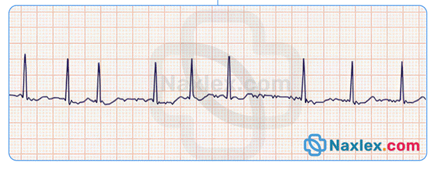
Atrial Flutter
A macro-reentrant circuit within the atria causes a rapid, regular atrial rate.
- ECG Characteristics:
- Rhythm: Atrial rhythm is regular; ventricular rhythm may be regular or variable (depending on AV block conduction ratio, e.g., 2:1, 3:1).
- P Waves: Absent; replaced by distinctive, regular sawtooth waves (F-waves).
- Nursing Implications: Similar risks to AF, including potential for thrombus. Requires rate control and consideration for cardioversion or ablation.
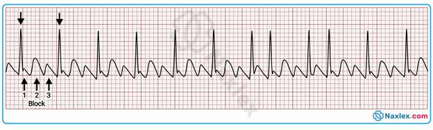
Supraventricular Tachycardia (SVT)
A general term for a tachycardia originating above the ventricles, commonly AV nodal reentrant tachycardia (AVNRT).
- ECG Characteristics:
- Rate: Rapid, typically 150–250 bpm.
- Rhythm: Strictly regular.
- P Waves: Often buried within the preceding T wave or invisible due to the rapid rate.
- QRS Complex: Usually narrow.
- Nursing Implications: The rapid rate may compromise cardiac output, causing hypotension, chest pain, or syncope. Initial management may include vagal maneuvers (e.g., carotid massage performed by a physician, Valsalva maneuver) followed by IV adenosine if unstable or symptomatic.
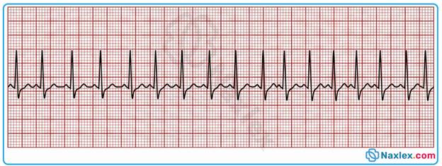 Ventricular Tachycardia (VT)
A life-threatening rhythm originating from an ectopic focus in the ventricles.
- ECG Characteristics:
- Rate: Rapid, 100–250 bpm.
- Rhythm: Regular or slightly irregular.
- P Waves: Absent (AV dissociation may be present but hard to see).
- QRS Complex: Wide (≥ 0.12s) and bizarre in morphology.
- Nursing Implications:
- Assess for a Pulse: If the patient is pulseless, initiate immediate defibrillation (Code Blue).
- If the patient has a pulse but is unstable (hypotension, altered mental status), prepare for synchronized cardioversion.
- If stable, manage with antiarrhythmics (e.g., amiodarone, lidocaine) as per ACLS protocols.
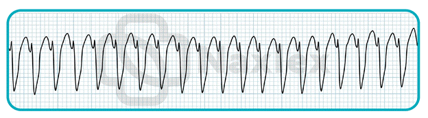 Torsades de Pointes (TdP)
A specific, polymorphic form of VT associated with a prolonged QT interval.
- ECG Characteristics: QRS complexes appear to twist around the isoelectric baseline, changing in amplitude and morphology.
- Nursing Implications: This rhythm frequently degrades into ventricular fibrillation. It is often precipitated by hypokalemia, hypomagnesemia, or QT-prolonging drugs. Treatment involves immediate defibrillation if pulseless, and administration of IV magnesium sulfate.
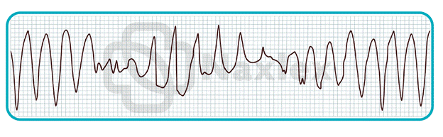 Ventricular Fibrillation (VF)
A chaotic, disorganized rhythm resulting in quivering of the ventricles with no effective cardiac output.
- ECG Characteristics:
- Rate: Not discernible.
- Rhythm: Chaotic and irregular.
- Waveforms:
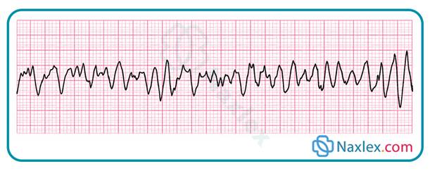
ECG Changes In Important Clinical Conditions
Cardiac rhythms often reflect systemic pathology. Recognizing these patterns aids in rapid diagnosis and collaborative clinical management.
- Myocardial Infarction (MI):
- ST Elevation (STEMI): Indicates transmural injury. Look for "tombstone" ST-segment elevation in contiguous leads.
- Reciprocal Changes: ST depression in leads opposite the site of infarction (e.g., inferior leads showing ST depression during an anterior MI).
- Evolving Patterns: Appearance of new pathological Q waves (indicates completed necrosis) and T wave inversions (indicates reperfusion or chronic ischemia).
- Pulmonary Embolism (PE):
- Sinus Tachycardia: The most common finding.
- S1Q3T3 Pattern: A classic but infrequent sign featuring a deep S wave in lead I, a Q wave in lead III, and an inverted T wave in lead III.
- Right Heart Strain: Right axis deviation or RBBB may appear as the right ventricle struggles against high pulmonary pressures.
- Pericarditis:
- Diffuse ST Elevation: Upward, concave ST segment elevation in nearly all leads except aVR and V1.
- PR Depression: Often seen early in the course of the disease.
- Cardiac Tamponade:
- Electrical Alternans: A phenomenon where the amplitude of the P, QRS, or T waves varies from beat to beat, caused by the heart swinging within a large pericardial effusion.
- Low Voltage: Often present due to the fluid dampening the electrical signal.
- Electrolyte Abnormalities:
- Hyperkalemia: Peaked, "tented" T waves are the first sign. As levels rise, the PR interval lengthens, the P wave flattens, and the QRS complex widens significantly.
- Hypokalemia: ST-segment depression, flattened T waves, and the appearance of prominent U waves.
- Genetic Syndromes:
- Long QT Syndrome: Characterized by a QTc interval exceeding 0.48s, predisposing the patient to Torsades de Pointes.
- Brugada Syndrome: Notable for ST elevation in V1–V3 with a characteristic "coved" appearance and a right bundle branch block pattern.
Ambulatory ECG Monitoring
When symptoms are transient, standard 12-lead ECGs often fail to capture the pathology. Ambulatory monitoring provides extended diagnostic windows.
- Holter Monitoring:
- Continuous recording of the heart's electrical activity, typically for 24 to 48 hours.
- Used to correlate daily symptoms with cardiac rhythm changes.
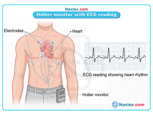
- Event Monitoring:
- Device is worn for several days to weeks.
- Recording is initiated by the patient pressing a button when they feel symptoms (e.g., palpitations, lightheadedness).
- Loop Recorders:
- External (patch) or internal (subcutaneous, implantable) devices that continuously record and overwrite data.
- When the patient triggers the device, it saves the rhythm data from before, during, and after the event.
- Nursing Responsibilities:
- Patient Education: Teach the patient how to keep an accurate diary of activities and symptom onset times.
- Skin Care: Emphasize the importance of keeping the electrode sites clean and dry.
- Troubleshooting: Instruct the patient on how to check for lead detachment or battery alerts.
- Data Integrity: Ensure the patient understands the necessity of returning the device to the cardiology department for analysis, as the raw data provides the diagnostic insight.
The Nurse’s Role In Cardioversion
Cardioversion is a time-sensitive procedure requiring technical proficiency, patient safety, and collaborative communication.
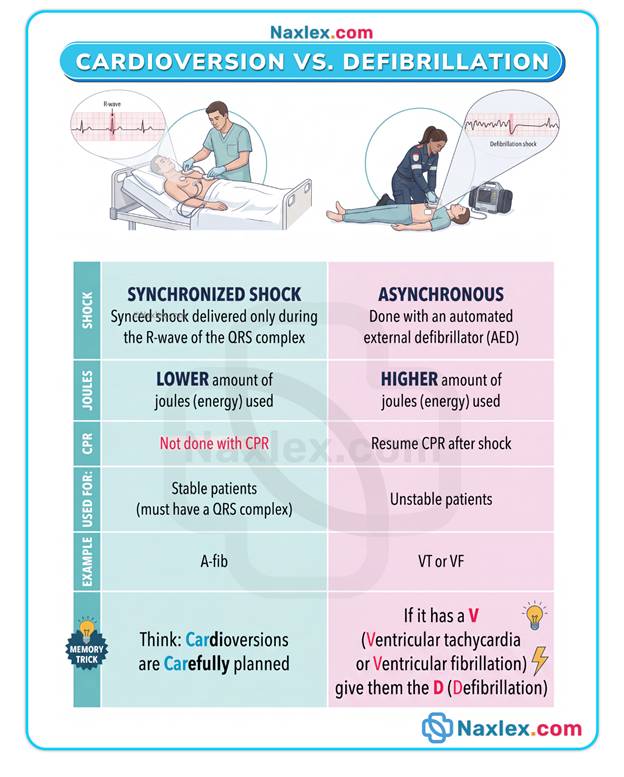
- Indications:
- Used for unstable tachyarrhythmias (e.g., AF, atrial flutter, or SVT) that are causing hypotension, acute heart failure, or ischemic chest pain.
- Preparation:
- Ensure a patent large-bore IV line and verify NPO status if elective.
- Obtain a baseline 12-lead ECG and perform a "timeout" to verify the patient and procedure.
- Verify the existence of a signed informed consent form.
- Synchronization:
- Crucial Step: The "SYNC" mode must be activated on the defibrillator.
- The machine detects the R wave, ensuring the shock is delivered during ventricular depolarization to avoid the "R-on-T" phenomenon, which could induce ventricular fibrillation.
- Sedation and Safety:
- Administer short-acting sedative/anxiolytic agents as ordered.
- Maintain continuous pulse oximetry and capnography.
- Have suction, airway adjuncts, and a crash cart immediately available at the bedside.
- Anticoagulation Considerations:
- For atrial arrhythmias lasting longer than 48 hours, there is a risk of dislodging an atrial thrombus.
- Confirm adequate anticoagulation (or perform a transesophageal echocardiogram) to Rule out thrombus before proceeding with elective cardioversion.
- Post-Procedure Care:
- Assess for a return to sinus rhythm.
- Monitor the skin for burns or irritation from the pads.
- Observe for potential complications such as transient bradycardia or new ectopy.
- Provide the patient with clear instructions on follow-up care, including continued medication management and signs to report after discharge.
Summary
- Success in ECG interpretation begins with rigorous adherence to standardized electrode placement protocols and proper machine calibration to ensure high-quality, diagnostic data.
- You must always utilize a structured, step-by-step approach when analyzing cardiac rhythms to evaluate rate, rhythm, intervals, and morphology to prevent the oversight of subtle abnormalities.
- Mastery of the ECG paper grid, where one small box represents 0.04 seconds, serves as the essential bedrock for accurately measuring conduction delays and determining heart rates.
- It is critical to remember that you should never treat the monitor in isolation; instead, you must always interpret ECG findings in the context of the patient’s clinical symptoms and hemodynamic stability.
- You must be able to rapidly identify lethal rhythms such as ventricular fibrillation and ventricular tachycardia, which mandate the immediate implementation of life-saving interventions and advanced cardiac life support.
- A solid understanding of conduction blocks is necessary because PR interval prolongation and missing QRS complexes are key diagnostic markers for various degrees of heart block that may require pacing.
- Recognizing ST-segment elevation and depression is vital, as these are the primary indicators of myocardial injury and ischemia that necessitate urgent notification of the medical team.
- You can utilize the ECG as a diagnostic tool for assessing systemic health, specifically by identifying peaked T waves for hyperkalemia and prominent U waves for hypokalemia.
- Diagnostic tools such as Holter monitors, event monitors, and loop recorders are essential for capturing transient or sporadic dysrhythmias that do not manifest during routine, short-term testing.
- Safety remains the top priority during cardioversion, which is why you must always verify the use of the synchronization function to avoid the life-threatening R-on-T phenomenon.