





 Video Library


Acquired Cardiovascular Disorders: Pediatric Cardiomyopathies
Lessons
 Naxlex
Just Now
Naxlex
Just Now
Notes Highlighting is available once you sign in. Login Here.
Objectives
- Analyze the epidemiology, etiology, and genetic predispositions associated with pediatric cardiomyopathies including dilated, hypertrophic, restrictive, arrhythmogenic right ventricular, and left ventricular non-compaction subtypes.
- Compare and contrast the underlying pathophysiological mechanisms of ventricular dysfunction, remodeling, and altered hemodynamics across the major classifications of pediatric cardiomyopathy.
- Identify early and late clinical manifestations of heart failure, outflow tract obstruction, impaired diastolic filling, and lethal dysrhythmias in infants, children, and adolescents.
- Interpret key findings from diagnostic modalities including electrocardiography, chest radiography, echocardiography, cardiac magnetic resonance imaging, serum biomarkers, and genetic testing.
- Formulate evidence-based therapeutic management strategies encompassing pharmacological interventions, mechanical circulatory support, surgical procedures, implantable devices, and pediatric heart transplantation.
- Develop a prioritized, individualized nursing care plan utilizing the nursing process to manage fluid volume status, optimize cardiac output, promote energy conservation, meet nutritional demands, and deliver psychosocial support to pediatric clients and their families.
- Recognize and anticipate life-threatening complications associated with pediatric cardiomyopathies, such as sudden cardiac death, thromboembolism, refractory acute heart failure, and lethal ventricular dysrhythmias.
Introduction
- Pediatric cardiomyopathies represent a complex and heterogeneous group of acquired and inherited disorders affecting the myocardium, resulting in mechanical or electrical dysfunction of the heart.
- Although relatively rare compared to adult myocardial disease, pediatric cardiomyopathy is a leading cause of severe heart failure, morbidity, and cardiac transplantation in infants and children.
- These disorders disrupt the structural integrity of cardiac myocytes, leading to progressive impaired ventricular contractility, compromised diastolic relaxation, or mechanical obstruction to systemic blood flow.
- Unlike congenital structural heart defects that feature abnormal anatomical communications or misplaced blood vessels, cardiomyopathies primarily impair the functional capability of the cardiac muscle tissue itself, often in an structurally intact heart.
- The etiology of pediatric cardiomyopathy encompasses a broad spectrum of clinical drivers, ranging from genetic mutations encoding sarcomeric or cytoskeletal proteins to acquired myocardial injury caused by viral myocarditis, anthracycline chemotherapy toxicity, metabolic storage diseases, or autoimmune inflammation.
- Pediatric clients present across a vast developmental spectrum, from neonates with severe, rapidly progressive acute pump failure to adolescents with silent hypertrophic changes who are at elevated risk for sudden cardiac death during strenuous physical exertion.
- Because infants and young children possess limited physiological reserve, compensatory mechanisms such as sinus tachycardia and myocardial hypertrophy can quickly become overwhelmed, rapidly transitioning a stable client into overt cardiogenic shock.
- From a pediatric nursing perspective, early clinical recognition requires high diagnostic vigilance and a subtle assessment approach.
- Early presentation in infants frequently mimics benign infant conditions, featuring subtle signs such as feeding intolerance, diaphoresis during bottle feeding, tachypnea, irritability, and failure to thrive.
- As myocardial compromise advances, children exhibit overt signs of systemic venous congestion and impaired forward tissue perfusion.
- Pediatric nurses play a critical role in early identification, managing continuous intravenous inotropic therapy, maintaining delicate fluid and electrolyte balance, preventing thromboembolic events, coordinating interdisciplinary care, providing end-of-life or transplant education, and supporting families navigating a unpredictable, life-altering diagnosis.
Epidemiology, Etiology, Risk Factors, And Pathophysiology
Epidemiology
Pediatric cardiomyopathy has an estimated incidence of 1.1 to 1.5 cases per 100,000 children under 18 years of age. The highest incidence occurs within the first year of life, primarily driven by severe dilated cardiomyopathy and metabolic or malformation syndromes. Dilated cardiomyopathy is the most frequent form encountered in pediatric practice, accounting for approximately 50% to 60% of all childhood cases, followed by hypertrophic cardiomyopathy, which accounts for roughly 35% to 40%. Restrictive cardiomyopathy, arrhythmogenic right ventricular cardiomyopathy, and left ventricular non-compaction represent the remaining small percentage of cases, with restrictive cardiomyopathy carrying the worst overall long-term prognosis and highest early transplantation rate.
Etiology
The causative origins of pediatric cardiomyopathy are divided into primary (idiopathic or genetic) and secondary (acquired, systemic, or toxic) categories:
- Idiopathic: No identifiable underlying cause is discovered after exhaustive diagnostic evaluation; accounts for nearly half of all presenting cases.
- Genetic and Inherited: Mutations in genes encoding sarcomeric proteins (such as MYH7, MYBPC3), cytoskeletal proteins (dystrophin), nuclear membrane proteins, or ion channels. Mitochondrial DNA mutations and inherited metabolic disorders (such as Pompe disease, fatty acid oxidation defects, and mucopolysaccharidoses) also directly impair myocyte energy production.
- Acquired Infectious and Inflammatory: Acute viral myocarditis caused by enteroviruses (Coxsackievirus B), adenovirus, parvovirus B19, human herpesvirus 6, or SARS-CoV-2 (including Multisystem Inflammatory Syndrome in Children [MIS-C]).
- Toxic and Infiltrative: Exposure to cardiotoxic antineoplastic agents, most notably anthracyclines (doxorubicin, daunorubicin), or iron overload secondary to chronic transfusion therapy in conditions such as thalassemia major.
- Autoimmune and Endocrine: Systemic lupus erythematosus, Kawasaki disease with coronary or myocardial involvement, maternal diabetes mellitus (causing transient hypertrophic cardiomyopathy in neonates), and thyroid dysfunction.
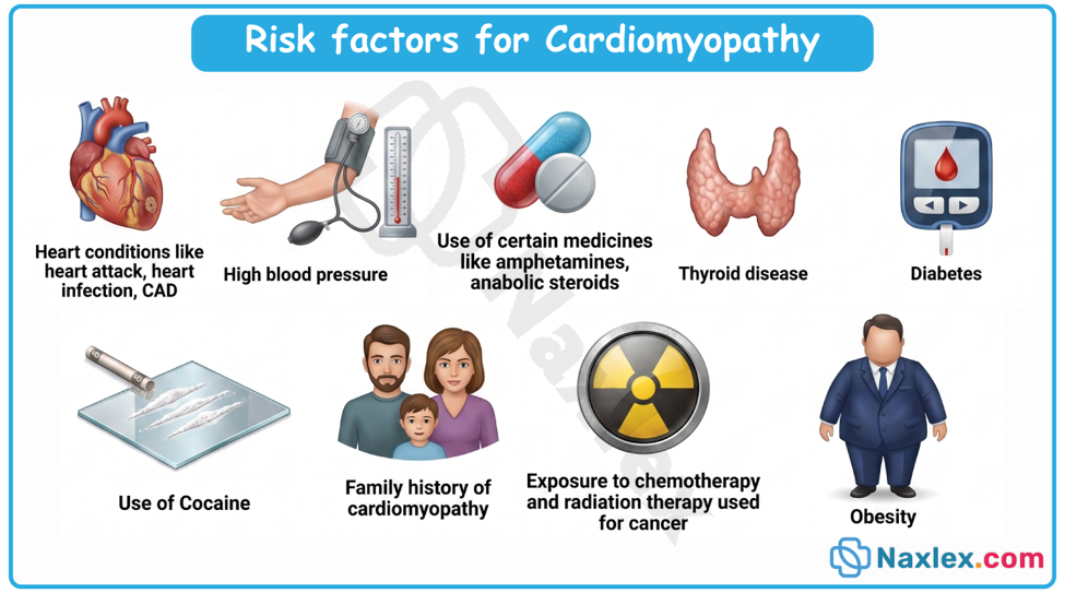
Risk Factors
- Family history of sudden unexplained cardiac death, early heart failure, or known cardiomyopathy in a first-degree relative.
- Personal history of severe viral illnesses, viral myocarditis, or systemic inflammatory conditions.
- Prior exposure to cardiotoxic chemotherapy regimens or mediastinal radiation therapy.
- Genetic syndromes such as Noonan syndrome, Friedrich ataxia, Duchenne or Becker muscular dystrophy, and Inborn Errors of Metabolism.
Neonatal exposure to maternal hyperinsulinemia or maternal autoimmune disease.
Pathophysiology
The central pathophysiological process in pediatric cardiomyopathy centers on structural failure of cardiac myocytes to effectively contract, relax, or maintain structural membrane integrity:
- In dilated cardiomyopathy, myocyte injury or genetic mutations impair systolic contractile force. To compensate for reduced stroke volume, the heart increases end-diastolic volume. Over time, progressive cardiac chamber dilation occurs with thin, stretched ventricular walls. This wall thinning increases ventricular wall stress according to Laplace's law, precipitating a downward spiral of increasing oxygen demand, progressive mitral or tricuspid valve regurgitation, elevated left ventricular end-diastolic pressure, pulmonary venous congestion, and diminished systemic cardiac output.
- In hypertrophic cardiomyopathy, disarray of cardiac muscle fibers and excessive myocardial hypertrophy occur without secondary cause. The thickened myocardium, particularly involving the interventricular septum, causes compliance failure (diastolic dysfunction) and elevated filling pressures. During exertion, dynamic left ventricular outflow tract obstruction can occur as the anterior leaflet of the mitral valve is pulled toward the hypertrophied septum during systole (systolic anterior motion [SAM]), drastically reducing forward cardiac output and increasing dynamic pressure gradients.
- In restrictive cardiomyopathy, severe myocardial fibrosis or infiltrative material creates a rigid, non-compliant ventricular myocardium. While systolic function and wall thickness initially remain near normal, impaired diastolic relaxation leads to elevated filling pressures, massive bi-atrial enlargement, systemic venous congestion, and pulmonary hypertension.
- In arrhythmogenic right ventricular cardiomyopathy, progressive fibrous and fatty tissue replacement of the right ventricular myocardium disrupts normal electrical conduction, predisposing the client to life-threatening ventricular tachyarrhythmias and right-sided heart failure.
- In left ventricular non-compaction, premature arrest of normal endomyocardial morphogenesis leaves prominent trabeculations and deep intertrabecular recess pockets within the ventricular apex, predisposing to localized thrombus formation, impaired contraction, and malignant dysrhythmias.