





 Video Library


Please set your exam date
Acquired Cardiovascular Disorders: Pediatric Hyperlipidemia
Study Questions
Practice Exercise 1
The nurse is assessing a 10-year-old client with a body mass index (BMI) in the 98th percentile. Which laboratory finding should the nurse identify as most characteristic of obesity-induced dyslipidemia?
Explanation
Pediatric obesity drives systemic insulin resistance, leading to increased hepatic release of free fatty acids, overproduction of very-low-density lipoproteins, and subsequent alterations in lipid clearance.
Rationale for correct answer:
2. Obesity-induced dyslipidemia is classically characterized by elevated triglyceride levels, reduced high-density lipoprotein cholesterol, and normal to mildly elevated low-density lipoprotein cholesterol. Insulin resistance alters adipocyte lipolysis and hepatic lipid processing, shifting the metabolic profile toward hypertriglyceridemia and low HDL. Recognizing this atherogenic triad is essential for guiding pediatric cardiovascular risk stratification and intervention.
Rationale for incorrect answers:
1. High-density lipoprotein cholesterol levels are typically reduced, not elevated, in clients with obesity and metabolic syndrome. Insulin resistance and inflammatory cytokine activity impair reverse cholesterol transport mechanisms and accelerate HDL clearance. Elevated HDL serves as a cardioprotective lipid component rather than an indicator of metabolic dysfunction. High HDL levels are inconsistent with obesity-induced dyslipidemia profiles.
3. Isolated elevation of low-density lipoprotein cholesterol with normal triglycerides strongly suggests familial hypercholesterolemia or genetic receptor defects rather than obesity-related metabolic dysfunction. Primary genetic dyslipidemias present with distinct lipid patterns compared to the triglyceride-predominant profile seen in pediatric obesity. Elevated triglycerides are a primary diagnostic hallmark of insulin resistance-driven lipid disorders. Attributing isolated high LDL solely to obesity misses potential primary genetic etiologies.
4. Total cholesterol measures the sum of all circulating lipid fractions and typically increases or remains normal during dyslipidemic states rather than decreasing. A decreased total cholesterol level accompanied by elevated LDL is biochemically contradictory unless HDL and triglycerides are severely depleted. Metabolic derangements associated with pediatric obesity elevate overall circulating atherogenic lipoproteins. Lowered total cholesterol does not reflect obesity-induced dyslipidemia.
Test-taking strategy:
- Analyze the scenario/question: The nurse is assessing a 10-year-old child with severe pediatric obesity (BMI at the 98th percentile). The goal is to identify the classic laboratory finding of obesity-induced dyslipidemia.
- Apply pathophysiological principles:
- Recall the metabolic effects of insulin resistance in pediatric obesity: elevated triglycerides and reduced HDL cholesterol.
- Differentiate secondary obesity-related lipid changes from primary genetic lipid disorders like familial hypercholesterolemia (isolated LDL elevation).
- Evaluate Options:
- Rule out Choice 1: HDL is reduced, not elevated, in metabolic syndrome and obesity.
- Choice 2: Elevated triglycerides paired with decreased HDL is the hallmark pattern of obesity-induced dyslipidemia.
- Rule out Choice 3: Isolated LDL elevation points toward genetic hypercholesterolemia rather than obesity.
- Rule out Choice 4: Total cholesterol does not characteristically drop in obesity-related dyslipidemia.
Take home points
- The classic lipid profile in pediatric obesity-induced dyslipidemia consists of elevated triglycerides, low HDL cholesterol, and normal to mildly elevated LDL cholesterol.
- Insulin resistance is the primary driver of obesity-induced dyslipidemia, leading to increased hepatic VLDL production and altered lipoprotein lipase activity.
- Screening for lipid abnormalities in pediatric clients with a BMI at or above the 95th percentile should begin between 2 and 10 years of age.
- Initial management of pediatric obesity-induced dyslipidemia focuses on lifestyle interventions, including increased physical activity, dietary modification, and weight stabilization.
The nurse is reviewing the pathophysiology of atherosclerosis with a group of parents. Which statement should the nurse include to explain the earliest visual stage of plaque development in children?
Explanation
Atherosclerosis is a chronic inflammatory disorder characterized by lipid accumulation within the arterial wall, initiated by endothelial dysfunction and lipid oxidation. Low-density lipoprotein particles cross damaged endothelium, undergoing oxidation that triggers monocyte recruitment and differentiation into macrophages. These cells engulf oxidized lipids to become foam cells, which aggregate to form early structural lesions. Fatty streaks represent the earliest grossly visible lesion, frequently emerging in children and young adults as precursor states to complex fibrous plaques.
Rationale for correct answer:
2. The aggregation of lipid-laden macrophages forms fatty streaks within the tunica intima. This initial stage of lesion formation begins in early childhood without immediate hemodynamic compromise. Smooth muscle cell migration and collagen deposition occur later in disease progression. Endothelial permeability changes drive this cellular accumulation.
Rationale for incorrect answers:
1. Fibrous plaques represent a mature, advanced atherosclerotic lesion rather than the initial developmental stage. Significant luminal narrowing from thick fibrous caps typically requires decades of progressive lipid accumulation. Infancy lesions remain limited to minimal intimal cellular infiltration. Advanced stenosis remains extremely rare during infant vascular development.
3. Medial layer arterial calcification describes Monckeberg arteriosclerosis, a process distinct from subendothelial atherosclerotic plaque formation. High dietary sugar intake contributes to metabolic dysfunction but does not directly cause isolated arterial medial calcification. Atherosclerotic calcification occurs within the intimal plaque matrix during late structural remodeling. Medial sclerosis leaves the intima and lumen uninvolved.
4. Acute plaque rupture and subsequent thrombus formation represent late-stage ischemic complications seen predominantly in adults. Pediatric lesions remain stable, non-thrombogenic fatty streaks without a thin vulnerable cap. Immediate arterial thrombosis requires severe structural plaque instability that is absent in school-aged children. Vascular occlusion in children usually stems from vasculitis or embolic phenomena rather than ruptured atheromas.
Test-taking strategy:
- Analyze the scenario/question: The nurse is identifying the earliest visual stage of pediatric atherosclerotic plaque development. The focus is on foundational vascular pathophysiology and child development.
- Evaluate pathophysiological progression:
- Choice 2 correctly identifies foam cell aggregation forming fatty streaks as the initial microscopic and macroscopic lesion visible during pediatric vascular screening.
- Rule out advanced or unrelated lesions:
- Rule out Choice 1: Fibrous plaques represent an advanced, late-stage structural change, not an early pediatric lesion.
- Rule out Choice 3: Medial calcification reflects Monckeberg sclerosis, which is pathologically distinct from intimal atherogenesis.
- Rule out Choice 4: Plaque rupture with thrombosis is a acute terminal event of adult coronary disease, not early pediatric pathology.
Take home points
- Fatty streaks are the earliest visible macroscopic lesions of atherosclerosis and frequently appear in early childhood.
- Foam cells form when vascular monocytes differentiate into macrophages and internalize oxidized low-density lipoproteins within the arterial intima.
- Advanced fibrous plaques and plaque ruptures require decades of systemic inflammation and are extremely rare in pediatric populations.
- Monckeberg arteriosclerosis involves calcification of the tunica media, whereas atherosclerosis strictly targets the tunica intima.
The nurse is obtaining a family health history for a 5-year-old client. Which finding should the nurse identify as a risk factor for primary hyperlipidemia?
Explanation
Primary hyperlipidemia is a group of genetic disorders characterized by abnormal lipid metabolism resulting in elevated systemic cholesterol or triglyceride levels. Endogenous mutations in cell surface receptors or key metabolic enzymes impair the clearance of circulating low-density lipoprotein particles. Inherited autosomal dominant conditions, such as familial hypercholesterolemia, cause severe premature vascular damage starting in early childhood. Universal lipid screening or targeted selective evaluation relies on identifying familial premature cardiovascular disease or severe maternal/paternal inherited dyslipidemias.
Rationale for correct answer:
3. A maternal history requiring pharmacotherapy for elevated low-density lipoprotein cholesterol indicates significant inherited monogenic or polygenic dyslipidemia. Familial hypercholesterolemia is an autosomal dominant disorder that impairs low-density lipoprotein receptor function, causing severe lipid elevation in offspring. First-degree relatives with severe hypercholesterolemia represent the strongest genetic predictor for primary pediatric hyperlipidemia.
Rationale for incorrect answers:
1. Gestational diabetes in a maternal aunt represents a transient, pregnancy-induced carbohydrate intolerance that does not confer a genetic risk for primary lipid disorders. Second-degree collateral relatives do not carry the same hereditary weight as first-degree parents or siblings. Transient endocrine alterations during gestation do not alter baseline lipid transport genome expression in offspring. Primary hyperlipidemia stems directly from inherited defects in apolipoprotein or receptor genes.
2. A paternal grandfather experiencing a myocardial infarction at age 72 reflects late-onset cardiovascular disease rather than premature coronary events. Premature cardiovascular disease is defined as an ischemic event occurring before age 55 in males or age 65 in females. Late-onset cardiac events in second-degree relatives usually stem from lifetime environmental lifestyle factors rather than high-penetrance monogenic lipid mutations. Age thresholds are critical when stratifying pediatric familial cardiovascular risk.
4. Mild paternal hypertension controlled exclusively by non-pharmacological lifestyle modifications does not establish a primary inherited lipid metabolism defect. Hypertension and hyperlipidemia are distinct cardiovascular risk factors with unique pathophysiological pathways and genetic origins. Mild blood pressure alterations managed without medication reflect low systemic vascular risk that does not warrant targeted pediatric lipid screening. Isolated essential hypertension does not indicate mutated low-density lipoprotein receptor alleles.
Test-taking strategy:
- Analyze the scenario/question: The nurse is assessing a 5-year-old client's family history to identify risk factors for primary pediatric hyperlipidemia.
- Apply screening criteria:
- Targeted pediatric lipid screening requires a positive family history of premature cardiovascular disease (males < 55 years, females < 65 years) or severe primary dyslipidemia in a first-degree relative.
- Evaluate first-degree vs. second-degree relatives:
- Choice 3 identifies a first-degree relative (mother) with severe dyslipidemia requiring pharmacotherapy, which directly signals autosomal dominant primary hypercholesterolemia.
- Rule out non-diagnostic findings:
- Rule out Choice 1: A maternal aunt (second-degree relative) with gestational diabetes is an endocrine finding unrelated to primary lipid gene mutations.
- Rule out Choice 2: A grandfather experiencing an event at age 72 is not premature cardiovascular disease (threshold is < 55 years for males).
- Rule out Choice 4: Mild paternal hypertension is an isolated vascular risk factor, not an inherited primary lipid defect.
Take home points
- Targeted pediatric lipid screening is indicated for children with a family history of premature cardiovascular disease or severe parental dyslipidemia.
- Premature cardiovascular disease is defined as coronary events occurring before age 55 in first-degree male relatives and before age 65 in first-degree female relatives.
- Familial hypercholesterolemia is an autosomal dominant genetic disorder causing elevated low-density lipoprotein levels from birth.
- Second-degree relatives with late-onset disease or non-lipid metabolic conditions do not meet the risk threshold for primary hyperlipidemia screening.
The nurse is preparing a presentation on pediatric cardiovascular health. Which modifiable risk factors should the nurse include as accelerators of the atherosclerotic process? Select all that apply
Explanation
Atherosclerosis is a progressive vascular disease characterized by lipid deposition, chronic inflammation, and structural remodeling within the arterial intimal wall. Endothelial injury triggered by metabolic or mechanical stressors facilitates low-density lipoprotein retention and oxidation, driving macrophage recruitment and foam cell accumulation. Addressing pediatric endothelial dysfunction, persistent vascular inflammation, systemic dyslipidemia, and accelerated atherogenesis through lifestyle management prevents premature cardiovascular morbidity in adulthood.
Rationale for correct answers:
1. Exposure to environmental tobacco smoke introduces toxic chemical constituents that directly induce endothelial cell dysfunction and oxidative stress. Passive smoking impairs nitric oxide bioavailability, promoting vascular cell adhesion and accelerating foam cell formation in pediatric arterial walls. Endothelial impairment from second-hand smoke represents a direct, preventable environmental driver of early vascular lesions. Childhood smoke exposure significantly accelerates arterial stiffening and premature atherogenic progression.
3. Chronic physical inactivity contributes significantly to systemic metabolic dysfunction, insulin resistance, and adverse lipid profile alterations in children. Regular physical exertion enhances endothelial nitric oxide synthase expression, optimizing vascular tone and suppressing vascular inflammatory cascades. Sedentary behavior reduces high-density lipoprotein activity, facilitating continuous lipid accumulation within the vascular subendothelial space. Increasing physical activity restores endothelial protective mechanisms and attenuates early atherosclerotic changes.
4. Elevated dietary intake of trans fatty acids severely alters pediatric lipid profiles by increasing circulating low-density lipoprotein levels while suppressing high-density lipoproteins. Trans fats induce systemic inflammatory cytokine release, enhancing leukocyte adhesion to damaged arterial endothelial cells. Increased subendothelial retention of oxidized lipid particles accelerates fatty streak maturation and early plaque development.
Rationale for incorrect answers:
2. A biological parent with a history of premature coronary artery disease represents a major non-modifiable genetic risk factor. Genetic inheritance influences endogenous receptor expression, lipid clearance pathways, and vascular structural susceptibility that cannot be directly altered through lifestyle or environmental interventions. While familial history mandates targeted screening and aggressive modification of secondary factors, the genetic predisposition itself remains non-modifiable.
5. Biological male sex is an unalterable non-modifiable demographic risk factor associated with higher baseline cardiovascular event rates. Chromosomal architecture and endogenous sex hormone profiles influence baseline lipid transport patterns and endothelial biology without opportunity for lifestyle alteration.
Test-taking strategy:
- Analyze the scenario/question: The nurse is identifying modifiable risk factors that accelerate pediatric atherosclerotic progression. This is a select all that apply question requiring differentiation between modifiable and non-modifiable variables.
- Differentiate risk factor categories:
- Modifiable factors involve behaviors, environment, or diet that can be altered or eliminated.
- Non-modifiable factors include innate biological traits, genetics, age, and biological sex.
- Evaluate each option:
- Choice 1 is modifiable (environmental tobacco smoke exposure can be eliminated).
- Rule out Choice 2: Parent history of premature CAD is non-modifiable (inherited genetics).
- Choice 3 is modifiable (physical inactivity can be remediated with exercise).
- Choice 4 is modifiable (dietary intake of trans fats can be restricted).
- Rule out Choice 5: Biological male sex is non-modifiable (innate biological attribute).
Take home points
- Modifiable pediatric risk factors for atherosclerosis include environmental smoke exposure, physical inactivity, unhealthy dietary fats, hypertension, and obesity.
- Non-modifiable cardiovascular risk factors comprise age, biological sex, race, and inherited genetic conditions or familial disease history.
- Pediatric atherosclerosis is a reversible process during its early stages through targeted lifestyle modifications and risk reduction.
- Second-hand smoke exposure directly causes endothelial toxicity and impairs nitric oxide-mediated vasodilation in pediatric vascular beds.
Practice Excercise 2
The nurse is reviewing universal lipid screening guidelines with a group of clinic nurses. At which age ranges should the nurse state that universal lipid screening is recommended for all children, regardless of risk factors?
Explanation
Universal pediatric lipid screening identifies covert hypercholesterolemia before early atherosclerosis progresses into complex fibrous lesions. Pubertal hormonal surges induce transient physiological fluctuations in serum lipid concentrations, making mid-childhood and late adolescence optimal diagnostic windows. Systemic guidelines establish screening protocols during periods of lipid stability to detect familial hypercholesterolemia, prevent premature coronary artery disease, optimize endothelial nitric oxide, and arrest early atherogenic plaque development.
Rationale for correct answer:
3. Universal lipid screening is specifically recommended for all pediatric clients once between ages 9 and 11 years and once again between ages 17 and 21 years. These designated developmental windows avoid peak pubertal maturation, during which serum low-density lipoprotein levels transiently drop, potentially yielding false-negative screening results. Universal screening in late childhood and late adolescence catches severe inherited dyslipidemias that selective, risk-based screening protocols miss.
Rationale for incorrect answers:
1. Screening children aged 2 to 4 years is restricted strictly to high-risk selective screening rather than universal population testing. Children under 9 years without identified family risk factors or personal metabolic conditions do not undergo routine universal lipid panel testing. The 12 to 14 years range coincides with early puberty, when fluctuating gonadotropin levels induce unreliable lipid variations. Universal testing during early puberty increases false-negative diagnostic errors.
2. The age ranges of 5 to 7 years and 14 to 16 years fall outside established universal pediatric screening algorithms. Children aged 5 to 7 years only receive targeted selective lipid testing if they possess significant family history of premature cardiovascular disease. Ages 14 to 16 years represent mid-pubertal maturation, a developmental phase associated with physiological drops in total cholesterol and low-density lipoprotein concentrations. Universal screening is deliberately postponed until post-pubertal stability.
4. The age range of 12 to 14 years occurs during peak pubertal transition, making it unsuitable for universal screening due to hormonal alterations in lipid transport molecules. Testing adults aged 22 to 25 years falls under standard adult preventative health protocols rather than pediatric and adolescent screening frameworks. Postponing initial universal screening until age 12 misses early diagnostic opportunities during stable late childhood. Pediatric universal screening terminates at the transition to young adulthood.
Test-taking strategy:
- Analyze the scenario/question: The nurse is presenting universal pediatric lipid screening guidelines, identifying specific age ranges recommended for all children without requiring pre-existing risk factors.
- Apply clinical guidelines:
- Recall National Heart, Lung, and Blood Institute (NHLBI) and American Academy of Pediatrics (AAP) recommendations for universal pediatric screening.
- Target ages represent stable pre-pubertal (ages 9 to 11) and post-pubertal/late-adolescent (ages 17 to 21) periods.
- Evaluate age options:
- Rule out Choice 1: Ages 2 to 4 are reserved for selective risk-based screening; ages 12 to 14 are during pubertal lipid drops.
- Rule out Choice 2: Ages 5 to 7 are selective screening only; ages 14 to 16 are during mid-puberty.
- Choice 3 accurately reflects the recommended universal screening windows: ages 9 to 11 and 17 to 21.
- Rule out Choice 4: Age 12 to 14 is peak puberty; age 22 to 25 is outside pediatric guidelines.
Take home points
- Universal pediatric lipid screening is recommended once between ages 9 and 11 years and once between ages 17 and 21 years.
- Universal screening is avoided between ages 12 and 16 due to pubertal hormonal fluctuations that lower serum low-density lipoprotein levels.
- Selective lipid screening begins as early as age 2 for children with a high-risk family history or personal cardiovascular risk factors.
- Identifying dyslipidemia in late childhood enables early lifestyle and pharmacological interventions that prevent adult coronary artery disease.
The nurse is performing a physical assessment on a 12-year-old client with suspected homozygous familial hypercholesterolemia. Which physical finding should the nurse expect if the client has severely elevated cholesterol levels?
Explanation
Homozygous familial hypercholesterolemia is a rare, life-threatening autosomal recessive disorder characterized by severe low-density lipoprotein receptor gene mutations. Absent or non-functional receptors lead to extreme, uninhibited circulating low-density lipoprotein accumulation exceeding 500 mg/dL in early childhood. Macrophages ingest excess plasma lipoproteins, forming foam cells that deposit within dense connective tissue and subendothelial matrix spaces to create tendon xanthomas, premature corneal arcus, accelerated coronary atherogenesis, and rapidly progressive aortic stenosis.
Rationale for correct answer:
2. Tendon xanthomas present as firm, nodular, yellowish-to-reddish cholesterol deposits infiltrating tendon sheaths, particularly the Achilles tendon and extensor tendons of the hands. In homozygous familial hypercholesterolemia, extreme low-density lipoprotein elevations cause early tissue macrophage uptake and cutaneous lipid deposition during childhood. Tendinous xanthomas are pathognomonic physical markers of severe, long-standing primary hypercholesterolemia.
Rationale for incorrect answers:
1. Prominent bilateral temporal wasting and pedal edema reflect severe protein-calorie malnutrition, advanced end-stage hepatic disease, or nephrotic syndrome rather than familial lipid receptor defects. Temporal muscle atrophy indicates systemic catabolism and somatic protein depletion. Lower extremity edema stems from decreased plasma oncotic pressure or fluid retention, not localized lipid tissue accumulation.
3. Blue-gray pigmentation of the buccal mucosa is a classic physical finding of Peutz-Jeghers syndrome or systemic heavy metal toxicity such as argyria. Endogenous mucosal melanin or metal deposition produces distinct focal pigmentation without altering vascular lipid concentrations. Primary familial lipid disorders manifest cutaneously as lipid-laden xanthomas or xanthelasma rather than mucosal pigmented macules. Oral mucosal hyperpigmentation does not correlate with serum cholesterol levels.
4. A cherry-red spot on the macula during ophthalmoscopic examination is the hallmark finding of lysosomal storage diseases such as Tay-Sachs disease and Niemann-Pick disease. Ganglioside or sphingomyelin accumulation within retinal ganglion cells spares the fovea, producing a distinct red spot contrast. Primary lipid receptor mutations cause corneal arcus or lipemia retinalis rather than macular lipid-storage spots. Retinal ganglion degeneration is absent in familial hypercholesterolemia.
Test-taking strategy:
- Analyze the scenario/question: The nurse is assessing a 12-year-old client with suspected homozygous familial hypercholesterolemia, looking for physical manifestations of extremely elevated low-density lipoprotein levels.
- Correlate pathology to clinical findings:
- Severe, long-standing hypercholesterolemia causes lipid deposition in skin, tendons, and vascular structures (xanthomas, xanthelasma, corneal arcus).
- Evaluate option specificity:
- Rule out Choice 1: Temporal wasting and pedal edema reflect malnutrition or organ failure, not lipid receptor gene mutations.
- Choice 2 accurately identifies firm, yellowish Achilles tendon plaques (tendon xanthomas), which are classic markers of severe primary dyslipidemia.
- Rule out Choice 3: Buccal pigmentation is characteristic of Peutz-Jeghers syndrome or Addison's disease, not dyslipidemia.
- Rule out Choice 4: Cherry-red macular spots indicate Tay-Sachs or Niemann-Pick lysosomal storage disease, not hypercholesterolemia.
Take home points
- Homozygous familial hypercholesterolemia causes extreme low-density lipoprotein elevations (> 500 mg/dL) from birth, leading to early childhood cardiovascular disease.
- Tendon xanthomas, particularly of the Achilles tendon, are nodular lipid deposits pathognomonic for severe inherited hypercholesterolemia.
- Other classic physical signs include planar xanthomas, xanthelasma, and premature corneal arcus (arcus senilis appearing before age 45).
- Without aggressive intervention such as apheresis or novel biologic agents, homozygous clients often develop fatal coronary disease before age 30.
The nurse is reviewing the medical record of an 8-year-old child. Which clinical indicators should prompt the nurse to recommend initiating selective lipid screening? Select all that apply
Explanation
Selective pediatric lipid screening targets children aged 2 to 8 years and 12 to 16 years who possess personal or familial risk factors for premature cardiovascular disease. Metabolic dysfunction, systemic inflammation, and inherited lipid receptor defects accelerate early atherogenesis when unmanaged during childhood growth. Identifying pediatric obesity, severe maternal or paternal inherited dyslipidemias, and a documented family history of premature coronary events enables targeted early intervention to preserve endothelial nitric oxide and prevent premature vascular damage.
Rationale for correct answers:
1. A body mass index at the 97th percentile meets the threshold for pediatric obesity, which significantly elevates risk for secondary dyslipidemia and metabolic syndrome. Excess adiposity drives insulin resistance, increased free fatty acid flux, and hepatic low-density lipoprotein overproduction. Pediatric obesity impairs endothelial vasodilation and accelerates subendothelial lipid retention. Obesity represents a clear clinical indication for targeted selective lipid testing in an 8-year-old child.
2. A biological mother with a total cholesterol of 260 mg/dL exceeds the 240 mg/dL high-risk clinical threshold for parental hypercholesterolemia. High maternal serum cholesterol suggests an underlying monogenic or polygenic primary lipid clearance disorder such as familial hypercholesterolemia. Offspring of parents with severe hypercholesterolemia face an increased probability of inheriting mutated lipid transport alleles. This elevated parental cholesterol level mandates targeted selective lipid panel evaluation.
4. A biological father experiencing a myocardial infarction at age 42 represents a clear positive family history of premature cardiovascular disease. Premature ischemic heart disease is defined as a coronary event occurring before age 55 in first-degree male relatives. Early paternal coronary artery thrombosis indicates severe, high-penetrance cardiovascular risk factors transmitted to offspring.
Rationale for incorrect answers:
3. Mild seasonal allergies reflect an IgE-mediated type I hypersensitivity reaction restricted to the upper respiratory tract mucosa without systemic vascular implications. Histamine release and localized allergic mucosal inflammation do not alter hepatic lipid metabolism, systemic cholesterol transport, or arterial endothelial biology. Atopic conditions carry no hereditary correlation with primary hypercholesterolemia or premature atherogenesis. Seasonal allergies do not constitute an indication for selective lipid screening.
5. A history of acute streptococcal pharyngitis is a self-limiting bacterial upper respiratory infection caused by Streptococcus pyogenes. While untreated streptococcal infections can trigger immune-mediated complications such as acute rheumatic fever or post-streptococcal glomerulonephritis, they do not induce chronic lipid metabolism defects. Transient bacterial inflammation does not promote subendothelial apolipoprotein deposition or inherited dyslipidemia.
Test-taking strategy:
- Analyze the scenario/question: The nurse is evaluating an 8-year-old child's medical record to identify indications for selective lipid screening before the universal 9-to-11-year screening window.
- Recall selective lipid screening criteria:
- Indications include a positive family history of premature cardiovascular disease (males < 55 years, females < 65 years), parental hypercholesterolemia (total cholesterol ≥ 240 mg/dL), or personal health factors (obesity BMI ≥ 95th percentile, hypertension, diabetes mellitus).
- Evaluate each option:
- Choice 1: BMI at the 97th percentile indicates obesity (≥ 95th percentile), a personal risk factor for selective screening.
- Choice 2: Maternal total cholesterol of 260 mg/dL meets the ≥ 240 mg/dL threshold for selective screening.
- Rule out Choice 3: Seasonal allergies are an localized atopic condition with no lipid metabolism involvement.
- Choice 4: Paternal MI at age 42 is premature CAD (< 55 years for males), a strong hereditary indication for selective screening.
- Rule out Choice 5: Streptococcal pharyngitis is a transient bacterial infection with no relation to lipid transport disorders.
Take home points
- Selective lipid screening is indicated for children aged 2 to 8 years who have high-risk personal health factors or a positive family history.
- Personal indications include pediatric obesity (BMI ≥ 95th percentile), hypertension, diabetes mellitus, or chronic inflammatory diseases.
- Familial indications include a parent with total cholesterol ≥ 240 mg/dL or a first-degree male relative with cardiovascular disease before age 55.
- Transient pediatric infections and localized allergic conditions do not alter lipid transport mechanisms or warrant lipid testing.
The nurse is assessing a child with extremely high triglyceride levels. Which life-threatening complication should the nurse monitor for closely?
Explanation
Severe pediatric hypertriglyceridemia, defined as serum triglyceride concentrations exceeding 1,000 mg/dL, poses an immediate risk for severe systemic toxicity and organ damage. Free fatty acids generated by pancreatic lipase overload local binding capacity, triggering acinar cell membrane lysis, intense autoinflammation, and pancreatic acinar necrosis. Prompt recognition of severe epigastric distress prevents catastrophic systemic inflammatory response syndrome, multiorgan failure, pancreatic necrosis, and capillary leak syndrome.
Rationale for correct answer:
3. Acute pancreatitis is a severe, life-threatening complication directly triggered by extreme hypertriglyceridemia when serum levels exceed 1,000 mg/dL. Pancreatic lipase hydrolyzes excess circulating triglycerides within pancreatic capillaries, releasing toxic unbound free fatty acids that induce acinar cell injury and microvascular thrombosis. Affected children present with severe, sudden-onset epigastric abdominal pain radiating directly to the back, intractable nausea, and persistent vomiting.
Rationale for incorrect answers:
1. Acute glomerulonephritis is an immune-mediated inflammatory disorder of the renal glomeruli typically triggered by immune complex deposition following group A beta-hemolytic streptococcal infections. Circulating lipid molecules do not deposit within the glomerular basement membrane to cause acute nephritic inflammation, hematuria, or red blood cell casts. Primary renal glomerular damage stems from antigen-antibody complex activation of the complement cascade rather than lipotoxicity. Hypertriglyceridemia does not directly induce acute post-infectious glomerulonephritis.
2. Diabetic ketoacidosis is an acute, life-threatening metabolic complication of insulin deficiency characterized by severe hyperglycemia, metabolic acidosis, and elevated ketone bodies. While uncontrolled diabetic ketoacidosis can cause secondary transient hypertriglyceridemia due to impaired lipoprotein lipase activity, elevated triglycerides themselves do not cause pancreatic beta-cell destruction or ketoacidosis. Diabetic ketoacidosis stems directly from absolute or relative insulin deficiency and counter-regulatory hormone excess.
4. Pulmonary embolism involves the occlusion of the pulmonary arterial bed by a thromboembolus originating from deep venous thrombosis, air, fat, or amniotic fluid. Although hypertriglyceridemia promotes mild prothrombotic state alterations, it does not cause acute, direct venous thromboembolism or pulmonary arterial occlusion. The primary immediate microvascular damage from chylomicronemia occurs within the pancreatic capillary network rather than systemic deep venous beds.
Test-taking strategy:
- Analyze the scenario/question: The nurse is assessing a pediatric client with extremely high triglyceride levels and evaluating for a specific, life-threatening complication.
- Correlate pathophysiology:
- Recall that extreme hypertriglyceridemia (> 1,000 mg/dL) leads to chylomicronemia, pancreatic lipase cleavage of lipids into toxic free fatty acids, and localized acinar cell injury.
- Evaluate life-threatening complications:
- Rule out Choice 1: Glomerulonephritis is an immune-complex renal disorder, not a lipotoxic condition.
- Rule out Choice 2: Diabetic ketoacidosis stems from insulin deficiency; hypertriglyceridemia is a downstream result, not a primary cause of DKA.
- Choice 3 accurately identifies acute pancreatitis, the classic, immediate life-threatening complication of severe hypertriglyceridemia.
- Rule out Choice 4: Pulmonary embolism arises from deep vein thrombosis, not direct pancreatic capillary lipotoxicity.
Take home points
- Severe hypertriglyceridemia (serum levels > 1,000 mg/dL) carries a high risk for acute hypertriglyceridemia-induced pancreatitis.
- Pancreatic lipase breaks down excessive triglycerides into unbound free fatty acids that cause pancreatic acinar cell cytotoxicity and local ischemia.
- Clinical manifestations include sudden severe epigastric abdominal pain radiating to the back, vomiting, and elevated serum lipase and amylase.
- Immediate treatment includes bowel rest, intravenous hydration, and insulin infusions or plasmapheresis to lower triglyceride levels rapidly.
The nurse is assessing an adolescent client during a routine physical exam. The nurse notes a thin, white, opaque ring surrounding the outer edge of both corneas. How should the nurse document this finding?
Explanation
Corneal arcus is a clinical manifestation of peripheral corneal lipid deposition, presenting as a grayish-white, opaque ring along the outer corneal margin. Circulating plasma low-density lipoproteins extravasate from limbal capillaries, selectively accumulating within the corneal stromal matrix. While common and benign in elderly populations, the presence of corneal arcus in children or adolescents indicates severe underlying familial hypercholesterolemia, requiring urgent evaluation for premature vascular disease, metabolic impairment, and structural atherosclerotic plaque development.
Rationale for correct answer:
1. Corneal arcus refers specifically to a thin, white or gray opaque ring encircling the outer perimeter of the cornea due to lipid deposition. In an adolescent client, finding corneal arcus is abnormal and highly suggestive of severe primary dyslipidemia, such as familial hypercholesterolemia. The ring forms when excess circulating low-density lipoprotein particles leak from perilimbal blood vessels into the stromal layer. Documenting this specific ocular finding prompts necessary diagnostic lipid panel evaluation and cardiovascular risk stratification.
Rationale for incorrect answers:
2. Xanthelasma describes soft, yellow, slightly elevated lipid plaques that develop on or around the cutaneous surface of the eyelids. While xanthelasma also indicates elevated systemic lipid concentrations, it affects the dermal layers of the periorbital skin rather than the clear corneal tissue. Dermal lipid deposition on eyelid skin represents a distinct anatomical finding from stromal corneal lipid accumulation.
3. Cataract formation involves progressive opacification of the crystalline lens of the eye, located posterior to the iris, rather than the anterior, peripheral corneal tissue. Lens opacities cause central visual acuity loss, glares, or blurs due to protein denaturation within the lens fibers. A clear peripheral ring on the outer corneal surface does not involve lens architecture or capsular opacification.
4. A Kayser-Fleischer ring is a golden-brown or greenish-copper ring deposited in Descemet's membrane of the cornea, characteristic of Wilson disease. Wilson disease is an inherited disorder of copper metabolism leading to toxic systemic tissue copper accumulation in the liver, brain, and eyes. Copper deposition produces a distinct brownish discoloration rather than a white or gray lipid ring.
Test-taking strategy:
- Analyze the scenario/question: The nurse observes a thin, white, opaque ring surrounding the outer edge of both corneas in an adolescent client and must select the correct medical documentation term.
- Correlate anatomy and appearance:
- White/gray ring around the peripheral cornea = corneal arcus (lipid accumulation in corneal stroma).
- Differentiate ocular and periorbital signs:
- Choice 1 correctly identifies corneal arcus based on its physical description and anatomical location.
- Rule out Choice 2: Xanthelasma affects periorbital eyelid skin, not the cornea itself.
- Rule out Choice 3: Cataract formation involves opacification of the internal ocular lens, not a peripheral corneal ring.
- Rule out Choice 4: Kayser-Fleischer rings present as golden-brown copper deposits in Wilson disease, not white lipid rings.
Take home points
- Corneal arcus is a white, gray, or opaque ring surrounding the outer corneal margin caused by lipid deposition.
- Corneal arcus occurring in an adolescent or adult under age 45 strongly suggests severe primary hypercholesterolemia.
- Xanthelasma refers to yellowish lipid plaques on the eyelids, whereas corneal arcus involves stromal corneal tissue.
- Kayser-Fleischer rings are golden-brown copper deposits associated with Wilson disease, distinctly different from lipid-induced corneal arcus.
Practice Excercise 3
A client with heterozygous familial hypercholesterolemia is not meeting lipid goals after 6 months of dietary modification. The nurse should anticipate that the healthcare provider will prescribe medication from which first-line class?
Explanation
Heterozygous familial hypercholesterolemia is an autosomal dominant disorder that causes significantly elevated low-density lipoprotein levels from birth, placing clients at severe risk for premature coronary artery disease. When therapeutic lifestyle changes fail to achieve target cholesterol thresholds after 6 months, pharmacological therapy becomes necessary. Pharmacotherapy aims to restore endothelial cell function, upregulate hepatic low-density lipoprotein clearance, attenuate persistent vascular inflammation, and arrest progressive atherosclerotic plaque development.
Rationale for correct answer:
3. HMG-CoA reductase inhibitors, commonly known as statins, are the first-line pharmacological therapy for clients with heterozygous familial hypercholesterolemia who do not achieve lipid goals through lifestyle modifications. Statins competitively inhibit HMG-CoA reductase, the rate-limiting enzyme in hepatic cholesterol synthesis, leading to compensatory upregulation of cell surface low-density lipoprotein receptors. Increased receptor availability enhances systemic clearance of circulating low-density lipoprotein particles.
Rationale for incorrect answers:
1. Fibric acid derivatives, such as fenofibrate and gemfibrozil, primarily activate peroxisome proliferator-activated receptor-alpha to stimulate triglyceride clearance and increase high-density lipoprotein synthesis. Fibrates are indicated as primary therapy for severe hypertriglyceridemia to prevent acute pancreatitis rather than isolated elevated low-density lipoprotein cholesterol. Their low-density lipoprotein lowering capacity is modest compared to statin therapy. Fibrates do not represent first-line pharmacotherapy for primary hypercholesterolemia.
2. Bile acid sequestrants, such as cholestyramine and colesevelam, bind bile acids in the intestinal lumen to prevent reabsorption, forcing the liver to consume cholesterol to synthesize replacement bile acids. While historically used as initial therapy in pediatric populations or as adjunctive therapy, bile acid sequestrants are considered second-line or add-on options due to lower efficacy and frequent gastrointestinal side effects. They are not the primary first-line choice when initiating monotherapy for heterozygous familial hypercholesterolemia.
4. Nicotinic acid, or niacin, inhibits hepatic synthesis of triglycerides and low-density lipoproteins while decreasing peripheral free fatty acid flux. Niacin is rarely used in initial dyslipidemia management due to significant adverse effect profiles, including cutaneous flushing, hepatotoxicity, hyperuricemia, and impaired glucose tolerance. Modern clinical guidelines do not recommend niacin as a first-line agent for primary hypercholesterolemia. It remains reserved for refractory cases under specialist supervision.
Test-taking strategy:
- Analyze the scenario/question: The client has heterozygous familial hypercholesterolemia and has failed to meet lipid goals after 6 months of dietary modifications. The nurse must identify the anticipated first-line medication class.
- Apply first-line pharmacological guidelines:
- Recall that HMG-CoA reductase inhibitors (statins) are universally established as first-line pharmacotherapy for elevated low-density lipoprotein cholesterol in familial hypercholesterolemia.
- Evaluate option classes:
- Rule out Choice 1: Fibrates target high triglycerides, not primary low-density lipoprotein elevation.
- Rule out Choice 2: Bile acid sequestrants are second-line or adjunctive options due to GI side effects and lower potency.
- Choice 3 accurately identifies HMG-CoA reductase inhibitors (statins) as the gold standard first-line agent.
- Rule out Choice 4: Niacin is limited by adverse effects and is not recommended as first-line therapy.
Take home points
- HMG-CoA reductase inhibitors (statins) are the first-line pharmacological treatment for heterozygous familial hypercholesterolemia.
- Statins work by inhibiting the rate-limiting enzyme in cholesterol synthesis, upregulating hepatic low-density lipoprotein receptors to clear circulating lipids.
- Pharmacotherapy is initiated when lifestyle and dietary modifications fail to achieve low-density lipoprotein targets after 3 to 6 months.
- Second-line or adjunctive agents include ezetimibe, bile acid sequestrants, and PCSK9 inhibitors if statin monotherapy is insufficient.
The nurse is teaching the parent of a 10-year-old child about the Step I diet. Which statement by the parent indicates an understanding of the dietary instructions?
Explanation
The Step I diet is an initial therapeutic dietary intervention designed to lower elevated low-density lipoprotein levels and reduce cardiovascular risk in pediatric clients. Dietary restructuring reduces exogenous saturated fatty acid intake to decrease hepatic low-density lipoprotein particle assembly and upregulate clearance pathways. Establishing balanced nutrition controls pediatric hypercholesterolemia, attenuates vascular endothelial injury, optimizes systemic lipid transport, and halts early atherogenesis.
Rationale for correct answer:
2. Restricting saturated fat intake to less than 10% of total daily calories represents a core nutritional parameter of the Step I diet. Decreasing saturated fatty acid consumption reduces hepatic low-density lipoprotein synthesis and increases cell surface low-density lipoprotein receptor expression. The Step I diet also limits total dietary fat to 30% or less of total daily energy intake and caps daily cholesterol intake at less than 300 mg. Parental understanding of these calorie percentages ensures proper home dietary execution.
Rationale for incorrect answers:
1. Limiting daily cholesterol intake to less than 100 mg per day represents an overly restrictive modification that exceeds standard pediatric Step I diet guidelines. The Step I diet allows up to 300 mg of dietary cholesterol daily, whereas the more restrictive Step II diet limits daily dietary cholesterol to less than 200 mg. Restricting cholesterol to less than 100 mg is nutritionally unnecessary and difficult to maintain during childhood development.
3. Completely avoiding all forms of dietary carbohydrates is medically contraindicated and nutritionally unsafe for a growing 10-year-old child. Complex carbohydrates serve as a crucial metabolic fuel source, providing essential vitamins, minerals, and dietary soluble fiber necessary for lipid clearance. Eliminating carbohydrates causes metabolic ketosis, micronutrient deficiencies, and growth impairment without offering cardiovascular benefits. Pediatric dietary therapy emphasizes replacing refined sugars with complex whole-grain carbohydrates.
4. Replacing dietary fats with high-fructose corn syrup alternatives significantly exacerbates metabolic dysfunction, hepatic steatosis, and atherogenic dyslipidemia. Excess refined fructose consumption stimulates hepatic de novo lipogenesis, driving elevated production of Very Low-Density Lipoprotein particles and triglycerides. High-fructose intake increases systemic insulin resistance and contributes directly to pediatric metabolic syndrome. Cardioprotective dietary management restricts refined sugars and emphasizes unsaturated healthy dietary fats.
Test-taking strategy:
- Analyze the scenario/question: The nurse is evaluating parental understanding of the Step I diet parameters for a 10-year-old child with elevated cholesterol.
- Recall Step I diet guidelines:
- Saturated fat: < 10% of total daily calories.
- Total fat: ≤ 30% of total daily calories.
- Dietary cholesterol: < 300 mg per day.
- Evaluate parental statements:
- Rule out Choice 1: Restricting cholesterol to < 100 mg is overly restrictive (Step I limit is < 300 mg; Step II is < 200 mg).
- Choice 2 accurately states the standard Step I parameter of limiting saturated fat to < 10% of total calories.
- Rule out Choice 3: Complete carbohydrate avoidance is dangerous and nutritionally unsound for pediatric growth.
- Rule out Choice 4: High-fructose corn syrup promotes hepatic de novo lipogenesis and worsens dyslipidemia.
Take home points
- The Step I diet limits saturated fat to less than 10% of total daily calories and total fat to 30% or less of total daily calories.
- Daily dietary cholesterol on the Step I diet is restricted to less than 300 mg per day.
- The Step II diet further restricts saturated fat to less than 7% of total daily calories and cholesterol to less than 200 mg per day.
- Pediatric dietary modifications must maintain adequate total caloric and micronutrient intake to support normal physical growth and development.
A client has been prescribed atorvastatin once daily. At which time of day should the nurse instruct the family to administer this medication?
Explanation
HMG-CoA reductase inhibitors reduce hepatic cholesterol synthesis by competitively inhibiting the rate-limiting enzyme in the mevalonate pathway. Inhibiting endogenously produced cholesterol upregulates cell surface low-density lipoprotein receptors, increasing systemic clearance of circulating atherogenic particles. Evening administration maximizes drug efficacy during peak nocturnal hepatic enzyme activity, aiding in controlling pediatric hypercholesterolemia, preserving endothelial cell integrity, optimizing plasma lipoprotein clearance, and preventing atherosclerotic plaque development.
Rationale for correct answer:
3. Administering atorvastatin in the evening or at bedtime optimizes therapeutic efficacy because hepatic cholesterol synthesis follows a circadian rhythm, peaking during early morning hours. Providing the medication prior to sleep aligns peak plasma drug concentrations with maximal endogenous HMG-CoA reductase enzymatic activity. Evening dosing results in greater reductions in circulating low-density lipoprotein levels compared to morning administration. Instructing families to give bedtime doses ensures maximum lipid-lowering benefits.
Rationale for incorrect answers:
1. Morning administration on an empty stomach results in sub-optimal therapeutic timing because HMG-CoA reductase activity remains low during daytime hours. While atorvastatin can be taken with or without food without altering systemic bioavailability, taking statins in the morning fails to sync peak drug levels with peak nocturnal cholesterol biosynthesis. Empty stomach administration provides no absorption benefit and increases the risk of gastrointestinal distress.
2. Administering atorvastatin concurrently with the mid-day lunch meal fails to match the nocturnal peak of hepatic cholesterol synthesis. Although some short-acting statins or specific formulations show enhanced absorption when taken with meals, lunchtime administration results in peak drug concentrations hours before maximal nighttime mevalonate pathway activation. Mid-day dosing offers no pharmacokinetic advantage and reduces total low-density lipoprotein clearance efficacy.
4. Timing statin administration exclusively around intense physical exercise is medically unnecessary and clinically inappropriate. Statins do not function as acute exercise-induced metabolic modulators and require consistent daily dosing to maintain steady-state tissue levels. Furthermore, administering statins immediately following strenuous exercise can complicate clinical monitoring for statin-induced myopathy or elevated serum creatine kinase levels.
Test-taking strategy:
- Analyze the scenario/question: The nurse is instructing a family on the optimal timing of daily atorvastatin administration.
- Correlate Pharmacokinetics and Physiology:
- Endogenous cholesterol synthesis occurs predominantly at night under circadian control of HMG-CoA reductase.
- Statins should be administered in the evening or at bedtime to align peak drug concentration with peak enzyme activity.
- Evaluate Option Timing:
- Rule out Choice 1: Morning dosing on an empty stomach does not match nocturnal hepatic cholesterol synthesis peaks.
- Rule out Choice 2: Mid-day lunch dosing provides peak drug levels hours before nocturnal enzyme upregulation.
- Choice 3 accurately identifies evening or bedtime administration as the optimal timing for statin therapy.
- Rule out Choice 4: Exercise-contingent administration is inappropriate for long-term daily lipid management.
Take home points
- Statins should generally be administered in the evening or at bedtime to coincide with peak nocturnal hepatic cholesterol synthesis.
- HMG-CoA reductase enzyme activity follows a physiological circadian rhythm, reaching maximum activation during early morning hours.
- Atorvastatin has a longer half-life than older statins but remains most effective when taken consistently in the evening.
- Consistent daily timing promotes medication adherence and ensures steady-state inhibition of the mevalonate pathway.
The nurse is reviewing laboratory results for an 11-year-old child taking pravastatin. The nurse notes that the creatine kinase (CK) level is significantly elevated above normal limits. Which symptom should the nurse expect the client to report?
Explanation
HMG-CoA reductase inhibitors impair endogenous hepatic cholesterol biosynthesis by competitively inhibiting the rate-limiting mevalonate pathway. In rare instances, statin-induced cellular toxicity disrupts myocytes, causing membrane destabilization, mitochondrial dysfunction, and leakage of intracellular enzymes into systemic circulation. Unchecked intracellular breakdown promotes statin-induced myopathy, progressive rhabdomyolysis, potential acute kidney injury, and significant creatine kinase elevation.
Rationale for correct answer:
2. Generalized muscle weakness, muscle tenderness, and severe muscle pain represent classic clinical manifestations of statin-induced myopathy and early rhabdomyolysis. Elevated creatine kinase levels reflect skeletal muscle membrane breakdown and intracellular enzyme leakage into systemic circulation. Statins can impair mitochondrial coenzyme Q10 synthesis within skeletal muscle tissue, precipitating cellular injury and severe myalgias. Identifying early muscle aching and weakness prompts immediate drug discontinuation to prevent acute renal failure.
Rationale for incorrect answers:
1. Severe abdominal pain radiating directly to the back is a characteristic presentation of acute pancreatitis rather than statin-induced skeletal myopathy. While extreme hypertriglyceridemia can trigger acute pancreatic acinar necrosis, elevated creatine kinase specifically isolates skeletal muscle tissue injury rather than pancreatic tissue breakdown. Pancreatic cell inflammation releases serum amylase and lipase rather than intramuscular enzymes.
3. Frequent watery diarrhea and abdominal cramps reflect acute gastroenteritis, inflammatory bowel disease, or common intestinal side effects associated with bile acid sequestrants. Skeletal muscle enzyme clearance does not irritate intestinal mucosal membranes or alter gastrointestinal transit times. Diarrhea causes serum electrolyte imbalances and dehydration without causing direct rhabdomyolysis or marked creatine kinase release.
4. Visual changes such as diplopia and halos around light sources indicate ocular pathology, optic nerve inflammation, or elevated intraocular pressure seen in glaucoma. Statin-induced skeletal muscle toxicity does not affect extraocular muscle physiology or corneal light refraction pathways. Creatine kinase elevation reflects skeletal muscle sarcoplasmic membrane rupture rather than optic pathway dysfunction. Visual disturbances carry no pathophysiological connection to statin-induced myopathy.
Test-taking strategy:
- Analyze the scenario/question: An 11-year-old taking pravastatin has significantly elevated creatine kinase levels, and the nurse must correlate this laboratory finding with expected clinical symptoms.
- Correlate biomarkers with pathophysiology:
- Creatine kinase (CK) is a specific biomarker for skeletal muscle breakdown (myopathy or rhabdomyolysis).
- Statin toxicity targets skeletal muscle cells, leading to muscle pain, weakness, and tenderness.
- Evaluate option symptoms:
- Rule out Choice 1: Abdominal pain radiating to the back indicates acute pancreatitis (monitored via lipase/amylase).
- Choice 2 accurately identifies generalized muscle weakness and muscle pain, which correlate directly with elevated creatine kinase.
- Rule out Choice 3: Watery diarrhea indicates gastrointestinal irritation, not skeletal muscle breakdown.
- Rule out Choice 4: Visual changes reflect ocular or intraocular pressure pathology, unrelated to creatine kinase.
Take home points
- Elevated creatine kinase in a client taking statins indicates skeletal muscle injury, ranging from myalgia to severe rhabdomyolysis.
- Clients experiencing statin-induced myopathy typically present with generalized muscle weakness, tenderness, and muscle pain.
- Unchecked rhabdomyolysis can cause myoglobinuria and acute kidney injury, evidenced by dark brown (tea-colored) urine.
- If a client reports severe muscle pain accompanied by elevated creatine kinase, the statin must be discontinued immediately.
The nurse is teaching the parents of a child about the Step II diet. Which dietary modifications should the nurse explain as being unique to the Step II diet compared to Step I? Select all that apply
Explanation
The Step II diet is an advanced therapeutic nutritional intervention initiated when the standard Step I diet fails to achieve target low-density lipoprotein goals after 3 months of trial. Restricting saturated fatty acid intake and exogenous cholesterol suppresses hepatic low-density lipoprotein particle production and upregulates cell surface receptor clearance mechanisms. Proper nutritional management controls pediatric hypercholesterolemia, mitigates vascular endothelial damage, reduces circulating apolipoprotein levels, and halts early atherogenesis.
Rationale for correct answers:
1. Restricting saturated fat further to less than 7% of total daily calories represents a key modification unique to the Step II diet. The initial Step I diet permits saturated fat intake up to 10% of total daily calories, whereas Step II tightens this restriction to less than 7% to further downregulate hepatic low-density lipoprotein synthesis. Decreasing saturated fatty acid intake upregulates cell surface low-density lipoprotein receptors, increasing systemic lipid clearing capacity.
2. Limiting daily dietary cholesterol to less than 200 mg is a hallmark modification that differentiates the Step II diet from Step I. While the Step I diet allows up to 300 mg of dietary cholesterol per day, Step II restricts exogenous cholesterol intake to less than 200 mg daily. Suppressing dietary cholesterol absorption decreases chylomicron remnant transport to the liver, further reducing endogenous low-density lipoprotein production.
5. Maintaining total fat at 30% or less of total daily calories while tightening specific fat sub-categories represents a primary structural principle of the Step II diet. Total fat intake limits remain identical between Step I and Step II diets (30% or less of daily caloric intake), but Step II selectively alters internal fat composition by further restricting saturated fats. Preserving adequate total fat ensures essential fatty acid intake and supports normal pediatric caloric and growth requirements.
Rationale for incorrect answers:
3. Severely reducing total daily carbohydrate intake to less than 10% of total calories is medically contraindicated and nutritionally unsafe for growing pediatric clients. Carbohydrates provide essential glucose for metabolic cellular function and brain development and should comprise 50% to 60% of total daily energy intake under both Step I and Step II dietary frameworks. Carbohydrate restriction induces metabolic ketosis, micronutrient deficiencies, and growth impairment without offering cardiovascular benefits.
4. Increasing saturated fat intake to 15% of daily calories is directly contrary to therapeutic lipid-lowering principles. Elevating saturated fatty acid consumption increases hepatic low-density lipoprotein assembly and suppresses cell surface low-density lipoprotein receptor expression, accelerating atherogenesis. While adequate essential fatty acids support infant brain development, therapeutic pediatric diets targeting dyslipidemia strictly restrict saturated fats to under 7%.
Test-taking strategy:
- Analyze the scenario/question: The nurse is identifying unique dietary modifications that distinguish the Step II diet from the Step I diet for a child with dyslipidemia.
- Recall dietary parameters:
- Step I Diet: Saturated fat < 10% of total calories, cholesterol < 300 mg/day, total fat ≤ 30% of total calories.
- Step II Diet: Saturated fat < 7% of total calories, cholesterol < 200 mg/day, total fat ≤ 30% of total calories.
- Evaluate each option:
- Choice 1: Restricting saturated fat to < 7% is correct (Step I is < 10%).
- Choice 2: Limiting cholesterol to < 200 mg is correct (Step I is < 300 mg).
- Rule out Choice 3: Carbohydrate reduction to < 10% is unsafe; carbohydrates should remain 50% to 60%.
- Rule out Choice 4: Increasing saturated fat to 15% is harmful and contradicts dyslipidemia management.
- Choice 5: Maintaining total fat at ≤ 30% while restricting saturated types is correct for both Step I and Step II.
Take home points
- The Step II diet restricts saturated fat to less than 7% of total daily calories (compared to less than 10% in Step I).
- The Step II diet caps daily cholesterol intake at less than 200 mg (compared to less than 300 mg in Step I).
- Total daily fat intake remains unchanged between Step I and Step II diets at 30% or less of total calories.
- Step II dietary modifications are implemented when Step I interventions fail to achieve target low-density lipoprotein levels after 3 months.
The nurse is preparing to administer the first dose of a prescribed statin to a pediatric client. Which baseline laboratory tests should the nurse confirm are completed before administering the medication?
Explanation
HMG-CoA reductase inhibitors impair endogenous hepatic cholesterol biosynthesis by competitively inhibiting the rate-limiting mevalonate pathway. Because statins undergo extensive hepatic metabolism and can rarely induce hepatocellular injury or skeletal muscle cell membrane destabilization, baseline biomarker clearance is essential before initiating therapy. Establishing baseline laboratory parameters ensures safe management of pediatric dyslipidemia, prevents statin-induced hepatotoxicity, monitors hepatic enzyme elevation, and mitigates risks associated with systemic lipid clearance.
Rationale for correct answer:
3. Alanine aminotransferase (ALT) and aspartate aminotransferase (AST) are essential baseline transaminase tests required before initiating statin therapy in pediatric clients. Statins can cause transient or persistent elevations in hepatic transaminases, reflecting localized hepatocellular inflammation. Obtaining baseline ALT and AST levels allows clinicians to establish a pre-treatment baseline to rule out underlying hepatic dysfunction and serve as a comparison point if hepatic toxicity occurs during treatment.
Rationale for incorrect answers:
1. Complete blood count and basic metabolic panel evaluate hematologic status, electrolyte balance, and renal function rather than specific targets of statin safety monitoring. Statins do not directly cause bone marrow suppression, anemia, leukopenia, or primary renal tubular impairment under normal therapeutic conditions. While general health screening is routine, complete blood count and metabolic panel are not the mandatory baseline safety markers required specifically for statin initiation.
2. Prothrombin time and international normalized ratio assess hepatic synthesis of extrinsic coagulation factors and monitor systemic anticoagulation therapy. Statins do not acutely impair hepatic coagulation factor synthesis or alter baseline coagulation cascades in non-cirrhotic clients. Monitoring prothrombin time is reserved for clients receiving oral vitamin K antagonists such as warfarin rather than routine statin initiation.
4. Serum amylase and lipase levels serve as diagnostic biomarkers for pancreatic acinar cell injury and acute pancreatitis. While extreme hypertriglyceridemia can trigger acute pancreatitis, statin therapy targets elevated low-density lipoprotein cholesterol rather than hypertriglyceridemia, and statins do not cause direct acinar toxicity. Pancreatic enzyme monitoring is indicated for severe hypertriglyceridemia or acute abdominal distress, not as a standard baseline requirement prior to statin administration.
Test-taking strategy:
- Analyze the scenario/question: The nurse is preparing to administer the initial dose of a statin to a pediatric client and must identify mandatory baseline laboratory assessments.
- Correlate pharmacotherapy and organ safety:
- Statins undergo hepatic metabolism and carry risk for elevated liver transaminases and muscle toxicity.
- Mandatory baseline testing includes liver function tests (ALT/AST) and creatine kinase (CK).
- Evaluate option biomarkers:
- Rule out Choice 1: CBC and BMP monitor blood counts and electrolytes, not hepatic or muscular statin toxicity.
- Rule out Choice 2: PT and INR assess coagulation pathways, which are unaffected by initial statin administration.
- Choice 3 accurately identifies alanine aminotransferase (ALT) and aspartate aminotransferase (AST) as mandatory baseline hepatic tests.
- Rule out Choice 4: Amylase and lipase assess pancreatic inflammation, unrelated to routine statin safety protocols.
Take home points
- Baseline liver function tests (ALT and AST) must be drawn before initiating statin therapy in pediatric clients.
- Baseline creatine kinase (CK) levels should also be obtained to evaluate for pre-existing myopathy prior to drug administration.
- Statin therapy should be held if baseline liver transaminases are greater than 3 times the upper limit of normal.
- Routine monitoring of ALT/AST is repeated if clinical symptoms of hepatotoxicity (such as jaundice, dark urine, or right upper quadrant pain) occur.
Comprehensive Questions
A sexually active female adolescent has been diagnosed with hyperlipidemia. After several months of lifestyle changes, the levels have not significantly decreased, and a statin medication is prescribed. A nurse is educating the client about this class of medications. Which instruction should be included in the nurse’s teaching plan for this client?
Explanation
HMG-CoA reductase inhibitors impair endogenous hepatic cholesterol biosynthesis by competitively inhibiting the rate-limiting mevalonate pathway. In rare instances, statin-induced cellular toxicity disrupts myocytes, causing membrane destabilization, mitochondrial dysfunction, and leakage of intracellular enzymes such as creatine kinase and myoglobin into systemic circulation. Educational planning must emphasize recognizing early signs of severe statin-induced myopathy, mitigating rhabdomyolysis, preventing myoglobinuric renal failure, and addressing teratogenic drug risks.
Rationale for correct answer:
1. New-onset muscle aches, tenderness, or weakness accompanied by dark or tea-colored urine are classic clinical indicators of statin-induced myopathy and acute rhabdomyolysis. Muscle breakdown releases myoglobin into systemic circulation, which precipitates within renal tubules and causes acute kidney injury. Instructing the client to discontinue the medication immediately and contact the prescriber prevents progressive renal damage and life-threatening systemic toxicity. Early reporting of muscle toxicity remains a critical safety instruction for all clients receiving statin therapy.
Rationale for incorrect answers:
2. Statins are strictly contraindicated during pregnancy because cholesterol and its metabolic intermediates are essential for embryonic and fetal organ development, neural tube formation, and steroid hormone synthesis. Inadvertent fetal statin exposure can cause severe congenital anomalies or fetal demise. Sexually active adolescent females prescribed statin therapy must receive comprehensive contraception counseling and instructions to discontinue the medication immediately if pregnancy is suspected or confirmed.
3. Morning administration provides sub-optimal therapeutic timing because endogenous hepatic cholesterol synthesis follows a circadian rhythm, peaking during early morning hours while sleeping. Administering statins in the evening or at bedtime aligns peak drug availability with maximal HMG-CoA reductase enzyme activity. While long-acting statins possess extended half-lives, evening dosing consistently optimizes plasma low-density lipoprotein clearance and therapeutic efficacy.
4. Discontinuing therapeutic lifestyle modifications is incorrect because diet, physical activity, and weight management serve as the foundational bedrock of all cardiovascular risk reduction strategies. Pharmacotherapy is designed to complement, rather than replace, dietary saturated fat restrictions and regular physical exertion. Discontinuing healthy behaviors undermines lipid-lowering drug efficacy and worsens secondary metabolic risk factors. Lifestyle modifications must be maintained continuously alongside prescribed statin therapy.
Test-taking strategy:
- Analyze the scenario/question: The nurse is educating a sexually active adolescent female about statin therapy and must select the correct instruction to include in the teaching plan.
- Correlate medication safety and adverse effects:
- Statins carry major risks of muscle toxicity (myopathy/rhabdomyolysis) and teratogenicity.
- Skeletal muscle breakdown causes dark urine (myoglobinuria) and requires immediate drug discontinuation.
- Evaluate each option:
- Choice 1 correctly identifies the need to stop therapy and report new-onset muscle aches or dark urine immediately.
- Rule out Choice 2: Statins are contraindicated in pregnancy; therapy must be stopped if pregnancy occurs.
- Rule out Choice 3: Statins are most effective when administered in the evening or at bedtime, not in the morning.
- Rule out Choice 4: Lifestyle modifications must be continued concurrently with statin therapy.
Take home points
- Clients taking statins must immediately report new-onset muscle aches, weakness, or dark (tea-colored) urine and hold the medication.
- Dark urine in the context of statin-induced myopathy indicates myoglobinuria, which can lead to acute kidney injury.
- Statins are contraindicated during pregnancy due to high risks of fetal harm and teratogenicity.
- Therapeutic lifestyle modifications must be continued alongside pharmacological treatment for optimal lipid management.
The nurse is educating an adolescent client about the long-term systemic complications of untreated hyperlipidemia. Which vascular conditions should the nurse explain as being directly accelerated by chronic dyslipidemia? Select all that apply
Explanation
Chronic dyslipidemia accelerates systemic atherogenesis by causing sustained low-density lipoprotein accumulation within arterial intimal walls. Endothelial dysfunction and lipid oxidation recruit monocyte-derived macrophages, transforming them into foam cells that form early fatty streaks and progressive fibrous plaques. Over time, persistent arterial wall inflammation and luminal compromise lead to coronary artery disease, impaired peripheral arterial perfusion, heightened risk of ischemic cerebrovascular events, and widespread atherosclerotic vascular stenosis.
Rationale for correct answers:
1. Coronary artery disease is a primary systemic complication directly accelerated by chronic dyslipidemia. Elevated low-density lipoprotein particles infiltrate the subendothelial space of coronary arteries, undergoing oxidation and driving foam cell formation. Over time, progressive fibrous plaque growth restricts myocardial blood flow, predisposing the client to myocardial ischemia and infarction. Early identification and management of pediatric dyslipidemia directly attenuate long-term coronary atherogenesis.
2. Peripheral artery disease results directly from systemic atherosclerotic plaque accumulation within peripheral arterial beds, most commonly affecting the lower extremities. Sustained hypercholesterolemia promotes intimal thickening, luminal narrowing, and reduced peripheral perfusion, leading to claudication and ischemic tissue injury in adulthood. Chronic dyslipidemia affects the entire arterial tree rather than isolated central vessels. Managing lipid levels during adolescence preserves peripheral arterial elasticity and patency.
3. Ischemic stroke is a critical cerebrovascular complication directly linked to long-term systemic dyslipidemia. Atherosclerotic plaque development within the carotid arteries or large cerebral vessels leads to progressive luminal stenosis, plaque rupture, and thromboembolism. Compromised cerebral blood flow causes acute focal ischemic neural injury. Aggressive control of elevated low-density lipoprotein levels slows carotid atherogenesis and significantly reduces lifetime ischemic stroke risk.
Rationale for incorrect answers:
4. Hypertrophic cardiomyopathy is a primary genetic disease of the myocardium, most commonly caused by autosomal dominant mutations in cardiac sarcomere protein genes. The condition is characterized by asymmetric ventricular septal hypertrophy, myofibrillar disarray, and impaired diastolic filling, independent of vascular plaque deposition. Elevated circulating lipid levels do not alter sarcomere gene expression or induce primary myocardial hypertrophy.
5. Mitral valve prolapse is a structural valvular abnormality characterized by myxomatous degeneration of the mitral valve leaflets and chordae tendineae. This connective tissue redundancy causes the valve leaflets to balloon retrogradely into the left atrium during ventricular systole. The underlying pathology stems from abnormal collagen and glycosaminoglycan matrix remodeling rather than intimal lipid deposition or atherogenesis.
Test-taking strategy:
- Analyze the scenario/question: The nurse is educating an adolescent client regarding long-term vascular complications directly accelerated by untreated chronic dyslipidemia.
- Correlate pathophysiology:
- Dyslipidemia directly drives systemic atherosclerosis (arterial lipid plaque formation).
- Atherosclerotic diseases include coronary artery disease, peripheral artery disease, and ischemic stroke.
- Evaluate each option:
- Choice 1: Coronary artery disease is caused by coronary artery atherosclerosis.
- Choice 2: Peripheral artery disease is caused by peripheral systemic atherosclerosis.
- Choice 3: Ischemic stroke is caused by cerebral or carotid atherosclerotic thrombosis/embolism.
- Rule out Choice 4: Hypertrophic cardiomyopathy is a genetic sarcomeric myocardial disease, not an atherosclerotic vascular disease.
- Rule out Choice 5: Mitral valve prolapse is a structural collagen/myxomatous valvular defect, unrelated to dyslipidemia.
Take home points
- Chronic dyslipidemia directly accelerates systemic atherosclerosis throughout the arterial vasculature.
- Major vascular complications of untreated hyperlipidemia include coronary artery disease, peripheral artery disease, and ischemic stroke.
- Conditions involving primary cardiac muscle structure (hypertrophic cardiomyopathy) or valvular collagen architecture (mitral valve prolapse) are not caused by dyslipidemia.
- Early intervention during childhood and adolescence halts plaque progression and reduces lifetime risk of major adverse cardiovascular events.
The nurse is performing a physical assessment on a 15-year-old client with severe hypertriglyceridemia. Which abdominal assessment finding should the nurse identify as indicating a potentially life-threatening complication?
Explanation
Severe hypertriglyceridemia (typically serum triglyceride levels exceeding 1,000 mg/dL) significantly increases the risk of acute pancreatitis. Pancreatic lipase hydrolyzes excess circulating triglycerides within the pancreatic vascular bed, generating high concentrations of toxic free fatty acids. These unbound fatty acids cause localized capillary ischemia, acinar cell toxicity, and premature intracellular activation of zymogens, triggering self-perpetuating hypertriglyceridemia-induced pancreatitis, pancreatic auto-digestion, retroperitoneal systemic inflammation, and potential multiorgan failure.
Rationale for correct answer:
2. Severe, acute epigastric tenderness radiating directly to the back is the classic clinical hallmark of acute pancreatitis, a life-threatening complication of severe hypertriglyceridemia. Intracapillary accumulation of free fatty acids causes pancreatic inflammation, edema, and parenchymal necrosis. Pain radiates retroperitoneally to the back due to the pancreas's anatomical location. Acute pancreatitis can rapidly progress to systemic inflammatory response syndrome, shock, pancreatic necrosis, and acute respiratory distress syndrome, making this finding an immediate emergency.
Rationale for incorrect answers:
1. Hyperactive bowel sounds in all four quadrants typically reflect heightened intestinal peristalsis, seen in acute gastroenteritis, early mechanical bowel obstruction, or high-fiber dietary changes. In contrast, acute pancreatitis causes localized retroperitoneal inflammation and paralytic ileus, which typically results in hypoactive or completely absent bowel sounds rather than hyperactive bowel motility. Hyperactive bowel sounds are not indicative of a life-threatening vascular or pancreatic complication of hypertriglyceridemia.
3. Mild, dull aching localized to the right lower quadrant is a characteristic finding associated with early acute appendicitis, cecal inflammation, or mesenteric lymphadenitis. Although appendicitis requires timely surgical evaluation, it does not represent a direct pathophysiological complication of severe hypertriglyceridemia. Triglyceride-induced organ toxicity specifically targets the retroperitoneal pancreas rather than the right lower quadrant bowel structures.
4. A soft, non-distended abdomen without palpable masses represents a normal, reassuring abdominal physical assessment. This finding indicates the absence of acute peritoneal irritation, severe organomegaly, abdominal distension, or intra-abdominal hemorrhage. While a normal assessment is the desired finding, it does not indicate the presence of a life-threatening complication.
Test-taking strategy:
- Analyze the scenario/question: A 15-year-old with severe hypertriglyceridemia presents for physical assessment; the nurse must identify an abdominal finding indicating a life-threatening complication.
- Correlate pathophysiology:
- Severe hypertriglyceridemia (> 1,000 mg/dL) is a primary cause of acute pancreatitis.
- Classic signs of acute pancreatitis include severe epigastric pain/tenderness radiating retroperitoneally to the back.
- Evaluate each option:
- Rule out Choice 1: Hyperactive bowel sounds indicate gastrointestinal hypermotility, whereas pancreatitis causes paralytic ileus (hypoactive/absent sounds).
- Choice 2 correctly identifies severe epigastric tenderness radiating to the back as acute pancreatitis.
- Rule out Choice 3: Right lower quadrant pain suggests appendicitis, not hypertriglyceridemia-induced toxicity.
- Rule out Choice 4: A soft, non-distended abdomen is a normal physical assessment finding.
Take home points
- Severe hypertriglyceridemia (triglycerides > 1,000 mg/dL) directly triggers acute pancreatitis due to toxic free fatty acid accumulation.
- Severe epigastric pain radiating to the back is the classic presentation of acute pancreatitis and requires immediate medical intervention.
- Acute pancreatitis can cause hypoactive or absent bowel sounds due to secondary paralytic ileus.
- Key laboratory parameters for evaluating suspected pancreatitis include elevated serum lipase and amylase levels alongside a lipid panel.
A client taking simvastatin reports experiencing dark, amber-colored urine and severe muscle weakness in the calves. Which action should the nurse take immediately?
Explanation
Dark, amber-colored (or tea-colored) urine paired with severe muscle weakness in a client taking a statin indicates rhabdomyolysis, a severe, potentially life-threatening complication. Disruption of skeletal muscle cell membranes leads to myocyte necrosis and massive release of intracellular contents, including myoglobin and creatine kinase (CK), into the systemic circulation. Filtered myoglobin precipitates within renal tubules, leading to tubular obstruction, acute tubular necrosis, and acute kidney injury (AKI).
Rationale for correct answer:
2. Instructing the client to withhold the simvastatin immediately and seek emergency medical evaluation is the priority life-saving intervention. Dark, amber-colored urine combined with localized or generalized muscle weakness signifies myoglobinuria from rapid skeletal muscle breakdown. Stopping the statin prevents further myocyte degradation, while emergency evaluation allows for prompt aggressive intravenous hydration, alkalinization of urine if indicated, and monitoring of renal function and electrolyte imbalances, such as hyperkalemia.
Rationale for incorrect answers:
1. Reassuring the client that dark urine is a harmless side effect is incorrect and dangerous. Dark urine is an alarm signal for myoglobinuria and impending acute renal failure, not a benign reaction. Dismissing this finding delays critical medical intervention.
3. Advising the client to increase dietary protein intake is contraindicated. Increasing protein places an additional nitrogenous waste load on kidneys that are already at immediate risk of acute tubular damage from circulating myoglobin casts. While intravenous fluid resuscitation is essential in a hospital setting, telling a client with active rhabdomyolysis to manage this at home with oral fluids and dietary changes is unsafe and delays emergency care.
4. Recommending grapefruit juice is unsafe. Grapefruit juice contains furanocoumarins, which potently inhibit the hepatic cytochrome P450 3A4 (CYP3A4) enzyme system, the main pathway responsible for metabolizing simvastatin. Inhibiting CYP3A4 increases plasma simvastatin concentrations, directly exacerbating statin toxicity and worsening muscle breakdown.
Test-taking strategy:
- Analyze the scenario/question: A client taking simvastatin presents with classic signs of statin-induced rhabdomyolysis (dark amber urine + severe calf muscle weakness). What is the immediate nursing action?
- Identify key pathophysiological red flags:
- Dark/tea-colored urine = Myoglobinuria.
- Severe muscle weakness = Myopathy / Rhabdomyolysis.
- Complication = Acute Kidney Injury (AKI).
- Evaluate options:
- Rule out Choice 1: False reassurance; dark urine is a severe warning sign.
- Choice 2 correctly instructs the client to withhold the medication and seek emergency medical care immediately.
- Rule out Choice 3: Managing rhabdomyolysis at home with protein/oral fluids is insufficient and unsafe; emergency IV hydration and monitoring are required.
- Rule out Choice 4: Grapefruit juice inhibits CYP3A4, increasing simvastatin levels and worsening toxicity.
Take home points
- Dark, tea-colored or amber-colored urine in a client taking statins signifies myoglobinuria, a key marker of rhabdomyolysis.
- The immediate action for suspected rhabdomyolysis is to hold the statin and instruct the client to seek emergency medical evaluation.
- Grapefruit juice inhibits CYP3A4 metabolism of statins such as simvastatin, lovastatin, and atorvastatin, increasing the risk of toxicity and myopathy.
- Emergency management of rhabdomyolysis focuses on immediate drug cessation, aggressive intravenous hydration to maintain renal perfusion, and monitoring for acute kidney injury and hyperkalemia.
The nurse is teaching a group of adolescents about the cardiovascular effects of cigarette smoking. Which physiological effect should the nurse explain as accelerating atherosclerotic plaque progression?
Explanation
Cigarette smoking is a major, independent, and modifiable risk factor for accelerating systemic atherogenesis and cardiovascular disease. Toxic components of tobacco smoke, specifically nicotine, carbon monoxide, and reactive oxygen species, exert direct noxious effects on the vascular system, promoting endothelial dysfunction, enhanced low-density lipoprotein (LDL) oxidation, accelerated foam cell creation, and systemic vascular inflammation.
Rationale for correct answer:
2. Nicotine and reactive oxygen species in tobacco smoke cause direct oxidative stress and physical injury to the vascular endothelial lining. Endothelial cell injury increases vascular permeability, allowing circulating LDL particles to penetrate the subendothelial intimal layer. Concurrently, tobacco toxins significantly accelerate the oxidation of LDL. Oxidized LDL is recognized as foreign by scavenger receptors on monocyte-derived macrophages, triggering massive macrophage uptake and foam cell formation, which accelerates atherosclerotic plaque development.
Rationale for incorrect answers:
1. Cigarette smoking directly decreases protective high-density lipoprotein (HDL) levels, rather than increasing them. HDL plays a vital role in reverse cholesterol transport, clearing excess cholesterol from peripheral tissues and vascular walls back to the liver. By lowering HDL levels, smoking impairs this clearing mechanism and further accelerates plaque accumulation within arterial walls.
3. Carbon monoxide in tobacco smoke increases (rather than decreases) the permeability of the vascular intima to circulating lipids. Carbon monoxide causes localized tissue hypoxia and destabilizes tight junction integrity between endothelial cells. Increased endothelial permeability facilitates greater entry of circulating LDL particles into the arterial wall, directly promoting atherogenesis.
4. Cigarette smoking does not slow down HMG-CoA reductase, the rate-limiting enzyme in hepatic cholesterol synthesis (which is the target mechanism of statins). Instead, smoking alters systemic lipid profiles by increasing hepatic production of very-low-density lipoproteins (VLDL), raising circulating triglycerides, and impairing systemic lipid clearance.
Test-taking strategy:
- Analyze the scenario/question: The nurse is teaching adolescents about how cigarette smoking accelerates atherosclerotic plaque progression.
- Correlate pathophysiology:
- Smoking introduces toxins that injure endothelial cells.
- Toxins increase LDL oxidation, driving macrophage uptake, foam cell formation, and plaque buildup.
- Evaluate option statements:
- Rule out Choice 1: Smoking decreases protective HDL levels.
- Choice 2 accurately states that nicotine causes endothelial cell injury and increases LDL oxidation, driving atherogenesis.
- Rule out Choice 3: Carbon monoxide increases vascular intimal permeability to lipids.
- Rule out Choice 4: Smoking does not inhibit hepatic HMG-CoA reductase.
Take home points
- Cigarette smoking causes direct endothelial cell injury and promotes the oxidation of low-density lipoproteins (LDL).
- Oxidized LDL triggers macrophage recruitment and foam cell formation, the foundational process of atherosclerotic plaque accumulation.
- Tobacco smoke lowers protective high-density lipoprotein (HDL) levels and increases endothelial permeability to lipids.
- Combining smoking cessation education with pediatric and adolescent dyslipidemia management is essential for long-term cardiovascular prevention.
The nurse is coordinating a community health fair. Which preventive strategies should the nurse promote to reduce the incidence of secondary hyperlipidemia in the community? Select all that apply
Explanation
Secondary hyperlipidemia results from underlying, modifiable systemic factors, metabolic disorders, or lifestyle behaviors, such as obesity, type 2 diabetes mellitus, poor dietary habits, and sedentary lifestyles, rather than primary genetic defects. Community-based primary and secondary prevention strategies focus on modifying these underlying risk factors to improve metabolic health, enhance systemic insulin sensitivity, lower circulating triglyceride and low-density lipoprotein levels, and prevent early-onset atherosclerotic cardiovascular disease, metabolic syndrome, and vascular endothelial dysfunction.
Rationale for correct answers:
1. School-based programs targeting obesity prevention and weight management address a major root cause of secondary hyperlipidemia in children and adolescents. Excessive adiposity, particularly visceral fat accumulation, promotes systemic insulin resistance, increases hepatic free fatty acid flux, and drives overproduction of very-low-density lipoproteins (VLDLs) while lowering protective high-density lipoproteins (HDLs). Community weight management programs directly reduce the incidence of metabolic syndrome and secondary dyslipidemia.
2. Replacing refined carbohydrates and trans fats with dietary fiber and unsaturated fats directly addresses diet-induced lipid abnormalities. Trans fats and simple, high-glycemic carbohydrates increase hepatic lipogenesis, elevate fasting triglycerides, and increase low-density lipoprotein particles. Conversely, soluble fiber binds luminal bile acids to increase intestinal lipid clearance, while monounsaturated and polyunsaturated fats improve systemic insulin sensitivity and optimize overall lipid profiles.
4. Routine monitoring of blood glucose and blood pressure in children with a high body mass index (BMI) identifies early metabolic syndrome and secondary dyslipidemia. Insulin resistance and type 2 diabetes impair lipoprotein lipase activity, preventing triglyceride breakdown and raising circulating VLDLs and low-density lipoproteins. Early detection of glycemic impairment and hypertension allows for timely lifestyle or pharmacological interventions before irreversible vascular damage occurs.
Rationale for incorrect answers:
3. Advising children to avoid aerobic exercise is completely incorrect and counterproductive. Regular aerobic physical activity stimulates lipoprotein lipase activity in skeletal muscle tissue, lowers serum triglycerides, raises protective HDL cholesterol, and enhances insulin sensitivity. Children and adolescents should engage in at least 60 minutes of moderate-to-vigorous physical activity daily to promote cardiovascular health and prevent secondary dyslipidemia.
5. Recommending 4 to 6 hours of daily screen time promotes a sedentary lifestyle, which significantly increases the risk of childhood obesity, insulin resistance, and secondary hyperlipidemia. Major health organizations recommend limiting recreational screen time to no more than 1 to 2 hours per day. High screen time displaces physical activity and is strongly linked to increased snacking on high-calorie, processed foods.
Test-taking strategy:
- Analyze the scenario/question: The nurse is identifying preventive community strategies to reduce the incidence of secondary hyperlipidemia (lipid elevation caused by external, modifiable metabolic/lifestyle factors).
- Differentiate secondary causes:
- Secondary causes include obesity, type 2 diabetes, sedentary lifestyle, high-trans-fat diets, and metabolic syndrome.
- Evaluate each option:
- Choice 1: School obesity programs address a primary secondary cause of dyslipidemia.
- Choice 2: Substituting trans fats/refined carbs with fiber/unsaturated fats directly improves lipid clearance.
- Rule out Choice 3: Avoiding aerobic exercise worsens lipid profiles; physical activity is recommended.
- Choice 4: Monitoring blood glucose/blood pressure in high-BMI children captures early metabolic syndrome and secondary dyslipidemia.
- Rule out Choice 5: 4–6 hours of screen time encourages sedentariness; screen time should be restricted to ≤ 1–2 hours.
Take home points
- Secondary hyperlipidemia is caused by modifiable factors such as obesity, metabolic syndrome, type 2 diabetes, poor diet, and sedentariness.
- Effective community prevention includes school weight management programs, healthy dietary substitutions (high fiber, unsaturated fats), and regular aerobic physical activity.
- Screening high-risk (high BMI) children for co-existing conditions like hypertension and impaired blood glucose helps identify early metabolic syndrome.
- Recreational screen time should be limited to 1–2 hours daily to reduce sedentary behavior and prevent childhood obesity.
The nurse is caring for an adolescent female client who is being initiated on atorvastatin therapy. Which action is a priority for the nurse?
Explanation
Atorvastatin is a potent HMG-CoA reductase inhibitor that lowers serum cholesterol by blocking endogenous hepatic synthesis. Inhibiting cholesterol biosynthesis deprives developing embryological tissues of essential lipophilic building blocks required for cell membrane structural integrity, neural tube development, and steroid hormone production. Because statins are teratogenic, verifying non-pregnant status and ensuring effective contraceptive measures is the absolute priority when initiating therapy in females of childbearing potential to prevent congenital anomalies, fetal demise, and severe embryofetal toxicity.
Rationale for correct answer:
2. Obtaining a negative serum or urine pregnancy test and providing comprehensive counseling on highly effective contraception is the essential priority before initiating statin therapy in a sexually active female of childbearing potential. Statins disrupt essential fetal cholesterol biosynthesis pathways, posing severe teratogenic risks. Statins should be immediately discontinued if pregnancy occurs. Verifying non-pregnant status prior to the first dose and establishing a reliable contraceptive plan protects against drug-induced fetal harm.
Rationale for incorrect answers:
1. Statins do not exhibit ototoxic properties and do not impair cochlear hair cells, acoustic nerve pathways, or auditory processing centers. Performing a baseline hearing assessment is unnecessary because ototoxicity is not an adverse effect of HMG-CoA reductase inhibitors, unlike aminoglycoside antibiotics or loop diuretics.
3. Atorvastatin does not induce bronchospasm, reactive airway inflammation, or drug-induced asthma. Pulmonary function testing evaluates primary respiratory disorders (such as asthma or chronic obstructive pulmonary disease) or drugs known to cause pulmonary toxicity, such as amiodarone or bleomycin. Statins carry no indication for baseline pulmonary function monitoring.
4. Assessing baseline muscle strength and asking about baseline muscle pain are appropriate nursing actions prior to starting statin therapy given the risk of statin-induced myopathy. However, preventing teratogenic injury and embryonic damage in a female of childbearing potential is the critical priority intervention before drug administration.
Test-taking strategy:
- Analyze the scenario/question: An adolescent female is initiating atorvastatin; the nurse must select the priority action.
- Identify critical drug precautions:
- Statins inhibit cholesterol synthesis, making them dangerous in pregnancy due to high teratogenic potential.
- For any female of childbearing age, pregnancy screening and contraception counseling take absolute safety priority before drug initiation.
- Evaluate options:
- Rule out Choice 1: Statins do not cause ototoxicity.
- Choice 2 correctly identifies verifying a negative pregnancy test and providing contraceptive counseling as the priority.
- Rule out Choice 3: Statins do not cause asthma or require pulmonary function tests.
- Rule out Choice 4: While baseline muscle evaluation is helpful due to myopathy risks, preventing teratogenic harm takes priority.
Take home points
- Statins are strictly contraindicated during pregnancy due to high risks of teratogenicity and fetal harm.
- Prior to initiating statin therapy in females of childbearing potential, nurses must confirm a negative pregnancy test and counsel on effective birth control.
- Statins must be discontinued immediately if pregnancy is suspected or confirmed.
- While baseline muscle pain/strength and baseline liver transaminases (ALT/AST) should be evaluated, pregnancy prevention is the top priority for female clients of reproductive age.
The nurse is planning care for a pediatric client diagnosed with hyperlipidemia. Which nursing interventions should the nurse include to promote physical activity? Select all that apply
Explanation
Physical activity is a primary therapeutic lifestyle modification for pediatric hyperlipidemia. Regular physical exercise upregulates endothelial lipoprotein lipase, lowers serum triglyceride concentrations, increases protective high-density lipoprotein (HDL) levels, improves systemic insulin sensitivity, and promotes long-term weight management. Establishing sustainable movement habits and reducing sedentary behaviors reduce lifetime risk for early-onset atherosclerotic cardiovascular disease and vascular endothelial dysfunction.
Rationale for correct answers:
1. Recommending at least 60 minutes of daily moderate-to-vigorous physical activity aligns directly with clinical practice guidelines for pediatric cardiovascular health. Aerobic exercise stimulates skeletal muscle lipoprotein lipase activity, enhancing the clearance of circulating triglyceride-rich lipoproteins and boosting protective high-density lipoprotein levels. Meeting daily activity thresholds aids in reducing body fat percentage, improving arterial compliance, and optimizing overall lipid profiles.
2. Restricting non-academic screen time to less than 2 hours per day directly targets sedentary behavior, a primary independent risk factor for pediatric obesity, metabolic syndrome, and secondary dyslipidemia. Decreasing time spent sitting in front of televisions, computers, or mobile devices reduces opportunities for passive, high-calorie snacking and frees up daily time for physical movement. Sedentary time restriction serves as a vital environmental modification supporting exercise promotion.
4. Collaborating with the child to choose enjoyable activities fosters internal motivation, promotes self-efficacy, and ensures long-term adherence. Pediatric clients are significantly more likely to maintain consistent physical activity when it involves fun, developmentally appropriate sports, games, or outdoor play rather than forced or rigid exercise regimens. Tailoring activities to the child's personal interests supports lifelong healthy lifestyle habits.
Rationale for incorrect answers:
3. Advising 3 hours of daily high-intensity weightlifting is clinically inappropriate, developmentally unsafe, and unsustainable for a pediatric client. While age-appropriate resistance training can be a helpful component of an overall fitness routine, high-intensity weightlifting for 3 hours daily poses severe risks for acute musculoskeletal injury, overtraining syndrome, and physical exhaustion. Pediatric physical activity guidelines emphasize varied aerobic exercise over extreme weightlifting.
5. Advising bed rest during the first month of statin therapy is medically contraindicated. Statin therapy does not require physical immobility; in fact, immobilizing a child promotes rapid physical deconditioning, weight gain, worsening insulin resistance, and adverse lipid profile changes. Unless acute, severe statin-induced rhabdomyolysis or another acute medical complication occurs, physical activity and statin therapy should be maintained concurrently to achieve optimal cardiovascular risk reduction.
Test-taking strategy:
- Analyze the scenario/question: The nurse is selecting interventions to promote physical activity in a pediatric client with hyperlipidemia.
- Recall pediatric physical activity guidelines:
- Daily aerobic activity threshold: At least 60 minutes of moderate-to-vigorous physical activity.
- Sedentary screen time limit: Less than 2 hours per day.
- Behavioral strategies: Select fun, age-appropriate, family-centered activities.
- Evaluate each option:
- Choice 1: Recommending ≥ 60 minutes of daily aerobic activity is correct.
- Choice 2: Restricting non-academic screen time to < 2 hours per day is correct.
- Rule out Choice 3: 3 hours of high-intensity weightlifting is excessive and dangerous for a child.
- Choice 4: Identifying enjoyable activities promotes long-term adherence and is correct.
- Rule out Choice 5: Bed rest during statin initiation is harmful and contraindicated.
Take home points
- Pediatric clients with hyperlipidemia should engage in at least 60 minutes of moderate-to-vigorous aerobic physical activity every day.
- Non-academic screen time should be restricted to less than 2 hours daily to minimize sedentary behavior.
- Selecting fun, age-appropriate physical activities improves long-term compliance and behavioral retention.
- Statin initiation does not warrant bed rest; exercise and lipid-lowering pharmacotherapy act synergistically to lower cardiovascular risk.
The nurse is assessing a child with homozygous familial hypercholesterolemia. The nurse understands that this client is at extreme risk for which cardiovascular complication during childhood?
Explanation
Homozygous familial hypercholesterolemia (HoFH) is a rare, life-threatening autosomal recessive disorder caused by double-mutant alleles in lipid receptor pathways (most commonly the LDLR gene). Clients with HoFH have circulating low-density lipoprotein (LDL) levels often exceeding 500 to 1,000 mg/dL from birth. This massive lipid overload bypasses normal receptor-mediated clearance, driving accelerated subendothelial apolipoprotein deposition, continuous macrophage foam cell accumulation, and severe aortic root stenosis, leading to rapid atherosclerotic plaque development, severe coronary artery disease, and catastrophic premature myocardial infarction within the first two decades of life.
Rationale for correct answer:
2. Severe coronary artery disease and premature myocardial infarction represent the primary, life-threatening complications of homozygous familial hypercholesterolemia during childhood. Without functioning hepatic LDL receptors, serum LDL cholesterol reaches extremely high levels, accelerating coronary atherogenesis. Children with HoFH frequently develop extensive, calcified coronary artery lesions, supravalvular aortic stenosis, and angina in early childhood, often experiencing fatal myocardial infarctions before age 20 if aggressive therapeutic interventions (such as LDL apheresis or specialized biologics) are not initiated early.
Rationale for incorrect answers:
1. Left-to-right shunting of pulmonary blood flow occurs in acyanotic congenital heart defects characterized by intracardiac or extracardiac communications, such as atrial septal defects, ventricular septal defects, or patent ductus arteriosus. Structural septal or ductal shunts allow oxygenated blood to recirculate through the pulmonary vascular bed due to pressure gradients. Severe primary hypercholesterolemia alters vascular endothelial lipid deposition but does not create structural muscular or membranous septal defects that drive intracardiac shunting.
3. Chronic infective endocarditis is a microbial infection of the endocardial surface or heart valves, typically occurring when circulating bacteria adhere to damaged endothelial surfaces, prosthetic valves, or areas of high-velocity jet flow (such as uncorrected valvular defects). While severe HoFH can cause xanthomatous lipid infiltration at the base of the aortic valve leading to aortic stenosis, it does not cause primary infective endocarditis. Endocarditis stems from bacteremia and endocardial colonization rather than metabolic dyslipidemia.
4. Right ventricular hypertrophy caused by pulmonary over-circulation results from chronic pulmonary hypertension or large, uncorrected left-to-right intracardiac shunts that increase pulmonary vascular resistance. HoFH selectively affects systemic arterial beds, particularly the coronary arteries, aortic root, and carotid vessels, where systemic arterial pressures facilitate subendothelial lipid penetration. The low-pressure pulmonary circulation is generally spared from primary atherogenesis, making isolated right ventricular hypertrophy an unexpected finding in HoFH.
Test-taking strategy:
- Analyze the scenario/question: The nurse is evaluating a child with homozygous familial hypercholesterolemia (HoFH) and identifying the extreme cardiovascular risk specific to this condition in childhood.
- Correlate pathophysiology:
- Recall that HoFH involves two mutated LDL receptor genes, leading to extreme LDL elevations (> 500–1,000 mg/dL) from birth.
- Extreme LDL levels drive rapid, aggressive, widespread atherosclerosis in early childhood.
- Evaluate option complications:
- Rule out Choice 1: Left-to-right shunts are congenital structural defects (e.g., VSD, ASD), not lipid clearance disorders.
- Choice 2 accurately identifies severe coronary artery disease and premature myocardial infarction as the classic, life-threatening complication of HoFH in childhood.
- Rule out Choice 3: Infective endocarditis is an infectious disease, not a primary metabolic lipid disorder.
- Rule out Choice 4: Right ventricular hypertrophy from pulmonary over-circulation relates to pulmonary hypertension/shunts, whereas HoFH targets systemic arterial beds.
Take home points
- Homozygous familial hypercholesterolemia (HoFH) causes severe LDL elevations (> 500 to 1,000 mg/dL) starting in utero due to a near-total absence of functional LDL receptors.
- Children with HoFH are at extreme risk for aggressive coronary artery disease, aortic root stenosis, and fatal myocardial infarction in early childhood or adolescence.
- Standard oral statin monotherapy is often insufficient for HoFH; specialized treatments like LDL apheresis, PCSK9 inhibitors, or evinacumab are typically required.
- Unlike heterozygous FH (which usually presents with events in adulthood), HoFH manifests with severe, early-onset cardiovascular disease before age 20.
The nurse is preparing to obtain a fasting lipid panel for a pediatric client. Which fasting instructions should the nurse provide to the parent?
Explanation
To obtain an accurate, reliable baseline fasting lipid panel in pediatric clients, proper pre-test preparation is essential. Consuming food or carbohydrate-rich beverages triggers the release of postprandial chylomicrons from the intestinal mucosa and stimulates hepatic lipid output, which falsely elevates circulating serum triglyceride levels and skews the mathematical calculation for low-density lipoprotein (LDL) cholesterol.
Rationale for correct answer:
2. The standard protocol for a fasting lipid panel requires withholding all food and beverages, except plain water, for 9 to 12 hours (commonly structured as 12 hours) prior to blood collection. Water consumption is encouraged to prevent dehydration and hemoconcentration, which could artificially elevate blood solute levels. Strict adherence to this fasting window eliminates circulating postprandial chylomicrons, ensuring accurate measurement of baseline triglycerides and precisely calculated LDL levels via the Friedewald equation (LDL = Total cholesterol - HDL - [Triglycerides / 5]).
Rationale for incorrect answers:
1. Requiring a 24-hour clear liquid fast is unnecessary, clinically inappropriate, and increases the risk of pediatric hypoglycemia, dehydration, and poor compliance. Furthermore, many clear liquids (such as apple juice, popsicles, or sweetened gelatin) contain simple sugars that provoke insulin secretion and alter triglyceride dynamics, rendering the lipid panel results invalid.
3. Administering a high-fat snack right before starting the fast is counterproductive. High-fat ingestion severely delays gastric emptying and prolongs the systemic circulation of intestinal chylomicrons, which can result in lingering postprandial hypertriglyceridemia even after 12 hours, leading to falsely elevated triglyceride values.
4. Age does not eliminate the requirement for fasting when a formal fasting lipid panel is ordered. While universal pediatric lipid screening (conducted between ages 9–11) can utilize a non-fasting total cholesterol and HDL screen, any formal follow-up or targeted fasting lipid panel requires strict 9-to-12-hour fasting, regardless of whether the child is under or over 10 years of age.
Test-taking strategy:
- Analyze the scenario/question: The nurse is providing parent instructions for a pediatric fasting lipid panel.
- Identify diagnostic requirements:
- Fasting lipid panels require a 9–12 hour fast to clear postprandial chylomicrons and accurately assess triglycerides and calculated LDL.
- Water is permitted (and encouraged) to prevent hemoconcentration.
- Evaluate option statements:
- Rule out Choice 1: A 24-hour liquid fast is excessive and clear liquids with sugar disrupt lipid values.
- Choice 2 accurately states withholding all food and drink except water for 12 hours prior to testing.
- Rule out Choice 3: A high-fat snack right before fasting delays lipid clearance and skews results.
- Rule out Choice 4: Fasting is mandatory for a fasting lipid panel regardless of age.
Take home points
- A pediatric fasting lipid panel requires a 9 to 12-hour fast from all food and beverages except plain water.
- Water intake during the fast is permitted and recommended to avoid dehydration and hemoconcentration.
- Fasting clears intestinal chylomicrons, ensuring precise measurement of serum triglycerides and calculated LDL cholesterol.
- A non-fasting lipid screen only measures Total Cholesterol and HDL; any full diagnostic panel evaluating triglycerides and calculated LDL requires strict fasting.
Exams on Acquired Cardiovascular Disorders: Pediatric Hyperlipidemia
Custom Exams
Login to Create a Quiz
Click here to loginLessons
 Naxlex
Just Now
Naxlex
Just Now
Notes Highlighting is available once you sign in. Login Here.
Objectives
- Define pediatric hyperlipidemia and differentiate between primary (genetic) and secondary (acquired) etiologies.
- Explain the pathophysiological progression of early atherosclerosis, describing how fatty streaks form in the arterial walls during childhood.
- Classify pediatric lipid panel results into acceptable, borderline, and high-risk categories based on current clinical guidelines.
- Identify clinical manifestations of severe or genetic hyperlipidemia, including xanthomas, xanthelasmas, and corneal arcus.
- Summarize the national screening guidelines for pediatric dyslipidemia, explaining the difference between universal and targeted screening.
- Detail the dietary requirements of the Step I and Step II diets used to manage pediatric lipid disorders.
- Outline the pharmacologic management of pediatric hyperlipidemia, focusing on the mechanism of action, eligibility criteria, and monitoring protocols for statin therapy.
- Develop a comprehensive nursing care plan for pediatric clients diagnosed with hyperlipidemia and their families.
- Recognize potential complications of untreated pediatric dyslipidemia, including premature cardiovascular events and acute pancreatitis.
- Formulate family-centered teaching strategies to improve treatment adherence, physical activity levels, and dietary compliance.
Introduction
- Pediatric hyperlipidemia, an acquired and metabolic cardiovascular disorder characterized by abnormally elevated lipid levels in the blood, has emerged as a major public health concern due to its direct link to early-onset coronary artery disease in adulthood.
- Historically, pediatric cardiovascular nursing focused almost exclusively on congenital structural malformations, such as left-to-right shunting defects.
- However, contemporary nursing and medical practices recognize that the pathological foundation of atherosclerotic cardiovascular disease is established during childhood and adolescence.
- Sustained exposure to high circulating levels of low-density lipoprotein cholesterol (LDL-C) and triglycerides triggers the silent accumulation of lipids within the vascular intima. This process initiates a lifelong inflammatory response that eventually results in calcified arterial plaques.
- The role of the pediatric nurse is pivotal in shifting the healthcare paradigm toward primordial and primary prevention. Because pediatric hyperlipidemia is predominantly asymptomatic, it is frequently underdiagnosed and undertreated.
- Through diligent application of universal screening guidelines, evaluating lipid profiles first between ages 9 and 11, and again between ages 17 and 21, nurses can identify at-risk children before severe vascular damage occurs.
- Pediatric nurses must manage a delicate balance between genetic predispositions, such as familial hypercholesterolemia, and modifiable lifestyle factors, including dietary habits, physical inactivity, and the rising prevalence of childhood obesity.
- Nursing care for these patients extends beyond the clinic walls. It involves intensive family-centered education, behavioral counseling, and coordination with registered dietitians to implement sustainable lifestyle modifications.
- When lifestyle changes are insufficient, particularly in cases of genetic dyslipidemia, the nurse plays a central role in managing pharmacologic therapy, monitoring for adverse drug reactions, and reinforcing the importance of medication compliance.
- By recognizing that adult heart disease is a pediatric problem, nurses can intervene early, alter the trajectory of vascular disease, and significantly improve the long-term health outcomes of the pediatric population.
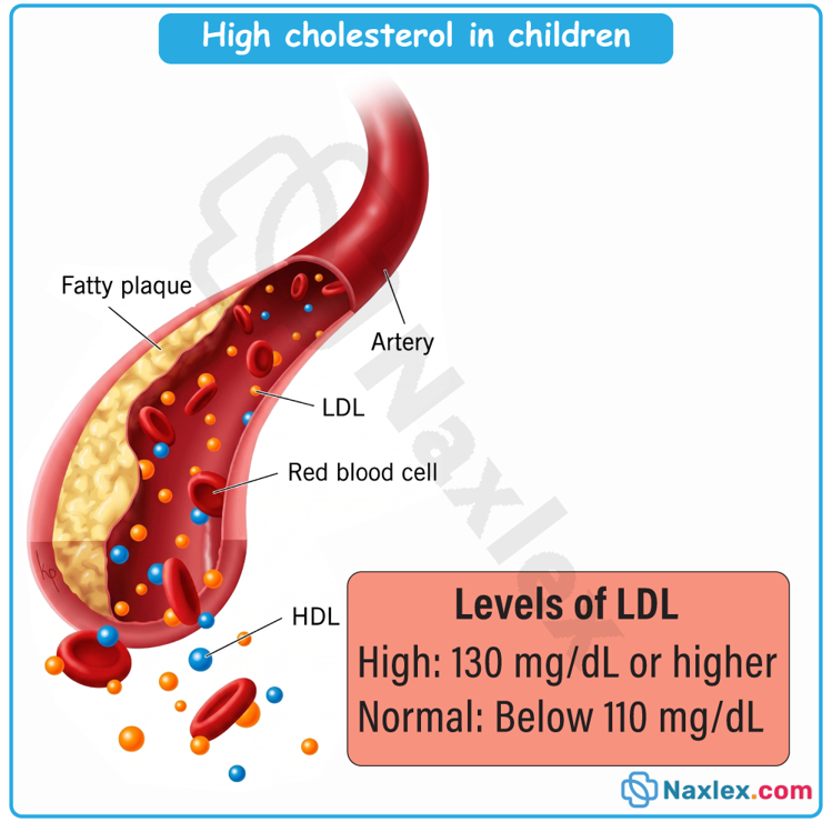
Epidemiology, Etiology, Risk Factors, And Pathophysiology
Epidemiology
Hyperlipidemia is highly prevalent in the pediatric population. It is estimated that approximately 20% of children and adolescents in the United States have some form of abnormal lipid level. This prevalence has climbed steadily alongside the rising rates of childhood obesity and sedentary lifestyles.
Etiology
Pediatric lipid disorders are classified into two broad categories based on their underlying cause:
- Primary (Genetic) Hyperlipidemia: Caused by specific genetic mutations that disrupt normal lipid synthesis, transport, or clearance.
- Familial Hypercholesterolemia (FH): An autosomal dominant disorder primarily caused by mutations in the LDL receptor gene (LDLR), apolipoprotein B gene (APOB), or proprotein convertase subtilisin/kexin type 9 gene (PCSK9). Heterozygous FH affects roughly 1 in 250 individuals, leading to severely elevated LDL-C levels from birth. Homozygous FH is rare (1 in 1 million) but leads to extreme LDL-C levels (greater than 500 mg/dL) and cardiovascular events in early childhood.
- Familial Hyperchylomicronemia (FCS): An autosomal recessive disorder resulting from a deficiency in lipoprotein lipase (LPL) or its cofactor apolipoprotein C-II (APOC2), leading to severe hypertriglyceridemia.
- Other Primary Disorders: Sitosterolemia (disruption in plant sterol excretion due to ABCG5 or ABCG8 mutations) and familial dysbetalipoproteinemia.
- Secondary (Acquired) Hyperlipidemia: Arises as a consequence of non-lipid medical conditions, environmental influences, or medications.
- Obesity and Insulin Resistance: The most common cause of pediatric dyslipidemia, typically presenting as elevated triglycerides, normal-to-high LDL-C, and low HDL-C.
- Endocrine and Metabolic Disorders: Poorly controlled Type 1 or Type 2 diabetes mellitus, hypothyroidism, and Cushing syndrome.
- Renal Diseases: Nephrotic syndrome (causes hepatic overproduction of lipids due to low oncotic pressure) and chronic kidney disease.
- Inflammatory and Other Conditions: Systemic lupus erythematosus, juvenile idiopathic arthritis, HIV, and Kawasaki disease with coronary aneurysms.
- Medications: Exogenous agents such as isotretinoin, beta-blockers, oral contraceptives, systemic corticosteroids, and immunosuppressants (cyclosporine, tacrolimus).
Risk Factors
To guide nursing interventions and risk stratification, risk factors are divided into modifiable and non-modifiable classes:
|
Modifiable Risk Factors |
Non-Modifiable Risk Factors |
|
Dietary Habits: High intake of saturated fats, trans fats, and simple sugars. |
Genetics: Family history of hyperlipidemia or premature cardiovascular disease (men younger than 55, women younger than 65). |
|
Sedentary Lifestyle: Lacking daily vigorous physical activity. |
Biological Sex: Normal physiological fluctuations in lipid values vary between males and females, particularly during puberty. |
|
Obesity: Elevated body mass index (BMI) equal to or greater than the 85th percentile. |
Age: Hormonal shifts during puberty temporarily lower total cholesterol and LDL-C levels by up to 10% before they stabilize. |
|
Tobacco Use: Active smoking or routine exposure to secondhand smoke. |
Ethnicity: Certain genetic backgrounds carry higher rates of metabolic syndrome and primary lipid gene mutations. |
|
Comorbidity Control: Management of secondary diseases such as diabetes or hypertension. |
|
Pathophysiology
- The development of cardiovascular disease is a progressive process that begins in early childhood. The process initiates with endothelial injury or dysfunction, which can be caused by mechanical shear stress from hypertension, circulating toxins from tobacco smoke, or chronic low-grade systemic inflammation.
- This damaged vascular endothelium becomes highly permeable, allowing circulating atherogenic lipoproteins, particularly low-density lipoprotein cholesterol, to penetrate and accumulate within the innermost layer of the arterial wall, known as the vascular intima.
- Once trapped inside the intima, these lipid particles undergo oxidative modifications and chemical changes to become oxidized LDL.
- The presence of oxidized lipids triggers an inflammatory response, signaling the overlying endothelial cells to express adhesion molecules.
- These molecules recruit circulating inflammatory white blood cells, primarily monocytes, which migrate across the endothelial barrier and differentiate into active macrophages.
- In an attempt to clear the foreign lipid debris, these macrophages use scavenger receptors to ingest the oxidized low-density lipoprotein particles. Because this uptake process is unregulated, the macrophages become progressively engorged with cholesteryl esters until they transform into bloated, lipid-laden cells called foam cells.
- As these foam cells aggregate in large numbers within the vascular intima, they form fatty streaks. These streaks represent the earliest visually identifiable pathological lesions of atherosclerosis and are commonly observed in the arteries of children, adolescents, and young adults.
- Over time, these foam cells undergo apoptosis and necrosis, releasing highly toxic lipids, inflammatory cytokines, and free radicals into the surrounding tissue.
- This chronic inflammation stimulates the migration and proliferation of vascular smooth muscle cells from the middle layer of the artery into the intima.
- These smooth muscle cells secrete extracellular matrix proteins, including collagen and elastin, which form a fibrous cap over the lipid core.
- This transformation turns the simple fatty streak into a complex, progressive fibrous plaque, which progressively narrows the arterial lumen, hardens the arterial walls, and sets the stage for premature ischemic cardiovascular events in adulthood.
Image Title: Pathophysiology of Hypercholesterolemia
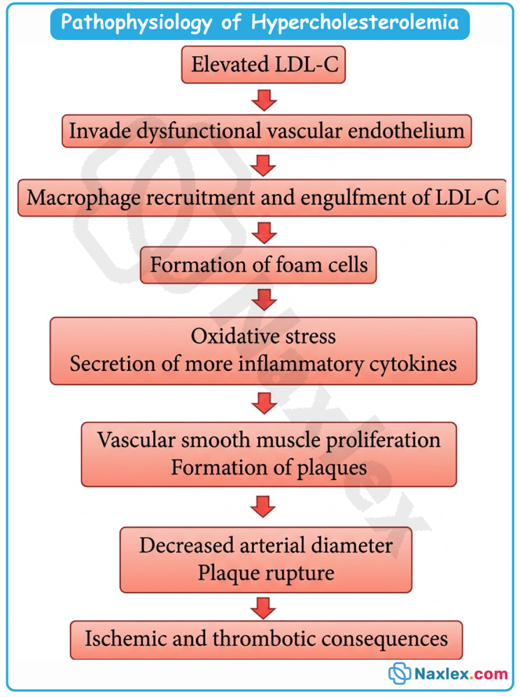
Clinical Manifestations
Pediatric hyperlipidemia is widely referred to as a silent disease because the vast majority of affected children do not present with obvious physical signs or symptoms. Instead, abnormalities are almost exclusively discovered through screening and laboratory evaluation.
However, in cases of severe, long-standing, or primary genetic hyperlipidemias (such as homozygous familial hypercholesterolemia or severe familial hypertriglyceridemia), several distinct physical signs can manifest:
- Xanthomas: Waxy, firm, yellowish-to-reddish plaques or nodules that result from localized lipid deposition within the skin and tendons.
- Tendon Xanthomas: Frequently found over the Achilles tendon and the extensor tendons of the hands, elbows, and knees. These are strongly suggestive of familial hypercholesterolemia.
- Eruptive Xanthomas: Small, itchy, yellow-papular lesions that appear suddenly in crops over the buttocks, back, and extensor surfaces of limbs. These are associated with severe hypertriglyceridemia.
- Tuberous Xanthomas: Larger, exophytic, nodules occurring over pressure points such as the elbows and knees.
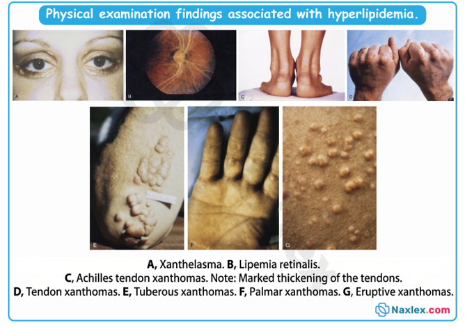
- Xanthelasma: Yellow, flat, raised plaques located on or around the upper and lower eyelids.
- Corneal Arcus: A white, gray, or blue opaque ring surrounding the outer margin of the cornea. While common in elderly adults, its presence in children or young adults is highly abnormal and indicates severe hypercholesterolemia.
- Abdominal Pain and Pancreatitis: Severe hypertriglyceridemia (triglycerides exceeding 1,000 mg/dL) can present with acute, severe abdominal pain, nausea, vomiting, and life-threatening acute pancreatitis.
- Hepatosplenomegaly: Enlargement of the liver and spleen due to macrophage accumulation of circulating chylomicrons, typically seen in severe chylomicronemia syndrome.
- Lipemia Retinalis: A creamy-white or milky appearance of the retinal blood vessels observed during ophthalmoscopic examination, resulting from extremely high triglyceride concentrations in the blood.
Diagnostic Evaluation
Screening Guidelines
The National Heart, Lung, and Blood Institute (NHLBI) and the American Academy of Pediatrics (AAP) recommend a dual approach to pediatric lipid screening:
- Universal Screening: Recommended for all children twice:
- Once between the ages of 9 and 11 years.
- Once between the ages of 17 and 21 years.
- Rationale: Universal screening identifies children with genetic dyslipidemias (like heterozygous FH) who do not have a family history of early heart disease, which occurs in up to 30-50% of cases. Universal screening can be performed using a non-fasting lipid panel, measuring non-HDL-C, which is highly predictive of cardiovascular risk.
- Selective (Targeted) Screening: Fasting lipid screening should be initiated starting at 2 years of age if the child has any of the following high-risk indicators:
- A parent or grandparent with a history of premature cardiovascular disease (males younger than 55, females younger than 65).
- A parent with a known history of total cholesterol greater than 240 mg/dL.
- The child has diabetes mellitus, hypertension, severe obesity (BMI greater than 95th percentile), or is a cigarette smoker.
- The child has a high-risk medical condition (e.g., chronic kidney disease, Kawasaki disease with coronary aneurysms, organ transplant).
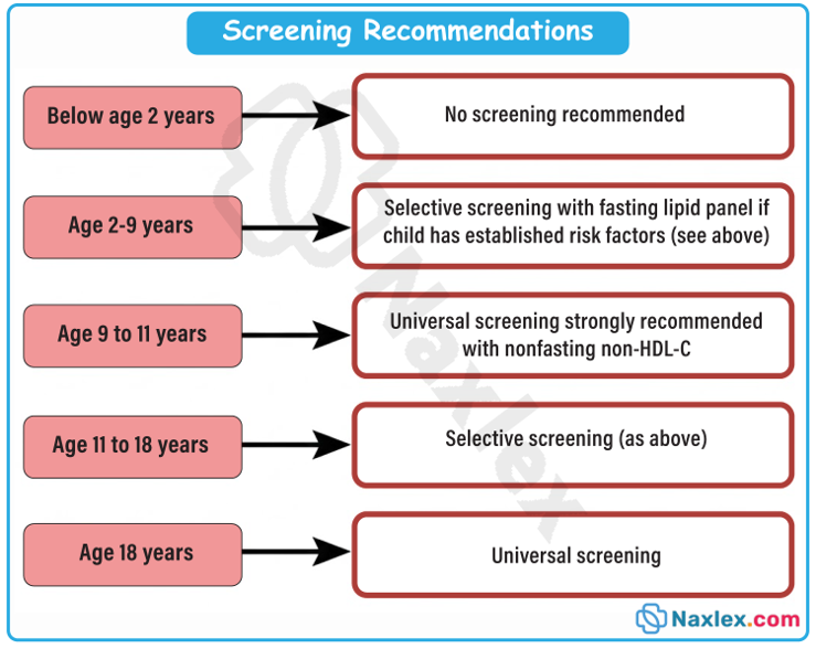
Laboratory Interpretation
If an initial non-fasting universal screening result is abnormal (non-HDL-C equal to or greater than 145 mg/dL, or HDL-C less than 40 mg/dL), a confirmatory fasting lipid panel (FLP) must be performed. Decisions regarding management must be based on the average of two fasting lipid panels obtained 2 weeks to 3 months apart.
The quantitative criteria for pediatric lipid values are defined below:
|
Lipid Parameter |
Acceptable Value |
Borderline Value |
High-Risk (Abnormal) Value |
|
Total Cholesterol |
Less than 170 mg/dL |
170 to 199 mg/dL |
Equal to or greater than 200 mg/dL |
|
LDL Cholesterol |
Less than 110 mg/dL |
110 to 129 mg/dL |
Equal to or greater than 130 mg/dL |
|
Non-HDL Cholesterol |
Less than 120 mg/dL |
120 to 144 mg/dL |
Equal to or greater than 145 mg/dL |
|
HDL Cholesterol |
Greater than 45 mg/dL |
40 to 45 mg/dL |
Less than 40 mg/dL (Low-Risk) |
|
Triglycerides (0-9 yrs) |
Less than 75 mg/dL |
75 to 99 mg/dL |
Equal to or greater than 100 mg/dL |
|
Triglycerides (10-19 yrs) |
Less than 90 mg/dL |
90 to 129 mg/dL |
Equal to or greater than 130 mg/dL |
Therapeutic Management
Dietary Interventions
Dietary modification is the primary therapeutic intervention for pediatric hyperlipidemia. The National Cholesterol Education Program (NCEP) recommends a two-step dietary approach:
- Step I Diet: Recommended for all children with borderline or high-risk lipid profiles.
- Total Fat: Less than 30% of daily total calories.
- Saturated Fat: Less than 10% of daily total calories.
- Dietary Cholesterol: Less than 300 mg per day.
- Trans Fats: Completely avoided.
- Step II Diet: Initiated if the Step I diet does not achieve therapeutic lipid goals after 3 months of strict compliance.
- Total Fat: Less than 30% of daily total calories.
- Saturated Fat: Restricted further to less than 7% of daily total calories.
- Dietary Cholesterol: Restricted further to less than 200 mg per day.
- Dietary Enhancements:
- Soluble Fiber: Increasing intake of soluble fiber (found in oats, barley, beans, and fruits) to 10-25 grams per day helps bind cholesterol in the intestine, reducing its absorption.
- Plant Sterols and Stanols: Recommended as dietary options for children with familial hypercholesterolemia to lower LDL-C levels by competing for intestinal absorption.
Physical Activity and Lifestyle Changes
- Exercise: Children should engage in at least 60 minutes of moderate-to-vigorous aerobic physical activity daily (e.g., running, swimming, cycling).
- Sedentary Time: Screen time (television, video games, computers) should be restricted to less than 2 hours per day.
- Weight Management: For overweight or obese children, structured lifestyle programs targeting a gradual reduction in BMI are essential to improve insulin sensitivity and resolve secondary dyslipidemia.
- Avoidance of Tobacco: Complete elimination of active smoking and exposure to environmental tobacco smoke is vital to protect the vascular endothelium.
Pharmacological Therapy
Medication is indicated only as an adjunct to lifestyle modifications when dietary changes fail to lower lipid levels to safe ranges after a 6-month trial.
- Indications for Pharmacotherapy:
- Initiated in children 8 to 10 years of age and older.
- Persistent LDL-C equal to or greater than 190 mg/dL despite dietary therapy.
- Persistent LDL-C equal to or greater than 160 mg/dL if there is a family history of premature cardiovascular disease or if the child has two or more cardiovascular risk factors (e.g., hypertension, obesity, smoking).
- Persistent LDL-C equal to or greater than 130 mg/dL if the child has diabetes mellitus.
- Statin Therapy (HMG-CoA Reductase Inhibitors):
- First-line therapy: Examples include atorvastatin, pravastatin, simvastatin, and rosuvastatin.
- Mechanism of Action: Statins inhibit the enzyme HMG-CoA reductase, which is the rate-limiting step in hepatic cholesterol synthesis. This reduction in hepatic cholesterol upregulates LDL receptors on the surface of hepatocytes, increasing the clearance of LDL-C from the bloodstream.
- Administration: Typically administered once daily, preferably in the evening, as hepatic cholesterol synthesis peaks during the night.
- Adverse Effects: Muscle pain (myalgia), myopathy, rhabdomyolysis (rare but severe muscle breakdown), and hepatotoxicity.
- Monitoring: Baseline and periodic liver function tests (ALT/AST) and creatine kinase (CK) levels must be monitored.
- Contraindications: Absolute contraindication in pregnancy due to teratogenic risks. Female adolescents must receive counseling on reliable contraception.
- Adjunctive Medications:
- Cholesterol Absorption Inhibitors (Ezetimibe): Inhibits cholesterol absorption at the brush border of the small
intestine. Often combined with low-dose statins. - Bile Acid Sequestrants (Colesevelam, Cholestyramine): Bind bile acids in the intestine, forcing the liver to use systemic LDL-C to synthesize new bile acids. Can cause gastrointestinal side effects (constipation, gas) and interfere with fat-soluble vitamin absorption.
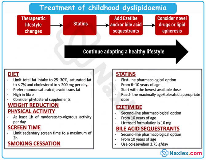
- Cholesterol Absorption Inhibitors (Ezetimibe): Inhibits cholesterol absorption at the brush border of the small
Nursing Care Plan
Developing a structured care plan is essential for organizing the nursing management of pediatric clients with hyperlipidemia.
Nursing Diagnosis 1: Deficient Knowledge
Related to: Lack of exposure to dietary, lifestyle, and pharmacological management guidelines for pediatric lipid disorders.
As Evidenced By: Patient and family reporting confusion over dietary fat restrictions, incorrect statements regarding physical activity requirements, or hesitation regarding the safety of prescribed medications.
- Desired Outcome:
- The parent and child will verbalize three key differences between saturated and unsaturated fats by the end of the teaching session.
- The parent and child will create a 3-day heart-healthy meal and exercise plan that aligns with Step I or Step II dietary guidelines.
- Nursing Interventions:
- Educate the family on the pathophysiology of pediatric hyperlipidemia, explaining that atherosclerotic plaque begins forming in childhood and can be slowed or reversed through early intervention.
- Provide a detailed list of allowed and restricted foods under the Step I and Step II diets, emphasizing the replacement of saturated/trans fats with unsaturated fats.
- Involve a registered pediatric dietitian to assist the family in modifying cultural dishes and favorite recipes to meet fat and cholesterol restrictions.
- Teach the family how to read nutrition facts labels, focusing on identifying hidden trans fats, saturated fats, and total carbohydrates.
- Rationales:
- Visual and age-appropriate educational models increase parent and child comprehension, promoting active engagement in the care plan.
- Saturated and trans fats increase LDL-C levels, whereas replacing them with monounsaturated and polyunsaturated fats (e.g., olive oil, fish) helps lower LDL-C.
- Dietary compliance increases when interventions are tailored to the family's socioeconomic, cultural, and personal preferences.
- The ability to interpret food labels empowers families to make informed purchasing and eating choices, avoiding accidentally elevated fat intake.
Nursing Diagnosis 2: Risk for Ineffective Tissue Perfusion (Cardiovascular)
Related to: Progressive lipid deposition and fatty streak formation within arterial walls secondary to chronic hyperlipidemia.
- Desired Outcome:
- The child will maintain blood pressure within normal limits for age, sex, and height percentiles throughout therapy.
- The child's average fasting LDL-C will decrease toward the target therapeutic level (less than 130 mg/dL, or less than 100 mg/dL if high-risk) within 6 months of initiating the treatment plan.
- Nursing Interventions:
- Monitor the child's serial blood pressure readings, weight, and BMI percentiles during each clinical visit.
- Assess for the presence of physical markers of advanced dyslipidemia, such as tendon xanthomas or corneal arcus.
- Reinforce the requirement of 60 minutes of daily moderate-to-vigorous aerobic exercise to improve vascular tone and raise protective HDL-C levels.
- For children prescribed statins, coordinate and review scheduled laboratory tests, including alanine aminotransferase (ALT), aspartate aminotransferase (AST), and creatine kinase (CK).
- Rationales:
- Hypertension and obesity act synergistically with hyperlipidemia to damage the vascular endothelium, accelerating plaque formation.
- Identifying physical manifestations helps track disease severity and guides the potential necessity for more aggressive therapeutic regimens.
- Physical activity promotes nitric oxide release, maintains endothelial health, lowers triglycerides, and increases anti-atherogenic HDL-C.
- Regular laboratory monitoring ensures early detection of potential adverse drug reactions, preventing drug-induced liver injury or severe myopathy.
Nursing Diagnosis 3: Non-adherence (or Risk for Non-adherence)
Related to: The asymptomatic nature of hyperlipidemia, long-term nature of treatment, and dietary restrictions that differ from peers.
- Desired Outcome:
- The child and family will demonstrate compliance by attending scheduled follow-up visits and completing required lipid panels.
- The child will report taking prescribed medications as ordered without missing doses.
- Nursing Interventions:
- Acknowledge the challenges of managing a chronic, "invisible" condition and validate the family's frustrations regarding permanent lifestyle alterations.
- Help the family set small, achievable goals (e.g., swapping soda for water three days a week, rather than an immediate complete ban).
- Incorporate the adolescent in decision-making, allowing them to select physical activities they enjoy and participate in meal planning.
- Establish a simple, visual routine for daily medication administration (e.g., taking medication immediately after brushing teeth at night).
- Rationales:
- Empathy and validation build a trusting relationship, which significantly enhances open communication and therapeutic compliance.
- Gradual, small behavioral changes are more likely to be integrated permanently into the family lifestyle than sudden, restrictive demands.
- Involving pediatric and adolescent clients in their own care fosters autonomy and increases long-term compliance with lifestyle changes.
- Pairing medication with established daily habits creates a behavioral trigger, reducing the likelihood of forgotten or missed doses.
Complications
Failure to manage pediatric hyperlipidemia can lead to several serious, irreversible health complications, both in childhood and later in life:
- Premature Atherosclerotic Cardiovascular Disease (ASCVD): The most significant long-term risk. Chronic exposure to high LDL-C accelerates the transition of fatty streaks into calcified plaques, leading to early-onset coronary artery disease, angina, and premature myocardial infarction.
- Ischemic Stroke: Advanced carotid artery plaque buildup can rupture or narrow blood flow, causing ischemic cerebrovascular events in young adulthood.
- Peripheral Artery Disease (PAD): Systemic atherosclerosis can compromise arterial flow to the extremities, leading to claudication and tissue ischemia.
- Acute Pancreatitis: This is a severe, life-threatening short-term complication of extreme hypertriglyceridemia (triglycerides greater than 1,000 mg/dL). High levels of circulating chylomicrons clog the pancreatic capillary bed, leading to ischemia and premature activation of digestive enzymes within the pancreas, causing autodigestion.
- Statin-Induced Hepatotoxicity: Elevated transaminases (ALT/AST) can indicate liver injury secondary to pharmacological therapy. This is usually reversible upon dose reduction or discontinuation of the medication.
- Myopathy and Rhabdomyolysis: Statins can cause skeletal muscle injury, ranging from benign muscle pain (myalgia) to life-threatening rhabdomyolysis. Rhabdomyolysis causes myoglobin release into the bloodstream, which can precipitate acute kidney injury.
Prognosis
The prognosis for pediatric hyperlipidemia depends heavily on the specific etiology of the lipid disorder and the timing of therapeutic intervention:
- Secondary Hyperlipidemia: Carry an excellent prognosis when the underlying primary medical condition (e.g., hypothyroidism, nephrotic syndrome) is successfully treated, or when lifestyle modifications result in weight reduction and normalized insulin sensitivity.
- Heterozygous Familial Hypercholesterolemia (HeFH): Prognosis is highly favorable if identified early in childhood (prior to puberty) and managed with a combination of the Step II diet and statin therapy. Early, long-term control of LDL-C can return the child's life expectancy and cardiovascular risk profile to near-normal levels.
- Homozygous Familial Hypercholesterolemia (HoFH): Carries a guarded-to-poor prognosis if left untreated. These children often develop severe, rapid atherosclerosis within the first decade of life and are at extreme risk for myocardial infarction, aortic stenosis, and sudden cardiac death before the age of 30. Advanced therapies such as LDL apheresis, liver transplantation, or novel biological agents are required.
- Adherence as the Key Determinant: In all cases, the primary prognostic indicator is long-term compliance with lifestyle modifications and prescribed pharmacological therapies.
Prevention
Nurses must promote primordial and primary prevention strategies within the pediatric population. The following ten specific preventive measures are recommended:
- Promote Universal Screening: Ensure every child undergoes a routine, non-fasting lipid profile check once between 9 and 11 years, and again between 17 and 21 years.
- Implement Early Targeted Screening: Perform selective lipid screening starting at age 2 for children with high-risk family history or predisposing chronic medical conditions.
- Encourage Daily Physical Activity: Educate families to ensure children obtain a minimum of 60 minutes of daily moderate-to-vigorous physical activity daily.
- Restrict Sedentary Screen Time: Advise parents to limit non-academic screen time to less than 2 hours daily to reduce sedentary behaviors.
- Optimize Saturated and Trans Fat Intake: Teach families to keep saturated fat intake below 10% of total daily calories and completely eliminate dietary trans fats.
- Ensure Adequate Dietary Fiber: Encourage a fiber-rich diet containing vegetables, fruits, and whole grains, aiming for 10-25 grams of soluble fiber daily.
- Avoid Sugar-Sweetened Beverages: Counsel families to eliminate sodas, sweet teas, and juice drinks, replacing them with water or low-fat dairy.
- Prevent Secondhand Smoke Exposure: Educate parents on the cardiovascular dangers of tobacco smoke and encourage smoking cessation programs to protect children's blood vessels.
- Support Weight Management: Monitor growth charts and BMI percentiles at every wellness visit to provide early nutritional intervention for children crossing into the overweight or obese categories.
- Establish Heart-Healthy Family Habits: Frame lifestyle modifications as a collective family effort rather than isolating the diagnosed child, ensuring long-term adherence.
Summary
- Pediatric hyperlipidemia represents an acquired cardiovascular disorder characterized by abnormally elevated blood concentrations of lipids and lipoproteins.
- Historically categorized as an adult concern, clinical and pathological research has established that atherosclerosis is a progressive, life-long process that begins during childhood with the accumulation of fatty streaks within the vascular intima.
- Dyslipidemia in children is divided into primary genetic etiologies, such as familial hypercholesterolemia (HeFH and HoFH), and secondary acquired etiologies, which are most commonly caused by childhood obesity, insulin resistance, sedentary lifestyles, and underlying systemic disorders like diabetes, hypothyroidism, or nephrotic syndrome.
- Because pediatric hyperlipidemia is almost completely asymptomatic, early and proactive screening is critical. Guidelines from the NHLBI and AAP dictate universal, non-fasting lipid screening for all pediatric clients twice: once between ages 9 and 11, and once between ages 17 and 21.
- High-risk children, such as those with a family history of premature cardiovascular disease or severe hypercholesterolemia, should begin targeted fasting lipid screening as early as age 2.
- Diagnosis and treatment decisions are based on the average of two fasting lipid panels.
- Therapeutic management begins with structured, family-centered lifestyle modifications. This includes implementing the Step I and Step II diets, ensuring 60 minutes of daily moderate-to-vigorous physical activity, limiting sedentary screen time, and eliminating tobacco exposure.
- Pharmacotherapy, primarily with HMG-CoA reductase inhibitors (statins), is indicated for children aged 8 to 10 and older who have persistent, high-risk LDL-C levels despite a 6-month trial of dietary changes.
- Nurses are responsible for managing the care plan, providing intensive family education, monitoring for medication complications (such as myopathy or hepatotoxicity), and advocating for long-term adherence to prevent future adult cardiovascular events. (309 words)
Naxlex
Videos
Login to View Video
Click here to loginTake Notes on Acquired Cardiovascular Disorders: Pediatric Hyperlipidemia
This filled cannot be empty
Join Naxlex Nursing for nursing questions & guides! Sign Up Now