





 Video Library


Please set your exam date
Pediatric Anatomy And Physiology Of The Gastrointestinal Tract
Study Questions
Practice Exercise 1
The nurse is providing dietary teaching to the parents of a 3-month-old infant client. The parents ask why solid foods such as rice cereal should not be introduced yet. Which physiological rationale should the nurse provide?
Explanation
Infant gastrointestinal maturity determines dietary readiness, as the pancreatic amylase enzyme remains inadequate for starch digestion until approximately 4 to 6 months of age. Early introduction of solid foods like rice cereal risks gastrointestinal distress and malabsorption due to immature digestive enzymes. Furthermore, the extrusion reflex persists in young infants, increasing the potential risk for asphyxiation if solids are introduced prematurely.
Rationale for correct answer:
2. Pancreatic amylase activity is insufficient in a 3-month-old infant to digest complex starches effectively. Unsplit starches pass into the colon, causing osmotic diarrhea and abdominal distension. Intestinal maturation typically achieves adequate carbohydrate hydrolyzing capability between 4 and 6 months. Early introduction compromises overall nutrient absorption.
Rationale for incorrect answers:
1. Gastric capacity in a 3-month-old infant is approximately 90 to 150 mL, not 10 mL. At birth, capacity starts around 5 to 7 mL and rapidly increases during the first month. Stating capacity is 10 mL reflects incorrect physiology. This limitation does not explain the specific barrier to complex starches.
3. Intestinal lactase levels peak during late gestation and are fully present at birth to digest human milk or milk-based formula lactose. Lactase deficiency is extremely rare in early infancy and does not spontaneously manifest or resolve at 6 months. Lactase handles disaccharides, whereas rice cereal contains complex carbohydrates. Premature solid introduction is unrelated to lactase activity.
4. Protein-digesting enzymes like trypsin, chymotrypsin, and carboxypeptidase are synthesized by the exocrine pancreas, not the liver. The liver synthesizes plasma proteins, bile, and clotting factors rather than digestive enzymes. Hepatic functional maturity relates to drug metabolism and bilirubin conjugation. This statement represents an inaccurate anatomical generalization.
Test-taking strategy:
- Analyze the scenario/question: The client is a 3-month-old infant whose parents are asking about the physiological rationale for withholding solid foods like rice cereal.
- Evaluate gastrointestinal physiology:
- Assess digestive enzyme maturation timelines in early infancy.
- Recall that pancreatic exocrine function matures gradually over the first year of life.
- Identify correct pathophysiological rationale:
- Choice 2 correctly identifies the lack of pancreatic amylase required to break down the alpha-1,4-glycosidic bonds in complex starches.
- Systematically Rule out options:
- Rule out Choice 1: The numerical value for gastric capacity at 3 months is inaccurate; capacity exceeds 90 mL.
- Rule out Choice 3: Lactase is necessary for milk digestion and is abundant at birth, not absent until 6 months.
- Rule out Choice 4: The liver does not produce protein-digesting enzymes; digestive enzymes are exocrine pancreatic products.
Take home points
- Pancreatic amylase production is insufficient in infants under 4 months of age, making complex starch digestion difficult.
- Infant gastric capacity expands from 5 to 7 mL at birth to over 90 mL by 3 months of age.
- Intestinal lactase is fully active at birth to facilitate the digestion of lactose present in breast milk and formula.
- Solid food introduction before 4 to 6 months increases the risks of aspiration, excessive calorie intake, and gastrointestinal distress.
A 1-month-old client is admitted to the pediatric unit with a GI infection. The nurse recognizes that the infant's increased intestinal mucosal permeability places the client at higher risk for which condition?
Explanation
Infant gastrointestinal architecture exhibits heightened intestinal mucosal permeability due to immature tight junctions and an underdeveloped mucosal barrier. This functional immaturity permits enhanced passage of un-degraded proteins, increasing susceptibility to macromolecular absorption and potential enterogenous sepsis. When a gastrointestinal infection compromises this fragile mucosal lining, the risk for bacterial translocation escalates significantly, predisposing the neonate to systemic inflammatory cascades and severe pathogen dissemination.
Rationale for correct answer:
A. Macromolecular absorption and systemic bacterial translocation occur because an infant's intestinal epithelial barrier lacks tight junction closure, known as gut closure. Gastrointestinal inflammation damages mucosal integrity, enabling viable bacteria and large antigenic proteins to penetrate the intestinal wall. Translocation into circulation leads to neonatal bacterial sepsis.
Rationale for incorrect answers:
B. Rapid hyper-synthesis of hepatic albumin does not occur in response to heightened intestinal permeability or acute infant gastrointestinal infections. Hepatic albumin synthesis is constrained by liver immaturity, and mucosal inflammation typically leads to protein loss rather than increased production. Protein-losing enteropathy causes hypoalbuminemia, which worsens systemic edema.
C. Severe hypertrophic pyloric stenosis involves congenital hypertrophy and hyperplasia of the smooth muscle surrounding the pyloric sphincter. This mechanical obstruction is mediated by environmental factors, genetic predisposition, and neurogenic muscular dysfunction, not increased mucosal permeability. Gastric outlet obstruction causes non-bilious projectile vomiting, unrelated to transmucosal absorption.
D. Irreversible loss of intestinal villi is an inaccurate pathological description of typical infant infectious gastroenteritis. Villous blunting or atrophy occurs transiently in viral infections like rotavirus, but the enterocytes regenerate rapidly once the acute infection resolves. Villar restoration depends on crypt cell proliferation, preventing permanent damage under standard nutritional support.
Test-taking strategy:
- Analyze the scenario/question: The client is a 1-month-old infant admitted with a gastrointestinal infection. The question asks about the physiological consequence of increased intestinal mucosal permeability.
- Evaluate pathophysiologic principles:
- Recognize that neonatal intestinal mucosa has large intercellular gaps (high permeability).
- Consider how mucosal injury from infection affects the passage of substances across the gut wall.
- Identify correct pathophysiological rationale:
- Choice 2 correctly identifies that high permeability allows intact proteins (macromolecules) and enteric pathogens to cross the intestinal epithelium into the bloodstream (translocation).
- Systematically Rule out options:
- Rule out Choice 1: Mucosal inflammation causes protein loss, not hyper-synthesis of albumin by the immature liver.
- Rule out Choice 3: Hypertrophic pyloric stenosis is a muscular hypertrophy issue of the pylorus, not a mucosal permeability defect.
- Rule out Choice 4: Villous atrophy caused by infectious gastroenteritis is reversible; crypt stem cells regenerate mucosal architecture.
Take home points
- Immature intestinal mucosa in infants permits macromolecular absorption, predisposing them to food protein allergies and systemic bacterial invasion.
- Infectious gastroenteritis disrupts the neonatal gut barrier, significantly increasing the risk of enterogenous sepsis via bacterial translocation.
- Hypertrophic pyloric stenosis is a muscular sphincter hypertrophy causing gastric outlet obstruction, independent of mucosal permeability.
- Intestinal villi possess high regenerative capacity, making villous loss from acute gastroenteritis temporary rather than irreversible.
The nurse is caring for a 4-day-old term neonate client. When reviewing the client's metabolic profile, which hepatic characteristic should the nurse consider when administering highly protein-bound medications?
Explanation
Neonatal hepatic function is characterized by immature protein synthesis, leading to low plasma concentrations of albumin and alpha-1-acid glycoprotein. When administering highly protein-bound medications, reduced binding capacity increases the unbound fraction of the drug in circulation. Consequently, neonates experience heightened drug activity, increased physiological sensitivity, and an elevated potential for drug toxicity.
Rationale for correct answer:
C. Immature liver enzyme synthesis limits the production of total plasma proteins, specifically albumin and circulating globulins. In a 4-day-old neonate, fewer available binding sites mean highly protein-bound drugs remain free in serum. The expanded unbound fraction increases pharmacodynamic activity and elevates the risk of drug toxicity.
Rationale for incorrect answers:
A. Adult-level drug metabolism rates are absent in a 4-day-old term neonate due to reduced hepatic blood flow and functional immaturity of metabolic pathways. Hepatic clearance mechanisms, including Phase I oxidation and Phase II conjugation reactions, take months to fully mature. Reduced clearance prolonged elimination half-lives, predisposing the neonate to drug accumulation.
B. Increased glycogen storage leading to drug entrapment is a pathophysiologically inaccurate mechanism. Neonatal glycogen stores are depleted rapidly after birth to support gluconeogenesis and baseline metabolic needs. Furthermore, glycogen functions as a glucose polymer within hepatocytes and does not bind or trap circulating pharmacological agents.
D. Hyper-synthesis of cytochrome P450 enzymes does not occur in early neonatal life. Phase I microsomal P450 enzymes are quantitatively reduced at birth, operating at less than half of adult capability. Expression of key isoenzymes like CYP3A4 increases gradually over the first year of life, causing diminished metabolic capacity.
Test-taking strategy:
- Analyze the scenario/question: The client is a 4-day-old neonate receiving highly protein-bound medications. The question asks for the relevant hepatic characteristic affecting drug administration.
- Evaluate neonatal hepatic pharmacology:
- Recall that hepatic protein synthesis (albumin) is reduced in newborns.
- Understand that highly protein-bound drugs require plasma albumin to carry them; low albumin means more free, active drug.
- Identify correct pathophysiological rationale:
- Choice 3 correctly links immature hepatic synthesis of plasma albumin to reduced drug-binding capacity and increased free drug levels.
- Systematically Rule out options:
- Rule out Choice 1: Metabolism rates are immature and significantly slower than adult rates, not equivalent.
- Rule out Choice 2: Glycogen is a carbohydrate source, not a protein binder or drug trap.
- Rule out Choice 4: Cytochrome P450 enzymes are deficient, not hyper-synthesized, in a 4-day-old infant.
Take home points
- Neonates have diminished serum albumin levels, leading to a higher fraction of unbound, active drug when highly protein-bound medications are given.
- Hepatic cytochrome P450 enzyme systems and Phase II conjugation pathways are functionally immature at birth, slowing drug clearance.
- Elevated free drug concentrations in neonates significantly increase the risk of tissue toxicity and adverse pharmacological reactions.
- Drugs that bind heavily to albumin can displace bilirubin, increasing the risk of hyperbilirubinemia and kernicterus in early infancy.
A 6-month-old infant client presents with watery diarrhea. The nurse calculates fluid resuscitation needs keeping in mind that infants are at a higher risk for rapid dehydration than adults due to which factor?
Explanation
Infant fluid dynamic balance is highly precarious due to a higher proportion of total body water residing within the extracellular fluid compartment. Total body water accounts for approximately 75% to 80% of an infant's weight, with extracellular fluid constituting nearly half of that volume. Consequently, gastrointestinal losses like watery diarrhea rapidly deplete this extracellular volume, predisposing the infant to swift hypovolemic shock and severe electrolyte imbalances.
Rationale for correct answer:
B. Higher proportion of total body water stored in the extracellular fluid compartment makes infants disproportionately vulnerable to fluid volume deficit. Extracellular fluid is lost rapidly through gastrointestinal disruptions like acute watery diarrhea. Because extracellular fluid turns over at a significantly higher rate in infants than adults, fluid depletion precipitates rapid dehydration.
Rationale for incorrect answers:
A. Lower basal metabolic rate is an physiologically inaccurate description of infant energetics. Infants possess a significantly higher basal metabolic rate relative to body weight to support rapid growth and thermoregulation. A elevated metabolic rate generates greater metabolic waste products, requiring higher fluid intake for renal clearance and contributing to accelerated metabolic turnover.
C. Decreased intestinal surface area relative to body mass is structurally incorrect regarding pediatric anatomy. Infants actually possess a larger intestinal surface area and a greater body surface area relative to body mass compared to adults. This expanded surface area increases fluid loss via insensible vaporization and fluid shifts during acute mucosal inflammation.
D. Slower intestinal peristalsis is not a characteristic of infant gastrointestinal physiology during acute infectious gastroenteritis. Peristalsis is typically accelerated in response to mucosal irritation, resulting in rapid intestinal transit and frequent watery stools. Hypermotility impairs fluid absorption, leading to progressive fluid loss.
Test-taking strategy:
- Analyze the scenario/question: The client is a 6-month-old infant with watery diarrhea. The question asks for the physiological factor that places infants at a higher risk for rapid dehydration compared to adults.
- Evaluate pediatric fluid and electrolyte differences:
- Recall that body water distribution changes significantly with age.
- Consider how fluid compartment distribution (extracellular versus intracellular) impacts fluid loss during illness.
- Identify correct pathophysiological rationale:
- Choice 2 correctly identifies that a higher percentage of total body water is located in the extracellular fluid compartment, which is readily lost during diarrheal illness.
- Systematically Rule out options:
- Rule out Choice 1: Infants have a higher, not lower, metabolic rate compared to adults.
- Rule out Choice 3: Relative to body mass, infants have a larger body surface area and intestinal mucosa, not a decreased area.
- Rule out Choice 4: Diarrhea involves hypermotility and rapid transit, not slower intestinal peristalsis.
Take home points
- Infants store up to 40% of their total body water in the extracellular compartment, which is subject to rapid depletion during illness.
- A higher metabolic rate and larger body surface area relative to weight accelerate fluid loss in pediatric clients.
- Immature renal function in infants impairs their ability to concentrate urine and conserve water during fluid restriction.
- Acute diarrhea leads to rapid volume contraction, requiring early assessment of hydration status and targeted rehydration therapy.
The nurse is reviewing the gastric emptying time of a 2-month-old breastfed infant client. The nurse expects that gastric emptying in this infant will typically occur within what timeframe?
Explanation
Infant gastric physiology is defined by reduced gastric capacity and altered motility patterns compared to older pediatric clients. In a 2-month-old breastfed infant, gastric motility is rapid because human milk forms soft, easily digestible curd structures that promote accelerated gastric emptying. Human milk empties from the stomach faster than bovine-based infant formulas, establishing a typical physiological window of 2.5 to 3 hours that directly influences normal infantile feeding frequency.
Rationale for correct answer:
D. Gastric emptying in a 2-month-old breastfed infant typically occurs within 2.5 to 3 hours due to the rapid breakdown of whey-predominant proteins in breast milk. Whey forms soft, soluble curds in the acidic gastric environment, allowing faster liquid phase emptying through the pylorus. This physiological processing time explains the frequent feeding intervals observed in exclusively breastfed neonates and young infants. Proper gastric clearance prevents chronic gastric distension.
Rationale for incorrect answers:
A. A timeframe of 30 to 40 minutes is unrealistically short for complete gastric emptying of a full liquid feeding in an infant. While the liquid phase of breast milk begins transit into the duodenum shortly after ingestion, full stomach clearance requires significantly more time. Expecting complete emptying in under an hour reflects physiologic overestimation. Transit this rapid would trigger osmotic dumping.
B. A timeframe of 6 to 8 hours represents pathologically delayed gastric emptying or the digestion time of complex solid foods in adults. In a young infant, holding gastric contents for 6 to 8 hours would indicate gastric outlet obstruction, severe gastroparesis, or ileus. Prolonged gastric retention causes excessive curd fermentation and persistent vomiting.
C. A timeframe of 10 to 12 hours is an erroneous measurement that exceeds normal pediatric upper gastrointestinal transit duration. Stomach contents pass into the small intestine long before 10 hours elapse in healthy infants. Holding breast milk in the stomach for this duration would compromise total daily caloric intake and cause severe dehydration.
Test-taking strategy:
- Analyze the scenario/question: The client is a 2-month-old breastfed infant. The nurse needs to identify the normal timeframe for gastric emptying.
- Evaluate pediatric gastrointestinal physiology:
- Consider the composition of human breast milk (whey-dominant, easily digested).
- Recall typical breastfed infant feeding schedules (every 2 to 3 hours).
- Identify correct pathophysiological rationale:
- Choice 4 accurately reflects the standard 2.5 to 3 hour gastric emptying window for a breastfed infant, aligning with known feeding patterns.
- Systematically Rule out options:
- Rule out Choice 1: 30 to 40 minutes is too short for complete emptying of a full volume feeding.
- Rule out Choice 2: 6 to 8 hours is abnormally prolonged and suggests gastroparesis or obstruction.
- Rule out Choice 3: 10 to 12 hours is pathologically delayed and inconsistent with pediatric gastrointestinal motility.
Take home points
- Gastric emptying in exclusively breastfed infants occurs within 2.5 to 3 hours due to the high whey-to-casein ratio of human milk.
- Commercial infant formulas contain higher casein ratios, forming denser curds that prolong gastric emptying to 3.5 to 4 hours.
- Infant gastric motility is influenced by meal volume, caloric density, position, and autonomic nervous system maturity.
- Understanding normal gastric emptying times guides appropriate feeding intervals and helps nurses identify pathologic delayed emptying.
A 5-month-old infant client is brought to the clinic. The mother asks why her baby has frequent bowel movements shortly after feeding. The nurse explains that this is due to which physiological reflex?
Explanation
Infant intestinal motility is heavily regulated by neurohumoral mechanisms that coordinate digestion and waste clearance. In a 5-month-old infant, feeding stimulates the gastrocolic reflex, an autonomic response where gastric distension triggers increased propulsive peristalsis throughout the colon. Mediated by gastrin and cholecystokinin, this physiological reflex clears the lower gastrointestinal tract to accommodate newly ingested nutrients, manifesting as frequent defecation patterns shortly after meals.
Rationale for correct answer:
B. Gastrocolic reflex is a physiological response where gastric stretching from a meal triggers mass colonic peristaltic movements. In infants, this autonomic neural pathway is highly pronounced due to an immature central nervous system and heightened myenteric plexus sensitivity. The resulting propulsive colonic contractions move stool into the rectum, causing prompt postprandial defecation.
Rationale for incorrect answers:
A. Esophagocardiological reflex is an inaccurate non-standard medical term that does not describe postprandial intestinal propulsion. Esophageal distension or irritation typically engages vagal pathways affecting heart rate, known as esophagocardiac reflexes, which cause bradycardia response. It plays no role in stimulating lower gastrointestinal tract peristaltic clearance.
C. Pyloroduodenal reflex controls the passage of chyme from the stomach through the pyloric sphincter into the duodenum based on duodenal pH and osmolarity. While it regulates gastric emptying, it does not directly stimulate lower colonic mass movements or defecation. Its primary action is to prevent duodenal overload by delaying gastric clearance.
D. Enterohepatic circulation refers to the continuous pathway where bile acids are synthesized by the liver, secreted into the duodenum, reabsorbed in the terminal ileum, and returned to the liver via the portal vein. This biochemical recycling process maintains the bile acid pool necessary for lipid digestion. It does not govern mechanical colonic motility.
Test-taking strategy:
- Analyze the scenario/question: The mother of a 5-month-old infant asks why her baby has frequent bowel movements shortly after eating. The nurse must identify the correct physiological reflex responsible.
- Evaluate gastrointestinal reflexes:
- Recall the physiological link between stomach stretching (gastro) and colonic peristalsis (colic).
- Recognize that mass movements in the colon after eating are driven by the gastrocolic reflex.
- Identify correct pathophysiological rationale:
- Choice 2 correctly identifies the gastrocolic reflex as the physiological trigger for defecation following gastric distension.
- Systematically Rule out options:
- Rule out Choice 1: Esophagocardiological reflex is not a functional gastrointestinal reflex for bowel clearance.
- Rule out Choice 3: Pyloroduodenal reflex regulates pyloric sphincter opening, not colonic evacuation.
- Rule out Choice 4: Enterohepatic circulation is a chemical recycling loop for bile acids, not a motor reflex.
Take home points
- The gastrocolic reflex causes mass colonic peristalsis and defecation in response to gastric distension following a meal.
- Infants exhibit a prominent gastrocolic reflex due to the physiological immaturity of inhibitory autonomic neural pathways.
- Reassuring parents that postprandial bowel movements are a normal physiological reflex prevents unnecessary dietary changes.
- Pathologic conditions like gastroenteritis or food allergies should be investigated if bowel movements become watery, bloody, or accompanied by poor weight gain.
The nurse is educating nursing students about fat absorption in a 2-month-old infant client. Which statement correctly describes lipid digestion in early infancy?
Explanation
Infant lipid processing relies on alternative digestive enzymes due to the developmental immaturity of the exocrine pancreas. In a 2-month-old infant, endogenous pancreatic lipase output is markedly deficient, and hepatic bile acid synthesis remains low. To compensate for these anatomical limitations, lipid emulsification and hydrolysis depend heavily on lingual lipase, gastric lipase, and bile salt-stimulated lipase present within human breast milk, ensuring adequate fat absorption.
Rationale for correct answer:
A. Digestion relies heavily on lingual and breast milk lipases due to low pancreatic lipase levels, which are severely depressed during the first few months of life. Lingual and gastric lipases initiate lipid hydrolysis in the stomach because they do not require bile salts for activation. Breast milk contributes bile salt-stimulated lipase, which completes triglyceride breakdown in the small intestine to maintain infant nutrition.
Rationale for incorrect answers:
B. Pancreatic lipase is present at three times adult levels is a physiologically false assertion regarding pediatric digestive development. Intestinal concentrations of pancreatic lipase in a 2-month-old infant are less than 5% to 10% of adult capacity. Adult enzyme activity levels are reached gradually around 1 to 2 years of age, making early fat digestion dependent on alternative enzymes.
C. The bile acid pool is twice as large as that of an adult is an inaccurate description of infantile hepatic synthesis. The intraluminal bile acid pool and duodenal bile acid concentrations in young infants are significantly smaller than in adults, often falling below the critical micellar concentration. Low bile concentrations impair fat emulsification.
D. Emulsification of fats occurs exclusively in the stomach is an anatomically and chemically incorrect statement. While mechanical churning in the stomach creates a preliminary lipid emulsion, the primary emulsification process occurs in the duodenum through the action of liver-derived bile salts and phospholipids.
Test-taking strategy:
- Analyze the scenario/question: The question asks for the statement that correctly describes lipid digestion and fat absorption in a 2-month-old infant.
- Evaluate pediatric gastrointestinal physiology:
- Recall that pancreatic exocrine enzymes (lipase, amylase) are low at birth and during early infancy.
- Consider how infants compensate for low pancreatic lipase to digest dietary fats (milk fats).
- Identify correct pathophysiological rationale:
- Choice 1 correctly identifies that early lipid hydrolysis relies on non-pancreatic sources, specifically lingual, gastric, and breast milk lipases.
- Systematically Rule out options:
- Rule out Choice 2: Pancreatic lipase levels are severely diminished (under 10% of adult levels), not elevated threefold.
- Rule out Choice 3: The bile acid pool is significantly smaller in infants, often limiting micelle formation.
- Rule out Choice 4: Emulsification requires bile salts in the duodenum; it does not occur exclusively in the stomach.
Take home points
- Pancreatic lipase production is significantly depressed in early infancy, requiring compensatory mechanisms for lipid digestion.
- Lingual lipase and gastric lipase initiate triglyceride digestion in the infant stomach without requiring bile acid activation.
- Human breast milk contains bile salt-stimulated lipase (BSSL), which aids fat breakdown in the infant duodenum.
- Infants have a reduced bile acid pool, which can limit efficient lipid emulsification and micellar solubilization.
A 10-day-old neonate client requires fluid management. The nurse notes that the neonate's anatomical gastric capacity at this stage is approximately:
Explanation
Infant upper gastrointestinal architecture undergoes rapid physiological expansion during the early neonatal period to support increasing metabolic demands and enteral intake. At birth, the neonate's anatomical gastric capacity is extremely limited, accommodating only 5 to 7 mL per feeding. By day 10 of life, progressive gastric tissue relaxation and physiological expansion increase functional holding capacity to approximately 75 to 90 mL, preventing gastric overdistension while supporting optimal enteral nutrition.
Rationale for correct answer:
C. Anatomical gastric capacity reaches approximately 75 to 90 mL by the tenth day of post-natal life. As feeding volumes increase over the first week, the gastric smooth muscle undergoes receptive relaxation, expanding to accommodate larger liquid boluses. Recognizing this standard volume guides appropriate fluid resuscitation and prevents accidental volume overload.
Rationale for incorrect answers:
A. A capacity of 10 to 20 mL reflects the anatomical stomach volume of a 1-day-old or 2-day-old neonate, not a 10-day-old neonate. During the first 48 hours of life, tiny gastric capacity accommodates low-volume, nutrient-dense colostrum. Expecting a 10-day-old infant to hold only 10 to 20 mL reflects physiologic underestimation. This underestimation could result in severe caloric deprivation.
B. A capacity of 250 to 300 mL corresponds to the gastric volume of an older infant or toddler aged 1 to 2 years. Administering a volume of 250 to 300 mL to a 10-day-old neonate would cause severe gastric rupture, mucosal laceration, or massive pulmonary aspiration. This excessive volume indicates
D. A capacity of 500 mL represents the functional gastric capacity of an older school-age child or an adult, not a neonate. The neonatal stomach cannot stretch to accommodate half a liter of liquid without catastrophic gastrointestinal perforation or severe respiratory compromise due to diaphragmatic elevation. Recommending this volume reflects an inaccurate baseline.
Test-taking strategy:
- Analyze the scenario/question: The client is a 10-day-old neonate requiring fluid management. The question asks for the approximate anatomical gastric capacity at this specific developmental stage.
- Evaluate pediatric gastrointestinal anatomy:
- Recall the progressive timeline of neonatal stomach growth from day 1 (5 to 7 mL), day 3 (22 to 27 mL), day 7 (45 to 60 mL), and day 10 (75 to 90 mL).
- Select the volume range that matches a 10-day-old neonate.
- Identify correct pathophysiological rationale:
- Choice 3 correctly identifies 75 to 90 mL as the physiological capacity of the stomach by the end of the first week to ten days of life.
- Systematically Rule out options:
- Rule out Choice 1: 10 to 20 mL is the capacity during the first 24 to 48 hours of life.
- Rule out Choice 2: 250 to 300 mL is far too large, representing toddler-stage gastric capacity.
- Rule out Choice 4: 500 mL represents adult or older pediatric gastric volume.
Take home points
- Neonatal gastric capacity expands rapidly from 5 to 7 mL at birth to approximately 75 to 90 mL by day 10 of life.
- Understanding gestational and post-natal gastric capacity prevents overfeeding, regurgitation, and aspiration events.
- Small, frequent enteral feedings align with natural gastric emptying times and limited anatomical holding capacity in neonates.
- Accurate gastric volume estimation is essential when calculating oral or tube feeding boluses and fluid maintenance schedules.
A 1-year-old client is admitted with fasting-induced hypoglycemia during a bout of gastroenteritis. The nurse recognizes that this occurs because of which hepatic limitation in young children?
Explanation
Pediatric glucose homeostasis is highly vulnerable during acute stress or prolonged fasting due to physiological metabolic constraints. In a 1-year-old child, the liver maintains a limited glycogen storage capacity relative to the high metabolic demand of a proportionally larger brain mass. When acute gastroenteritis limits oral caloric intake, hepatic glycogen stores are rapidly depleted within 8 to 12 hours, and immature gluconeogenesis pathways fail to sustain euglycemia, causing swift fasting hypoglycemia.
Rationale for correct answer:
C. Limited hepatic glycogen storage capacity and reduced gluconeogenesis prevent young children from maintaining adequate serum glucose levels during periods of poor intake. Children have a smaller hepatic mass and lower absolute glycogen reserves combined with a basal metabolic rate double that of adults. Once glycogen stores empty during illness, immature rate-limiting enzymes for hepatic gluconeogenesis cannot generate sufficient glucose, triggering fasting hypoglycemia.
Rationale for incorrect answers:
A. Absolute inability to store fat-soluble vitamins is a physiologically false statement that does not govern systemic carbohydrate metabolism. While hepatic storage of fat-soluble vitamins (A, D, E, and K) increases progressively with age, young children possess functional hepatic stellate cells that store these vitamins. Vitamin storage capacity is completely unrelated to short-term glucose regulation.
B. Oversaturation of hepatic insulin receptors is an incorrect pathophysiological mechanism for fasting-induced hypoglycemia. Insulin levels naturally drop during fasting state to allow counter-regulatory hormones to elevate blood glucose. Receptor oversaturation or hyperinsulinism occurs in specific endocrine disorders or insulin overdose, not as a normal pediatric limitation.
D. Accelerated breakdown of plasma proteins is an inaccurate primary driver of acute hypoglycemia in an ill toddler. Protein catabolism increases during prolonged starvation to supply amino acid substrates for gluconeogenesis, but it does not cause low blood sugar. Hypoglycemia stems from rapid glycogen depletion and inadequate gluconeogenesis rates.
Test-taking strategy:
- Analyze the scenario/question: The client is a 1-year-old child with fasting-induced hypoglycemia during gastroenteritis. The question asks for the physiological hepatic limitation responsible for this presentation.
- Evaluate hepatic carbohydrate metabolism:
- Recall that young children have high glucose consumption rates per unit of body weight.
- Consider how hepatic glycogen reserves and gluconeogenic enzyme activity differ between toddlers and adults during fasting.
- Identify correct pathophysiological rationale:
- Choice 3 correctly links limited hepatic glycogen stores and immature gluconeogenic capacity to rapid glucose depletion during fasting.
- Systematically Rule out options:
- Rule out Choice 1: Vitamin storage does not regulate blood glucose levels during acute illness.
- Rule out Choice 2: Insulin levels fall during fasting; insulin receptor oversaturation is not a normal physiological state in gastroenteritis.
- Rule out Choice 4: Protein breakdown occurs during starvation to provide substrates, but it is not the underlying limitation causing the failure to maintain euglycemia.
Take home points
- Young children have smaller hepatic glycogen reserves that can be completely depleted within 8 to 12 hours of fasting.
- Pediatric brain-to-body-weight ratio creates a significantly higher baseline glucose utilization rate compared to adults.
- Immature gluconeogenic enzyme activity in young children impairs the synthesis of glucose from non-carbohydrate substrates during acute illness.
- Intravenous dextrose solutions or frequent complex carbohydrate intake are required to prevent neuroglycopenia during pediatric illness.
The nurse caring for a 2-day-old neonate client monitors for the passage of meconium. Which statement describes normal meconium passage timing?
Explanation
Meconium clearance serves as an essential clinical indicator of neonatal gastrointestinal patency and neuromuscular functional integrity. Formed during fetal life, meconium is a viscous, dark-green substance composed of swallowed amniotic fluid, mucosal epithelial cells, lanugo, and biliary secretions. In healthy term infants, initial meconium passage typically occurs within 24 to 48 hours of birth, confirming anatomical continuity from the stomach to the anal sphincter and ruling out acute intestinal obstruction.
Rationale for correct answer:
A. Meconium should be passed within 24 to 48 hours of life in over 95% of healthy full-term neonates. The mechanical distension of the lower colon by accumulated fetal intestinal contents stimulates reflex peristalsis once extrauterine feeding commences. Timely expulsion confirms functional lower gastrointestinal continuity and rules out congenital anorectal malformations.
Rationale for incorrect answers:
B. Meconium passage delayed until 7 days post-birth represents a severe pathological delay that warrants immediate diagnostic investigation. Delayed passage beyond 48 hours raises high clinical suspicion for congenital conditions such as Hirschsprung disease, cystic fibrosis with meconium ileus, or intestinal atresia.
C. Meconium is passed only after the infant ingests 500 mL of formula is a statement that misinterprets neonatal feeding volume and gastrointestinal physiology. Meconium is formed entirely in utero prior to any enteral feeding and is frequently passed before significant oral intake occurs. Furthermore, a neonate cannot consume 500 mL of fluid within the first days of life due to stomach capacity. Stooling does not depend on formula threshold.
D. Meconium passage indicates abnormal intestinal malrotation is an incorrect statement because passing meconium is a normal, healthy physiological milestone. Intestinal malrotation is a congenital structural anomaly of fetal gut rotation that can lead to midgut volvulus, often presenting with bilious vomiting rather than meconium retention. Interpreting normal meconium passage as structural pathology demonstrates an inaccurate baseline.
Test-taking strategy:
- Analyze the scenario/question: The client is a 2-day-old (48-hour-old) neonate being monitored for meconium passage. The question asks for the statement that correctly describes normal meconium passage timing.
- Evaluate neonatal gastrointestinal milestones:
- Recall standard neonatal developmental timelines for initial bowel movements.
- Recognize that normal term infants clear meconium within 24 to 48 hours after birth.
- Identify correct pathophysiological rationale:
- Choice 1 correctly identifies the expected 24 to 48 hour window for initial meconium passage.
- Systematically Rule out options:
- Rule out Choice 2: A 7-day delay indicates intestinal obstruction (e.g., Hirschsprung disease or meconium ileus).
- Rule out Choice 3: Meconium is formed in utero and does not require a large volume of external formula intake to trigger passage.
- Rule out Choice 4: Meconium clearance is a normal physiological process, not an indicator of malrotation.
Take home points
- Over 99% of term neonates pass meconium within the first 24 to 48 hours of life.
- Failure to pass meconium within 48 hours requires immediate assessment for Hirschsprung disease, meconium ileus, or anorectal malformations.
- Meconium transition to seedy, yellow-green transitional stool occurs by day 3 to 4 as enteral feedings progress.
- Early meconium clearance helps eliminate conjugated bilirubin from the gut, reducing the risk of enterohepatic reabsorption and hyperbilirubinemia.
Practice Exercise 2
A 3-week-old infant client is diagnosed with infantile hypertrophic pyloric stenosis. The nurse understands that this condition involves hypertrophy of the smooth muscle located in which anatomical region?
Explanation
Infantile hypertrophic pyloric stenosis involves progressive hypertrophy and hyperplasia of the circular smooth muscle layers surrounding the pylorus. This localized muscular enlargement occurs at the junction of the stomach and duodenum, severely narrowing the pyloric canal and obstructing the egress of gastric contents. Gastric distension and high intraluminal pressure overcome sphincter resistance, manifesting classically as non-bilious projectile vomiting, hypokalemic hypochloremic metabolic alkalosis, and a palpable abdominal olive-shaped mass.
Rationale for correct answer:
B. Junction of the stomach and duodenum is the precise anatomical location of the pyloric sphincter. In hypertrophic pyloric stenosis, abnormal thickening of the pyloric circular muscle constricts the lumen, creating a physical gastric outlet obstruction. As the infant feeds, gastric contractions attempt to force milk through the narrowed channel, resulting in energetic non-bilious projectile vomiting.
Rationale for incorrect answers:
A. Lower esophageal sphincter hypertrophy is an anatomically incorrect description of pyloric stenosis. Incompetence or transient relaxation of the lower esophageal sphincter causes physiological gastroesophageal reflux or gastroesophageal reflux disease, presenting as effortless spitting up rather than forceful projectile vomiting. Lower esophageal sphincter pathology does not create a palpable epigastric olive-shaped mass.
C. Ileocecal valve hypertrophy describes a mechanical disruption at the junction of the terminal ileum and cecum, not the stomach. Obstructions at or distal to the ileocecal valve, such as ileal atresia or intussusception, impair lower intestinal transit and characteristically present with abdominal distension and bilious vomiting. Pathological changes at this junction do not cause primary gastric outlet obstruction.
D. Internal anal sphincter hypertrophy or spasm is associated with conditions like congenital megacolon or functional constipation, affecting distal bowel evacuation. Spasm of the internal anal sphincter impairs rectal emptying, leading to lower bowel distension, delayed meconium clearance, or severe constipation rather than upper gastrointestinal vomiting.
Test-taking strategy:
- Analyze the scenario/question: The client is a 3-week-old infant with infantile hypertrophic pyloric stenosis. The question asks for the anatomical region where smooth muscle hypertrophy occurs.
- Evaluate gastrointestinal anatomy:
- Break down the medical term: "pyloric" refers to the pylorus or pyloric sphincter.
- Recall that the pyloric sphincter regulates flow from the stomach into the duodenum.
- Identify correct pathophysiological rationale:
- Choice 2 correctly identifies the junction of the stomach and duodenum as the location of the pylorus.
- Systematically Rule out options:
- Rule out Choice 1: The lower esophageal sphincter is located between the esophagus and stomach (gastroesophageal junction).
- Rule out Choice 3: The ileocecal valve connects the small intestine (ileum) to the large intestine (cecum).
- Rule out Choice 4: The internal anal sphincter is located at the distal terminal end of the gastrointestinal tract.
Take home points
- Infantile hypertrophic pyloric stenosis involves hypertrophy of the pyloric sphincter at the junction of the stomach and duodenum.
- The primary clinical presentation is non-bilious projectile vomiting occurring shortly after feedings in an infant aged 2 to 8 weeks.
- Persistent vomiting causes hypochloremic, hypokalemic metabolic alkalosis and severe dehydration due to loss of gastric hydrochloric acid.
- Diagnosis is confirmed via abdominal ultrasound showing increased pyloric muscle thickness and length; treatment is surgical pyloromyotomy.
The nurse is assessing a 6-month-old infant. During the physical examination, the nurse palpates the liver edge 1.5 cm below the right costal margin. How should the nurse interpret this finding?
Explanation
Infant abdominal anatomy presents distinct physical differences compared to older children and adults due to a proportionally larger liver mass and underdeveloped abdominal musculature. In a 6-month-old infant, the normal hepatic border extends lower into the abdominal cavity, making a liver edge palpable 1.5 cm below the right costal margin. This finding represents a expected physiological baseline rather than structural pathology, confirming normal developmental anatomical alignment without indicating underlying hepatobiliary disease.
Rationale for correct answer:
D. Normal physical finding in an infant of this age reflects standard pediatric physical assessment parameters. In healthy infants and young children under 2 years of age, a soft, smooth, non-tender liver edge is routinely palpable 1 to 2 cm below the right costal margin. Recognizing normal anatomical variation prevents unnecessary diagnostic imaging and avoids generating caregiver anxiety.
Rationale for incorrect answers:
A. Pathological hepatomegaly requiring emergency referral is an inaccurate clinical interpretation of a normal 1.5 cm palpable liver edge. True pathological hepatomegaly in a 6-month-old infant is defined as a liver edge extending greater than 2 to 3 cm below the costal margin, accompanied by a firm, nodular texture or tenderness. Misinterpreting normal anatomy as an emergency leads to inappropriate resource utilization.
B. Early sign of acute biliary atresia is a clinically inappropriate interpretation of an isolated, normal liver palpation finding. Biliary atresia is a progressive fibro-inflammatory obliteration of the extrahepatic bile ducts that presents in early infancy (typically before 8 weeks of life) with persistent conjugated hyperbilirubinemia, acholic clay-colored stools, dark tea-colored urine, and severe hepatomegaly. A normal liver edge at 6 months does not indicate biliary atresia.
C. Manifestation of severe right-sided heart failure is an incorrect clinical conclusion for a solitary 1.5 cm liver edge without systemic symptoms. Right-sided heart failure causes venous congestion leading to significant, tender hepatomegaly (liver edge extending well below 3 cm), along with systemic tachypnea, peripheral edema, gallop rhythm, and jugular venous distension. Isolated normal liver size does not suggest cardiovascular compromise.
Test-taking strategy:
- Analyze the scenario/question: The client is a 6-month-old infant whose liver edge is palpated 1.5 cm below the right costal margin. The nurse must interpret this physical finding.
- Evaluate pediatric physical assessment standards:
- Recall that a liver edge palpable up to 2 cm below the right costal margin is normal in infants and young children.
- Differentiate between normal anatomical variation and true hepatomegaly (greater than 2 to 3 cm extension or abnormal consistency).
- Identify correct pathophysiological rationale:
- Choice 4 correctly identifies this finding as a normal physical assessment parameter for a 6-month-old infant.
- Systematically Rule out options:
- Rule out Choice 1: A 1.5 cm liver edge is not pathological hepatomegaly and does not require an emergency referral.
- Rule out Choice 2: Biliary atresia presents much earlier in infancy with severe cholestatic jaundice, acholic stools, and significant liver enlargement.
- Rule out Choice 3: Right-sided heart failure presents with marked hepatomegaly alongside systemic signs of fluid overload and respiratory distress.
Take home points
- A soft, smooth liver edge palpable 1 to 2 cm below the right costal margin is a normal physical assessment finding in infants and young children.
- Pathological hepatomegaly is suspected when the liver edge extends greater than 2 to 3 cm below the costal margin or presents with a firm, nodular, or tender consistency.
- Pediatric abdominal palpation should be performed with warm hands while the infant is calm to prevent voluntary abdominal guarding.
- Accurate physical assessment of normal pediatric anatomical variations prevents unnecessary diagnostic testing and clinical interventions.
A 2-year-old child client ingests a small marble. The nurse explains to the family that the marble will pass from the small intestine into the large intestine through which valve?
Explanation
The gastrointestinal tract relies on specialized muscular sphincters to regulate intestinal flow and prevent retrograde reflux. In a 2-year-old child, a swallowed foreign body that traverses the stomach and small intestine must pass through the ileocecal valve to enter the large intestine. Located at the junction of the terminal ileum and cecum, this anatomical barrier regulates the transit of chyme into the ascending colon while maintaining functional intestinal continuity and preventing fecal bacterial backflow.
Rationale for correct answer:
C. Ileocecal valve is the anatomical structure located between the terminal ileum of the small intestine and the cecum of the large intestine. Once a foreign body like a marble traverses the entire small bowel, it must pass through this valve to enter the large bowel for subsequent excretion. Ensuring passage through the ileocecal valve prevents distal small bowel obstruction.
Rationale for incorrect answers:
A. Cardiac sphincter, also known as the lower esophageal sphincter, is located at the gastroesophageal junction between the esophagus and stomach. It prevents the reflux of acidic gastric contents back into the thoracic esophagus. It governs upper gastrointestinal transit and plays no role at the junction of the small and large intestines.
B. Pyloric valve, or pyloric sphincter, is situated at the distal end of the stomach, connecting the gastric antrum to the duodenal bulb. It controls the rate of gastric emptying into the proximal small intestine rather than regulating passage into the large intestine. Foreign objects pass through the pyloric valve long before reaching the colonic junction.
D. Sphincter of Oddi is a muscular valve surrounding the ampulla of Vater that controls the flow of bile and pancreatic juices into the second portion of the duodenum. It does not regulate the passage of intraluminal bowel contents between major intestinal segments. Pathologies of this valve cause biliary dyskinesia or pancreatitis rather than intestinal obstruction.
Test-taking strategy:
- Analyze the scenario/question: A 2-year-old child swallowed a marble, which is moving from the small intestine into the large intestine. The question asks for the specific anatomical valve connecting these two regions.
- Evaluate Gastrointestinal Valve Anatomy:
- Recall the sequential order of the gastrointestinal tract: Esophagus -> Stomach -> Small Intestine (Duodenum, Jejunum, Ileum) -> Large Intestine (Cecum, Colon, Rectum).
- Identify the valve connecting the final part of the small intestine (ileum) to the first part of the large intestine (cecum).
- Identify correct pathophysiological rationale:
- Choice 3 correctly identifies the ileocecal valve as the junction between the small intestine (ileum) and large intestine (cecum).
- Systematically Rule out options:
- Rule out Choice 1: The cardiac sphincter connects the esophagus to the stomach.
- Rule out Choice 2: The pyloric valve connects the stomach to the small intestine (duodenum).
- Rule out Choice 4: The sphincter of Oddi controls bile and pancreatic enzyme secretions into the duodenum.
Take home points
- The ileocecal valve separates the terminal ileum of the small intestine from the cecum of the large intestine.
- Most smooth, small foreign objects (under 2 cm) that pass through the pylorus will traverse the ileocecal valve and pass spontaneously in the stool.
- The primary functions of the ileocecal valve are controlling intestinal transit time and preventing large intestinal bacteria from colonizing the small intestine.
- Objects trapped at narrowing sites such as the pylorus, C-loop of the duodenum, or ileocecal valve require serial radiographic monitoring or surgical intervention.
The nurse is teaching a parent of a 15-month-old client about toilet training readiness. The nurse explains that voluntary control of the external anal sphincter typically develops at what age?
Explanation
Neuromuscular maturation dictates toddler physiological readiness for voluntary bowel control. In a 15-month-old child, the neural pathways governing the external anal sphincter remain incompletely myelinated. Complete myelination of the spinal cord tract, specifically the corticospinal pathways, is typically achieved between 18 to 24 months of age. This physiological development allows the toddler to perceive rectal distension and consciously contract or relax the muscle, establishing the foundation for successful toilet training.
Rationale for correct answer:
C. Neuromuscular voluntary control of the external anal sphincter develops between 18 and 24 months of age due to progressive spinal cord myelination. Prior to this physiological milestone, sphincter contraction occurs purely via autonomic reflex pathways without conscious cortical inhibition. Initiating structured training once this myelination process completes prevents parent frustration and promotes pediatric continent.
Rationale for incorrect answers:
A. Voluntary control developing at 3 to 6 months of age is a physiological impossibility due to profound neuromuscular immaturity. During the first 6 months of life, infants exhibit uninhibited reflex defecation mediated entirely by the gastrocolic and defecation reflexes. Attempting toilet training at this age reflects developmental misinterpretation. It overlooks the absence of necessary corticospinal myelination.
B. Voluntary control occurring at 6 to 12 months of age represents an inaccurate developmental timeline. Although infants sit independently and improve gross motor control during this window, the somatic nervous system pathways regulating the pelvic floor muscles remain unmyelinated. Expecting conscious sphincter control during infancy indicates physiologic overestimation.
D. Development delayed until 4 to 5 years of age represents a significant delay beyond expected physiological norms. While nocturnal enuresis or occasional daytime accidents may persist in young children, basic voluntary control over the external anal sphincter is achieved much earlier. Attributing initial physiological capacity to 4 to 5 years demonstrates an inaccurate baseline. It confuses physiological capacity with behavioral encopresis.
Test-taking strategy:
- Analyze the scenario/question: The parent of a 15-month-old client is asking about toilet training readiness. The question asks for the age when voluntary control of the external anal sphincter typically develops.
- Evaluate pediatric neuromuscular development:
- Recall that voluntary sphincter control depends on complete spinal cord myelination.
- Identify the age range associated with myelination of the corticospinal tracts (18 to 24 months).
- Identify correct pathophysiological rationale:
- Choice 3 correctly identifies 18 to 24 months as the physiological threshold for voluntary external anal sphincter control.
- Systematically Rule out options:
- Rule out Choice 1: Spinal cord pathways are completely unmyelinated at 3 to 6 months.
- Rule out Choice 2: Sphincter control remains strictly autonomic and involuntary at 6 to 12 months.
- Rule out Choice 4: 4 to 5 years is far beyond the initial physiological capacity, which matures in late toddlerhood.
Take home points
- Voluntary control of the external anal sphincter requires complete myelination of the spinal cord, which occurs between 18 and 24 months of age.
- Physical readiness for toilet training includes the ability to walk, sit independently, pull pants up and down, and stay dry for at least 2 hours.
- Psychological readiness involves the child communicating the need to eliminate, expressing discomfort with dirty diapers, and showing interest in using the toilet.
- Forcing toilet training before 18 to 24 months can lead to stool withholding, chronic constipation, and emotional distress for the child.
The nurse is reviewing the digestive process of proteins in a school-age client. Which enzyme, secreted by gastric chief cells, converts to its active form in the presence of hydrochloric acid to break down proteins?
Explanation
Protein digestion begins in the stomach through the coordinated secretory activity of the gastric mucosa. In the gastric glands of the school-age client, gastric chief cells secrete the inactive zymogen pepsinogen into the lumen. Upon exposure to the acidic environment created by hydrochloric acid, pepsinogen undergoes an autocatalytic conformational change to form active pepsin, which initiates the cleavage of complex dietary proteins into smaller polypeptide chains.
Rationale for correct answer:
B. Pepsinogen is synthesized and stored within the zymogen granules of gastric chief cells. When released into the gastric lumen, the low pH environment created by parietal cell-derived hydrochloric acid cleaves the inhibitory peptide sequence from pepsinogen, activating it into pepsin. Pepsin then cleaves peptide bonds, initiating protein digestion.
Rationale for incorrect answers:
A. Trypsinogen is an inactive pancreatic zymogen synthesized and secreted by pancreatic acinar cells, not gastric chief cells. It is released into the duodenum via the pancreatic duct, where it contributes to luminal protein hydrolysis. Stating that trypsinogen is a gastric secretion represents an incorrect glandular origin. Trypsinogen activation requires duodenal enzymes.
C. Amylase is a carbohydrate-cleaving enzyme synthesized by the salivary glands and the exocrine pancreas, not by gastric chief cells. Salivary amylase begins starch digestion in the mouth, while pancreatic amylase operates within the neutral-to-alkaline pH of the duodenum. Amylase does not break down protein structures. It hydrolyzes alpha-glycosidic bonds.
D. Enterokinase, also known as enteropeptidase, is a brush-border enzyme anchored to the duodenal mucosal epithelium rather than a gastric cell secretion. Its specific physiological function is to convert pancreatic trypsinogen into active trypsin within the intestinal lumen. Enterokinase does not directly digest dietary proteins. It acts as an activator enzyme.
Test-taking strategy:
- Analyze the scenario/question: The question asks for the enzyme secreted by gastric chief cells that converts to its active form in the presence of hydrochloric acid to break down proteins.
- Evaluate gastric secretory physiology:
- Identify the cell type mentioned: Gastric chief cells (zymogenic cells).
- Recall the primary zymogen produced by chief cells (pepsinogen).
- Identify the activation mechanism: Hydrochloric acid converts pepsinogen to pepsin.
- Identify correct pathophysiological rationale:
- Choice 2 correctly identifies pepsinogen as the chief cell zymogen activated by gastric acid to digest proteins.
- Systematically Rule out options:
- Rule out Choice 1: Trypsinogen is secreted by the exocrine pancreas into the duodenum, not by gastric chief cells.
- Rule out Choice 3: Amylase digests carbohydrates (starches), not proteins, and originates from salivary and pancreatic glands.
- Rule out Choice 4: Enterokinase is a duodenal brush-border membrane enzyme that activates trypsinogen.
Take home points
- Gastric chief cells secrete pepsinogen, an inactive precursor that converts to active pepsin in an acidic environment (pH < 2.0).
- Pepsin initiates protein digestion in the stomach by cleaving interior peptide bonds, breaking proteins into smaller peptide fragments.
- Parietal cells secrete hydrochloric acid, which lowers gastric pH to activate pepsinogen and denature dietary proteins.
- Pancreatic zymogens (trypsinogen, chymotrypsinogen) continue protein hydrolysis in the small intestine after gastric processing.
A 4-year-old client is admitted with viral gastroenteritis. The nurse knows that the primary site for fluid and electrolyte reabsorption in the GI tract is which anatomical structure?
Explanation
Fluid and electrolyte balance requires precise regional specialization throughout the gastrointestinal tract. While the small intestine absorbs the bulk of fluid volume, the large intestine (colon) serves as the primary site for net, efficient, fine-tuned fluid reabsorption and final electrolyte conservation against steep concentration gradients. In a 4-year-old client with viral gastroenteritis, mucosal inflammation impairs colonic absorptive mechanisms, leading to rapid systemic water loss and severe dehydration.
Rationale for correct answer:
C. Large intestine (colon) functions as the definitive site for fluid and electrolyte conservation, absorbing remaining luminal water and sodium to convert fluid chyme into formed stool. Colonic enterocytes utilize active sodium-potassium ATPase pumps to pull sodium across the mucosal membrane, driving passive water reabsorption. Disruption of this barrier by viral gastroenteritis results in profuse watery diarrhea.
Rationale for incorrect answers:
A. Esophagus is a muscular conduit lined with stratified squamous epithelium designed exclusively to transport food boluses from the pharynx to the stomach. It lacks the specialized microvilli, transport proteins, and absorptive surface area necessary for electrolyte or fluid exchange. Pathologies here cause dysphagia or odynophagia, not systemic fluid imbalance.
B. Stomach functions primarily as a temporary reservoir for mechanical churning and initial enzymatic digestion of proteins and lipids. Its mucosal surface is protected by a tight, thick mucous-bicarbonate layer that renders it virtually impermeable to significant net fluid and electrolyte movement. Aside from absorbing small, lipophilic molecules like alcohol or aspirin, it does not contribute to fluid homeostasis.
D. Duodenum is the proximal segment of the small intestine dedicated to receiving gastric chyme, neutralizing stomach acid, and initiating major enzymatic breakdown via pancreatic and biliary secretions. While it transports solutes rapidly, its primary physiological role is chemical digestion and osmotic equilibrium rather than net volume conservation. It cannot perform distal electrolyte conservation.
Test-taking strategy:
- Analyze the scenario/question: The client is a 4-year-old with viral gastroenteritis. The question asks for the anatomical structure that serves as the primary site for net fluid and electrolyte reabsorption and stool concentration in the GI tract.
- Evaluate gastrointestinal absorptive physiology:
- Recall the anatomical roles: esophagus (conduit), stomach (mixing/digestion), duodenum (enzymatic breakdown/solute mixing), and colon (final water/electrolyte conservation and stool compaction).
- Identify the structure responsible for extracting the final fluid volume and electrolytes to prevent diarrhea.
- Identify correct pathophysiological rationale:
- Choice 3 correctly identifies the large intestine (colon) as the primary site for final fluid and electrolyte reabsorption and stool desiccation.
- Systematically Rule out options:
- Rule out Choice 1: The esophagus is a transit tube without absorptive capability.
- Rule out Choice 2: The stomach is impermeable to major fluid and electrolyte absorption.
- Rule out Choice 4: The duodenum focuses on enzymatic digestion and osmotic mixing rather than final fluid conservation.
Take home points
- The large intestine (colon) acts as the final site for net water and electrolyte reabsorption, converting liquid chyme into solid stool.
- Active sodium absorption via aldosterone-regulated channels drives osmotic water recovery across the colonic mucosal epithelium.
- Viral gastroenteritis damages colonic enterocytes, impairing sodium and fluid absorption and causing acute watery diarrhea.
- Rehydration therapy utilizing oral rehydration salts relies on intact sodium-glucose cotransporters in the intestinal mucosa to restore volume.
A 5-year-old client undergoes an abdominal ultrasound. The technician notes the duct entering the duodenum at the Ampulla of Vater. The nurse knows that this ampulla receives secretions from which two structures?
Explanation
The hepatopancreatic ampulla, commonly known as the Ampulla of Vater, is a critical anatomical junction within the second portion of the duodenum. In a 5-year-old child, this structure receives digestive secretions from both the biliary tree and the exocrine pancreas. The confluence of the common bile duct and the main pancreatic duct at this dilated sphincter mechanism allows bile and pancreatic enzymes to mix before entering the duodenal lumen to facilitate lipid emulsification and total nutrient digestion.
Rationale for correct answer:
B. Common bile duct and main pancreatic duct unite at the major duodenal papilla to form the Ampulla of Vater. Bile from the liver and gallbladder drains via the common bile duct, while digestive zymogens drain via the main pancreatic duct (duct of Wirsung). Flow through this shared orifice into the duodenum is regulated by the muscular sphincter of Oddi.
Rationale for incorrect answers:
A. Stensen's duct and Wharton's duct are major salivary gland ducts located within the oral cavity, completely outside the gastrointestinal tract. Stensen's duct drains the parotid gland opposite the upper second molar, while Wharton's duct drains the submandibular gland beneath the tongue. These structures regulate salivary secretion, not intestinal digestive entry.
C. Hepatic vein and portal vein represent major vascular structures involved in hepatic blood circulation, not digestive excretory ducts. The portal vein carries nutrient-rich venous blood from the gastrointestinal tract to the liver, while hepatic veins drain deoxygenated blood from the liver into the inferior vena cava. Vascular channels do not drain into the duodenal lumen.
D. Cystic duct and cardiac sphincter are two anatomically distinct, non-confluent gastrointestinal structures. The cystic duct connects the gallbladder to the common bile duct, whereas the cardiac sphincter (lower esophageal sphincter) is located at the gastroesophageal junction in the upper chest and abdomen.
Test-taking strategy:
- Analyze the scenario/question: A 5-year-old client undergoes an abdominal ultrasound showing the Ampulla of Vater entering the duodenum. The question asks which two structures drain into this ampulla.
- Evaluate hepatobiliary and pancreatic anatomy:
- Recall that the Ampulla of Vater is located at the major duodenal papilla in the second part of the duodenum.
- Identify the two primary digestive fluids delivered to the duodenum at this site: bile (from the biliary system) and pancreatic juice (from the exocrine pancreas).
- Identify correct pathophysiological rationale:
- Choice 2 correctly identifies the common bile duct and main pancreatic duct (duct of Wirsung) as the two structures forming the Ampulla of Vater.
- Systematically Rule out options:
- Rule out Choice 1: Stensen's and Wharton's ducts are oral salivary ducts.
- Rule out Choice 3: Hepatic and portal veins are blood vessels involved in hepatic circulation, not pancreaticobiliary ducts.
- Rule out Choice 4: The cystic duct joins the common hepatic duct to form the common bile duct; the cardiac sphincter is at the gastroesophageal junction.
Take home points
- The Ampulla of Vater (hepatopancreatic ampulla) is formed by the union of the common bile duct and the main pancreatic duct.
- It empties into the descending (second) portion of the duodenum at the major duodenal papilla.
- Flow of bile and pancreatic secretions through the Ampulla of Vater is controlled by the muscular Sphincter of Oddi.
- Obstruction of the Ampulla of Vater by a gallstone or mass can cause both obstructive jaundice and acute pancreatitis.
The nurse is explaining nutrient absorption to a nursing student. The majority of nutrient, carbohydrate, and amino acid absorption occurs in which segment of the pediatric GI tract?
Explanation
The small intestine is anatomically and physiologically structured to maximize nutrient assimilation. The jejunum, which forms the middle segment of the small intestine, possesses an expansive surface area lined with specialized plicae circulares, villi, and microvilli (the brush border). In pediatric and adult clients alike, the vast majority of hydrolyzed nutrients, including monosaccharides (carbohydrates), amino acids and small peptides (proteins), and water-soluble vitamins, are actively and passively absorbed across the jejunal mucosa into systemic circulation.
Rationale for correct answer:
C. Jejunum is the primary anatomical site for the absorption of carbohydrates, amino acids, water-soluble vitamins, and fats. Its mucosal lining features prominent mucosal folds and high concentrations of nutrient transport proteins (such as sodium-glucose cotransporters and amino acid transporters). Completing the bulk of nutrient recovery in the jejunum ensures efficient metabolic utilization before chyme reaches the distal ileum.
Rationale for incorrect answers:
A. Esophagus is a non-absorptive muscular transport conduit lined with stratified squamous epithelium. Its sole physiological purpose is to propel food boluses from the pharynx to the stomach via coordinated peristaltic waves. It contains no specialized microvilli or nutrient transport mechanisms necessary for macronutrient absorption.
B. Stomach functions primarily as a site for mechanical churning, chemical processing via hydrochloric acid and pepsin, and temporary food storage. Its mucosal lining is thick, tightly joined, and virtually impermeable to most nutrients, with absorption restricted to minor lipophilic compounds like alcohol and aspirin. It does not perform significant carbohydrate or amino acid absorption.
D. Rectum forms the terminal segment of the large intestine, serving primarily as a temporary storage reservoir for feces prior to defecation. While the proximal large intestine (colon) reabsorbs remaining water and electrolytes, the rectum does not absorb dietary nutrients or complex macromolecules.
Test-taking strategy:
- Analyze the scenario/question: The question asks for the specific segment of the pediatric GI tract where the majority of nutrient, carbohydrate, and amino acid absorption takes place.
- Evaluate intestinal absorptive anatomy:
- Recall the segments of the GI tract and their primary roles: Esophagus (conduit), Stomach (mixing/digestion), Duodenum (neutralization/enzymatic breakdown), Jejunum (primary nutrient absorption), Ileum (bile acid/vitamin B12 absorption), Colon (water/electrolyte recovery), Rectum (fecal storage).
- Identify the site dedicated to bulk macronutrient transport: The jejunum.
- Identify correct pathophysiological rationale:
- Choice 3 accurately identifies the jejunum as the principal site of nutrient, carbohydrate, and amino acid absorption.
- Systematically Rule out options:
- Rule out Choice 1: The esophagus is an organ of transit, not absorption.
- Rule out Choice 2: The stomach breaks down food mechanically and chemically but absorbs virtually no nutrients.
- Rule out Choice 4: The rectum is designed for fecal containment and elimination, not nutrient recovery.
Take home points
- The jejunum is the primary site for the absorption of the majority of nutrients, including carbohydrates, proteins (amino acids), lipids, and water-soluble vitamins.
- Structural adaptations, including plicae circulares, villi, and microvilli, dramatically expand the surface area of the jejunum to optimize nutrient transport.
- The duodenum prepares chyme through enzymatic mixing, while the ileum specializes in absorbing vitamin B12 and conjugated bile salts.
- Conditions causing jejunal mucosal damage (such as celiac disease or viral enteritis) lead to severe macronutrient malabsorption and pediatric growth faltering.
A 12-year-old client with Crohn's disease has extensive inflammation localized to the terminal ileum. The nurse monitors this client for deficiencies in which essential nutrient?
Explanation
The terminal ileum performs highly specialized transport functions that cannot be duplicated by other segments of the gastrointestinal tract. In a client with Crohn's disease localized to the terminal ileum, transmural inflammation impairs the specific receptors responsible for absorbing vitamin B12 (cobalamin bound to intrinsic factor) and reabsorbing bile salts (conjugated bile acids). Impaired B12 absorption leads to macrocytic/megaloblastic anemia and neurological deficits, while bile salt malabsorption disrupts the enterohepatic circulation, leading to fat malabsorption, steatorrhea, and deficiency in fat-soluble vitamins (A, D, E, K).
Rationale for correct answer:
B. Vitamin B12 and bile salts are uniquely absorbed in the terminal ileum. Cubam receptors located on the brush border of terminal ileal enterocytes bind the intrinsic factor-B12 complex for receptor-mediated endocytosis. Concurrently, sodium-dependent bile acid transporters (ASBT) reclaim conjugated bile salts to return them to the liver via portal circulation. Pathologies targeting the terminal ileum directly cause dual depletion of these essential compounds.
Rationale for incorrect answers:
A. Vitamin C (ascorbic acid) is a water-soluble vitamin absorbed primarily in the proximal small intestine (duodenum and jejunum) via sodium-dependent vitamin C transporters (SVCT1). Terminal ileal inflammation does not affect Vitamin C absorption unless severe, diffuse pan-enteritis is present.
C. Iron and calcium absorption occurs predominantly in the acidic environment of the duodenum and upper jejunum. Divalent metal transporter 1 (DMT1) for non-heme iron and calcium transport channels (TRPV6) are concentrated in the proximal intestine. Deficiencies in iron and calcium are characteristic of duodenal pathology or celiac disease rather than isolated terminal ileitis.
D. Simple glucose is absorbed rapidly across the mucosal brush border of the jejunum through sodium-glucose cotransporters (SGLT-1) and facilitated diffusion transporters (GLUT-2). By the time chyme reaches the terminal ileum, monosaccharide absorption is already complete under normal physiological conditions.
Test-taking strategy:
- Analyze the scenario/question: A 12-year-old client has Crohn's disease specifically localized to the terminal ileum. The question asks which nutrient deficiencies the nurse should monitor for based on this anatomical location.
- Recall specific anatomical absorption sites:
- Duodenum: Iron, calcium, folate.
- Jejunum: Carbohydrates (glucose), amino acids, fats, water-soluble vitamins.
- Terminal Ileum: Vitamin B12 (intrinsic factor complex) and conjugated bile salts.
- Identify correct rationale:
- Choice 2 matches the specific physiological absorption capabilities restricted to the terminal ileum.
- Rule out options:
- Rule out Choice 1: Vitamin C is absorbed proximally in the jejunum/duodenum.
- Rule out Choice 3: Iron and calcium require proximal duodenal absorption.
- Rule out Choice 4: Simple glucose absorption is completed in the jejunum.
Take home points
- The terminal ileum is the sole site for intrinsic factor-bound Vitamin B12 absorption and active bile salt reabsorption.
- Terminal ileal disease or resection leads to Vitamin B12 deficiency (macrocytic anemia, peripheral neuropathy) and bile acid malabsorption.
- Unabsorbed bile salts entering the colon act as an osmotic irritant, causing choleric (choleretic) watery diarrhea.
- Depletion of the circulating bile salt pool impairs mixed micelle formation, resulting in steatorrhea and fat-soluble vitamin (A, D, E, K) deficiencies.
The nurse assesses an infant client with suspected gastroesophageal reflux. The nurse identifies that anatomical stabilization of the infant's airway during swallowing is assisted by which feature unique to pediatric anatomy?
Explanation
Infants possess distinct anatomical adaptations designed to protect their respiratory tract during high-frequency feeding. In an infant client, the larynx is positioned high in the neck at the level of the C3-C4 vertebrae, allowing the epiglottis to interlock with or touch the soft palate (velum). This specialized epiglottic-palatal contact creates a functional separation between the respiratory and digestive pathways during swallowing, assisting in airway stabilization and reducing the risk of aspiration during feeding.
Rationale for correct answer:
B. A higher position of the larynx at C3-C4 allowing contact with the epiglottis is a high, anterior anatomical configuration unique to infants. The epiglottis overlaps with the soft palate, forming a anatomical barrier that routes liquids laterally down the pyriform sinuses into the esophagus while keeping the airway open for nasal breathing. As the child grows, the larynx descends to its adult position near C6-C7.
Rationale for incorrect answers:
A. A lower position of the larynx at C7 represents adult cervical anatomy. In mature adults, the lower anatomical position of the larynx increases the distance between the epiglottis and the soft palate, removing the interlocked structural protection present in early infancy and increasing reliance on vocal cord adduction and epiglottic deflection for airway protection.
C. Complete absence of the soft palate is a severe congenital structural defect (such as an uncorrected cleft palate) rather than a normal anatomical feature. The soft palate is fully present in healthy pediatric clients and works in tandem with the high larynx to seal off the nasopharynx during swallowing.
D. Elongated trachea measuring 20 cm is an adult tracheal dimension. The infant trachea is short, narrow, and pliable, measuring approximately 4 cm in length at birth and growing to about 7–8 cm by late childhood. An adult-length trachea in an infant would be anatomically impossible given the size of the pediatric chest cavity.
Test-taking strategy:
- Analyze the scenario/question: The question asks for the unique pediatric anatomical feature that assists in airway stabilization during swallowing in an infant.
- Evaluate pediatric airway and upper GI anatomy:
- Recall that infant upper airway anatomy differs significantly from adult anatomy to facilitate simultaneous breathing and suckling.
- Identify key infant characteristics: high larynx (C3-C4 level), large tongue relative to oral cavity, overlapping epiglottis and soft palate, narrowest point at the cricoid ring.
- Identify correct pathophysiological rationale:
- Choice 2 correctly describes the elevated position of the infant larynx (C3-C4) and its proximity to the soft palate.
- Systematically Rule out options:
- Rule out Choice 1: A larynx at C7 describes adult anatomy, not infant anatomy.
- Rule out Choice 3: The soft palate is present and functional in healthy infants.
- Rule out Choice 4: An infant trachea is around 4 cm long; 20 cm is adult length.
Take home points
- The infant larynx sits higher in the neck (C3-C4 level) compared to adults (C6-C7 level), allowing the epiglottis and soft palate to interlock.
- This structural configuration allows infants to breathe through their nose while liquid feeds flow laterally around the laryngeal inlet.
- By age 2 to 3, the larynx descends lower into the neck, altering the swallowing mechanics to match mature adult anatomy.
- Understanding these pediatric airway differences is critical when managing infants with gastroesophageal reflux, dysphagia, or respiratory distress.
Practice Excercise 3
The nurse is auscultating the abdomen of a 3-year-old child client who had abdominal surgery 12 hours ago. The nurse hears no bowel sounds after listening for 30 seconds in the right lower quadrant. What is the most appropriate action by the nurse?
Explanation
Postoperative paralytic ileus results from temporary disruption of gastrointestinal smooth muscle peristalsis due to surgical manipulation, anesthesia, and opioid analgesia. Diagnostic confirmation requires meticulous abdominal auscultation, as premature conclusions mask intestinal obstruction or peritonitis developments.
Rationale for correct answer:
C. Objective clinical determination of absent bowel sounds mandates continuous auscultation for a full 3 to 5 minutes across all quadrants. In the immediate postoperative period, bowel motility is naturally diminished, requiring prolonged listening to distinguish true paralytic ileus from transient hypoactivity.
Rationale for incorrect answers:
A. Declaring bowel sounds normal after merely 30 seconds of auscultation represents an improper assessment technique. Brief listening fails to capture infrequent peristaltic activity, potentially leading to inaccurate clinical documentation and missed postoperative complications.
B. Executing deep abdominal palpation on a pediatric client shortly after abdominal surgery poses severe clinical risks. Aggressive tissue compression can exacerbate incisional trauma, provoke intense discomfort, and potentially disrupt delicate surgical anastomoses.
D. Administering a high-dose oral laxative without confirming bowel function is strictly contraindicated. Intestinal administration during non-motile states increases intraluminal pressure, potentially causing severe vomiting, aspiration, or gastrointestinal perforation.
Test-taking strategy:
- Analyze the scenario/question: The pediatric client is 12 hours post-abdominal surgery. The nurse hears no bowel sounds after 30 seconds in 1 quadrant and must determine the most appropriate evidence-based assessment protocol.
- Evaluate Clinical Assessment Standards:
- Choice 3 follows standard diagnostic methodology. A full 3 to 5 minutes of total auscultation across all quadrants is required before declaring bowel sounds absent.
- Identify Hazards and Improper Techniques:
- Rule out Choice 1: 30 seconds is an insufficient duration. Documenting normal motility without full evaluation creates an unsafe assumption.
- Rule out Choice 2: Deep palpation on a fresh surgical site causes tissue injury and extreme pain. Inspection and auscultation must precede deep physical manipulation.
- Rule out Choice 4: Oral laxatives during unconfirmed bowel motility pose severe perforation risks. Interventions must never precede full physical assessment.
Take home points
- Continuous auscultation for 3 to 5 minutes across all four quadrants is required to verify completely absent bowel sounds.
- Postoperative paralytic ileus is a expected temporary cessation of bowel motility caused by anesthetic agents and bowel handling.
- Deep palpation is contraindicated on a newly operated pediatric abdomen due to risk of tissue disruption and severe pain.
- Enteral medications or laxatives should never be administered until active bowel motility or passage of flatus is confirmed.
A 3-week-old infant client presents with non-bilious projectile vomiting and severe hunger after feeds. During deep abdominal palpation, the nurse identifies a firm, mobile, olive-shaped mass in the right epigastrium. The nurse recognizes this finding as characteristic of which disorder?
Explanation
Infantile hypertrophic pyloric stenosis involves progressive pyloric sphincter hypertrophy, precipitating gastric outlet obstruction, non-bilious projectile emesis, severe dehydration, and hypochloremic metabolic alkalosis. Pathognomonic clinical features include a palpable olive-shaped epigastric mass and hyperperistaltic waves moving left to right.
Rationale for correct answer:
A. Identification of a firm, mobile, olive-shaped mass in the right epigastrium accompanied by non-bilious projectile vomiting is pathognomonic for pyloric stenosis. Smooth muscle hyperplasia obstructs the gastric outlet, impeding duodenal transit and producing persistent hunger following emesis.
Rationale for incorrect answers:
B. Intussusception manifests as invagination of an intestinal segment into an adjacent lumen, producing classic sausage-shaped masses in the right upper quadrant. Affected pediatric clients present with intermittent severe abdominal colic, knees drawn to chest, and currant jelly stools.
C. Hirschsprung disease stems from congenital absence of ganglion cells within the distal colon, impairing physiological intestinal relaxation. Infants present with delayed meconium passage beyond 48 hours, bilious vomiting, and progressive, marked abdominal distension.
D. Acute appendicitis represents acute inflammation of the vermiform appendix, presenting with periumbilical pain migrating to McBurney point. Affected clients exhibit localized tenderness, high-grade fever, involuntary abdominal guarding, and leukocytosis rather than upper epigastric masses.
Test-taking strategy:
- Analyze the scenario/question: The 3-week-old infant exhibits non-bilious projectile vomiting, post-emetic hunger, and a palpable olive-shaped mass in the right epigastrium. The nurse must identify the underlying congenital disorder.
- Evaluate Pathognomonic Physical Findings:
- Choice 1 directly matches the classic clinical triad of hypertrophic pyloric stenosis: non-bilious projectile emesis, post-feed hunger, and an olive-shaped epigastric mass.
- Identify Distinctive Clinical Signatures:
- Rule out Choice 2: Intussusception presents with currant jelly stools and a sausage-shaped mass, not an olive-shaped epigastric mass.
- Rule out Choice 3: Hirschsprung disease presents with delayed meconium passage, bilious emesis, and aganglionosis, rather than gastric outlet hypertrophy.
- Rule out Choice 4: Acute appendicitis causes right lower quadrant tenderness at McBurney point, accompanied by systemic signs of infection like fever.
Take home points
- Hypertrophic pyloric stenosis presents with non-bilious projectile vomiting, hunger, and a palpable olive-shaped right epigastric mass around 2 to 6 weeks of age.
- Persistent gastric emesis leads to a characteristic hypochloremic, hypokalemic metabolic alkalosis requiring preoperative fluid resuscitation.
- Intussusception is differentiated by severe intermittent abdominal pain, sausage-shaped abdominal masses, and red currant jelly stools containing blood and mucus.
- Hirschsprung disease is characterized by congenital aganglionosis resulting in distal bowel obstruction, delayed meconium passage, and bilious vomiting.
A 10-month-old infant client is brought to the emergency room with episodic severe abdominal pain, screaming, pulling knees to the chest, and passing "currant jelly-like" stools containing blood and mucus. Palpation reveals a sausage-shaped mass in the upper right quadrant. The nurse anticipates preparing the client for which initial diagnostic and therapeutic intervention?
Explanation
Intussusception represents the invagination of an intestinal segment into an adjacent lumen, causing mesenteric vascular compromise, progressive bowel wall edema, venous engorgement, and mucosal ischemia. Hydrostatic reduction using air or contrast pressure serves as the first-line intervention, successfully un-telescoping the bowel while simultaneously confirming the pathognomonic invagination.
Rationale for correct answer:
B. Non-surgical pneumatic or radiopaque contrast enema represents the gold-standard diagnostic and therapeutic procedure for acute intussusception. Exerting targeted hydrostatic pressure effectively reduces the telescoping bowel segment, immediately restoring intestinal perfusion and averting surgical intervention in most cases.
Rationale for incorrect answers:
A. Emergency exploratory laparotomy is reserved for clients exhibiting signs of intestinal perforation, peritonitis, or failed non-surgical reduction attempts. Immediate surgical resection is not the initial treatment when the client remains hemodynamically stable without peritoneal signs.
C. Upper GI endoscopy with biopsy evaluates upper gastrointestinal pathology such as peptic ulcer disease, eosinophilic esophagitis, or gastric mucosal lesions. Endoscopic visualization cannot access or reduce distal ileocolic invaginations characteristic of infantile intussusception.
D. Continuous 24-hour pH impedance monitoring quantifies acidic and non-acidic gastroesophageal reflux episodes. This diagnostic tool provides no therapeutic benefit and fails to address acute intestinal ischemia or bowel obstruction seen in intussusception.
Test-taking strategy:
- Analyze the scenario/question: The 10-month-old infant displays classic features of intussusception: severe episodic pain, knees drawn to chest, currant jelly stools, and a sausage-shaped mass. The nurse must identify the initial diagnostic and therapeutic intervention.
- Evaluate Non-Surgical First-Line Protocols:
- Choice 2 provides both definitive diagnosis and immediate therapeutic reduction through controlled hydrostatic pressure.
- Identify Incorrect Clinical Modalities:
- Rule out Choice 1: Surgical laparotomy is indicated only after contrast enema failure or if peritonitis and perforation are present.
- Rule out Choice 3: Endoscopy evaluates mucosal surfaces of the upper digestive tract, not distal ileocolic obstructions.
- Rule out Choice 4: pH impedance monitoring assesses gastroesophageal reflux, offering zero utility for acute bowel invaginations.
Take home points
- Air or radiopaque contrast enema is both the primary diagnostic test and the definitive first-line therapeutic intervention for intussusception.
- Passage of a normal brown stool indicates successful spontaneous or procedure-induced reduction and should be reported immediately.
- Intussusception is characterized by the classic clinical triad of intermittent colicky abdominal pain, a sausage-shaped mass, and currant jelly stools.
- Peritoneal signs, such as fever, severe abdominal rigidity, and shock, contraindicate non-surgical enema reduction due to high risk of perforation.
A 7-year-old client with suspected appendicitis is evaluated in the emergency department. The nurse assesses for tenderness at McBurney's point. Where should the nurse position their fingers to locate McBurney's point?
Explanation
Acute appendicitis arises from appendiceal lumen obstruction, leading to intraluminal pressure elevation, mucosal ischemia, bacterial overgrowth, and localized peritonitis. Accurate anatomical identification of McBurney's point is critical for detecting somatic pain caused by inflammation of the parietal peritoneum.
Rationale for correct answer:
A. McBurney's point is anatomically located one-third to one-half the distance along a line drawn from the umbilicus to the right anterior superior iliac spine. Palpation at this specific landmark evaluates for localized peritoneal irritation classic for acute appendicitis.
Rationale for incorrect answers:
B. The right upper quadrant directly beneath the costal margin corresponds to the anatomical location of the gallbladder. Palpation here assesses for Murphy sign, indicative of acute cholecystitis rather than vermiform appendicitis.
C. The left lower quadrant superior to the pubic bone overlies the sigmoid colon. Pain elicited in this region typically points toward acute diverticulitis, severe constipation, or left adnexal pathology.
D. The midline area located 2 cm above the umbilicus corresponds to the epigastric region. Tenderness in this area suggests peptic ulcer disease, acute pancreatitis, or gastric mucosal inflammation.
Test-taking strategy:
- Analyze the scenario/question: The 7-year-old client has suspected appendicitis. The nurse must identify the precise anatomical coordinates to locate McBurney's point.
- Evaluate Anatomical Landmarks:
- Choice 1 correctly defines McBurney's point as the junction between the umbilicus and the anterior superior iliac spine.
- Identify Alternative Anatomical Regions:
- Rule out Choice 2: The right upper quadrant below the costal margin evaluates gallbladder pathology.
- Rule out Choice 3: The left lower quadrant overlies the sigmoid colon, ruling out appendiceal involvement.
- Rule out Choice 4: The epigastric region above the umbilicus reflects upper gastrointestinal disease.
Take home points
- McBurney's point is located one-third of the distance from the right anterior superior iliac spine to the umbilicus.
- Pain at McBurney's point indicates parietal peritoneal inflammation secondary to acute appendicitis.
- Rovsing sign is identified when deep palpation of the left lower quadrant causes referred pain in the right lower quadrant.
- Sudden cessation of abdominal pain in a client with appendicitis indicates potential appendiceal perforation, requiring immediate surgical escalation.
A 6-year-old client undergoes an upper GI fluoroscopic barium swallow study. Which post-procedure nursing instruction should the nurse provide to the client's parents?
Explanation
Upper gastrointestinal fluoroscopic barium swallow studies utilize barium sulfate contrast, an insoluble radiopaque compound, to evaluate upper digestive anatomy. Post-procedure elimination requires aggressive fluid hydration to prevent contrast impaction, severe constipation, and potential intestinal obstruction.
Rationale for correct answer:
B. Administering copious fluids promotes prompt clearing of the dense, insoluble barium contrast through the lower gastrointestinal tract. White or chalky-colored feces represent expected, transient excretion of barium sulfate over the subsequent 24 to 48 hours.
Rationale for incorrect answers:
A. Enforcing NPO status for 48 hours post-procedure is unnecessary and strictly contraindicated. Prolonged fluid restriction accelerates barium inspissation, predisposing the pediatric client to severe fecal impaction and bowel obstruction.
C. Observing bright red hematochezia following a non-invasive fluoroscopic evaluation indicates active lower gastrointestinal hemorrhage. Hematochezia is never a normal post-procedure finding and requires immediate emergency medical evaluation.
D. Restricting physical activity for 7 days is clinically unjustified after simple fluoroscopy. Ambulation actually stimulates intestinal peristalsis, facilitating the natural propulsion and evacuation of barium.
Test-taking strategy:
- Analyze the scenario/question: A 6-year-old client completed a barium swallow study. The nurse must provide appropriate post-procedure discharge instructions regarding contrast elimination.
- Evaluate Evidence-Based Post-Contrast Care:
- Choice 2 correctly addresses the primary post-barium interventions: increasing fluids to prevent impaction and anticipating chalky white stools.
- Identify Incorrect Post-Procedure Protocols:
- Rule out Choice 1: Prolonged fluid restriction promotes barium solidification and intestinal impaction.
- Rule out Choice 3: Bright red blood indicates active hemorrhage, which is an abnormal and alarming finding.
- Rule out Choice 4: Activity restrictions are unwarranted and actually delay gastrointestinal motility.
Take home points
- Barium sulfate contrast causes stools to appear chalky white or light-colored for 24 to 48 hours following the study.
- Increasing oral fluid intake post-procedure is mandatory to prevent barium solidification and fecal impaction.
- Mild laxatives or stool softeners may be prescribed if the client fails to pass the contrast agent within 24 hours.
- Bright red rectal bleeding, severe abdominal pain, or absence of bowel movements requires immediate medical evaluation.
The nurse is caring for a premature infant client in the NICU. An abdominal radiograph reveals gas within the bowel wall (pneumatosis intestinalis). The nurse recognizes this radiological finding as pathognomonic for which life-threatening condition?
Explanation
Necrotizing enterocolitis involves severe ischemic bowel necrosis, intestinal mucosal breach, bacterial translocation, and pneumatosis intestinalis formation. Ischemia compromises epithelial integrity, allowing gas-producing enteric organisms to invade the intestinal submucosa, predisposing premature infants to fulminant sepsis and intestinal perforation.
Rationale for correct answer:
B. Pneumatosis intestinalis represents gas accumulation within the intestinal wall, serving as the hallmark pathognomonic radiological feature of necrotizing enterocolitis. Intestinal ischemia permits hydrogen gas produced by bacterial fermentation to infiltrate the submucosal tissue layer.
Rationale for incorrect answers:
A. Hirschsprung disease stems from congenital absence of intramural ganglion cells within the distal colon. Radiographs demonstrate marked proximal colonic dilation without gas entering the intestinal wall architecture.
C. Gastroesophageal reflux disease involves involuntary retrograde movement of gastric contents into the esophagus. Diagnosis relies on clinical history and pH impedance monitoring rather than radiographs demonstrating intramural intestinal gas.
D. Celiac disease is an autoimmune enteropathy triggered by gluten ingestion, causing small intestinal villous atrophy. Diagnostic confirmation requires serum antibody titers and duodenal biopsies, not emergency radiological imaging.
Test-taking strategy:
- Analyze the scenario/question: The premature infant in the NICU exhibits pneumatosis intestinalis on an abdominal radiograph. The nurse must identify the underlying life-threatening neonatal condition.
- Evaluate Pathognomonic Radiological Features:
- Choice 2 directly correlates pneumatosis intestinalis with necrotizing enterocolitis, driven by mucosal ischemia and submucosal gas accumulation.
- Identify Distinctive Disease Pathophysiologies:
- Rule out Choice 1: Hirschsprung disease displays distal obstruction and proximal dilation due to aganglionosis, not intramural gas.
- Rule out Choice 3: Gastroesophageal reflux involves esophageal acid exposure from an incompetent sphincter, lacking bowel wall changes.
- Rule out Choice 4: Celiac disease is a gluten-induced enteropathy diagnosed via antibody panels and intestinal mucosal tissue biopsy.
Take home points
- Pneumatosis intestinalis is the pathognomonic radiological sign of necrotizing enterocolitis in premature infants.
- Initial management of suspected necrotizing enterocolitis requires immediate NPO status, gastric decompression, and intravenous antibiotics.
- Serial abdominal girth measurements and frequent physical assessments are critical to detect impending intestinal perforation.
- Free air under the diaphragm (pneumoperitoneum) indicates bowel perforation and requires immediate surgical consultation.
A 9-month-old infant client with intussusception is scheduled for a therapeutic contrast enema. Just prior to sending the child to radiology, the infant passes a soft, fully formed brown stool. What is the priority nursing action?
Explanation
Spontaneous intestinal reduction occurs when elevated intraluminal pressure or active peristalsis successfully un-telescopes the invaginated bowel segment without mechanical intervention. Normalization of bowel architecture restores physiological fecal passage, rendering invasive therapeutic contrast enema unnecessary and changing the immediate surgical management plan.
Rationale for correct answer:
D. Passage of a normal brown stool indicates spontaneous reduction of the invaginated bowel segment. The nurse must immediately document this finding and notify the practitioner, as this signals resolution of the intestinal obstruction and precludes the need for hydrostatic reduction.
Rationale for incorrect answers:
A. Administering intravenous antibiotics is indicated for suspected intestinal ischemia, peritonitis, or systemic sepsis. It does not take priority over notifying the provider of a critical change in bowel status that alters the planned procedure.
B. Increasing the IV fluid rate to maximum risks volume overload unless severe dehydration or shock is present. Uncontrolled fluid administration is inappropriate without explicit provider orders or clinical signs of hypovolemic shock following bowel reduction.
C. Sending the child for an invasive procedure after signs of spontaneous resolution exposes the infant to unnecessary radiation exposure and procedural risks. Proceeding without provider notification demonstrates poor clinical judgment and improper patient advocacy.
Test-taking strategy:
- Analyze the scenario/question: An infant with intussusception passes a normal, fully formed brown stool immediately prior to a scheduled therapeutic contrast enema. The nurse must determine the priority clinical action.
- Evaluate Clinical Indicators of Resolution:
- Choice 4 correctly identifies that passing a normal brown stool signals spontaneous reduction, requiring immediate notification to cancel unnecessary procedures.
- Identify Inappropriate or Hazardous Interventions:
- Rule out Choice 1: Antibiotics do not address the acute change in procedure status resulting from spontaneous bowel un-telescoping.
- Rule out Choice 2: Arbitrarily maximizing IV fluid rates without clinical indications causes iatrogenic fluid overload.
- Rule out Choice 3: Proceeding with radiology exposes the client to unnecessary radiation hazards after the obstruction has resolved.
Take home points
- Passage of a normal, formed brown stool in an infant with intussusception indicates spontaneous reduction of the invaginated bowel.
- The nurse must immediately notify the healthcare provider to re-evaluate the child and likely cancel the planned therapeutic enema.
- Spontaneous resolution eliminates the need for invasive hydrostatic or surgical reduction procedures.
- Careful documentation and ongoing abdominal assessment remain critical to monitor for potential re-invagination.
A 12-year-old client with suspected Celiac disease is scheduled for tissue transglutaminase IgA (tTG-IgA) testing. Which dietary instruction must the nurse confirm the client has followed prior to blood collection?
Explanation
Celiac disease is an autoimmune enteropathy triggered by gluten ingestion, causing small intestinal villous atrophy and specific antibody production. Reliable serological detection of tissue transglutaminase IgA requires active mucosal immune exposure; removing gluten eliminates the circulating antibodies and yields false-negative results.
Rationale for correct answer:
C. Accurate diagnostic evaluation of tissue transglutaminase IgA requires active gluten ingestion to elicit an autoimmune response. Maintaining a gluten-containing diet ensures measurable circulating tTG-IgA antibodies, preventing diagnostic inaccuracies and false-negative screening results.
Rationale for incorrect answers:
A. Strict gluten elimination prior to diagnostic testing suppresses autoimmune antibody production, leading to falsely negative serology. Dietary restriction must be initiated only after diagnostic confirmation via duodenal mucosal biopsy.
B. Consuming a clear liquid diet for 72 hours is reserved for bowel preparation prior to colonoscopy or fluoroscopic imaging. Prolonged liquid fasting is unnecessary for serological antibody testing and fails to address gluten exposure requirements.
D. Restricting dairy products targets suspected secondary lactase deficiency or cow's milk protein allergy. Dairy elimination has no biological impact on tissue transglutaminase antibody titers or gluten-mediated enteropathy.
Test-taking strategy:
- Analyze the scenario/question: A 12-year-old client with suspected celiac disease is undergoing tTG-IgA serological testing. The nurse must identify the necessary pre-test dietary requirement.
- Evaluate Diagnostic Requirements:
- Choice 3 ensures continuous intestinal antigen exposure necessary to stimulate detectable anti-tTG IgA production.
- Identify Incorrect Testing Protocols:
- Rule out Choice 1: Premature gluten restriction resolves intestinal inflammation and produces false-negative serology.
- Rule out Choice 2: Clear liquid diets prepare the digestive tract for endoscopic visualization, not blood antibody assays.
- Rule out Choice 4: Dairy restriction addresses lactose intolerance, which is distinct from celiac autoantibody production.
Take home points
- Clients must continue consuming a regular gluten-containing diet prior to celiac serological testing and diagnostic endoscopy.
- Premature gluten elimination before complete testing leads to false-negative antibody titers and delayed diagnosis.
- Tissue transglutaminase IgA (tTG-IgA) is the primary, highly sensitive serological screening test for celiac disease.
- Definite confirmation of celiac disease requires an upper endoscopy with multiple duodenal mucosal biopsies showing villous atrophy.
A 5-year-old client is admitted with suspected acute gastrointestinal bleeding. The nurse performs a fecal occult blood test (Guaiac test) on a stool sample. Which color reaction on the testing paper indicates a positive result for occult blood?
Explanation
Guaiac-based fecal occult blood testing relies on the pseudoperoxidase activity of hemoglobin, which catalyzes the oxidation of alpha-guaiaconic acid by hydrogen peroxide. This chemical reaction yields a distinct blue quinone compound, confirming the presence of microscopic gastrointestinal bleeding.
Rationale for correct answer:
B. A positive guaiac fecal occult blood reaction produces a distinct deep blue color within 60 seconds of applying developer solution. The intensity of the blue discoloration directly correlates with hemoglobin oxidation, signaling active microscopic gastrointestinal blood loss.
Rationale for incorrect answers:
A. A bright yellow coloration represents a negative test result, reflecting the baseline pigment of fecal bilirubin and developer solution. Yellow indicates an absence of pseudoperoxidase activity and rules out detectable occult hemoglobin.
C. A chalky white reaction is characteristic of fecal stool containing ingested barium sulfate contrast following fluoroscopic imaging studies. Chalky white does not indicate peroxidase oxidation or gastrointestinal hemorrhage.
D. A dark green appearance in stool or testing substrate results from rapid intestinal transit containing unaltered biliverdin pigment. Green coloration is an insignificant chemical artifact rather than a positive marker for occult blood.
Test-taking strategy:
- Analyze the scenario/question: A 5-year-old client undergoes fecal occult blood testing using a guaiac reagent card. The nurse must identify the specific color change that confirms microscopic blood.
- Evaluate Chemical Reagent Indicators:
- Choice 2 correctly identifies deep blue as the universal color reaction indicating pseudoperoxidase-mediated guaiac oxidation.
- Identify Incorrect Visual Markers:
- Rule out Choice 1: Yellow reflects negative test paper or baseline fecal pigments.
- Rule out Choice 3: Chalky white indicates recent barium contrast administration.
- Rule out Choice 4: Dark green signifies rapid transit of bile pigments through the colon.
Take home points
- A deep blue color reaction on a guaiac test card within 60 seconds indicates a positive result for fecal occult blood.
- False-positive results can be caused by dietary ingestion of red meat, horseradish, broccoli, or vitamin C supplements within 3 days prior.
- Fecal occult blood testing is a non-invasive screening tool used to detect invisible microscopic gastrointestinal bleeding.
- Clients undergoing guaiac testing should avoid nonsteroidal anti-inflammatory drugs (NSAIDs) for 7 days prior to prevent mucosal bleeding artifacts.
A 3-year-old client is undergoing 24-hour esophageal pH monitoring for severe GERD. What is the key responsibility of the nurse and parents during the 24-hour testing period?
Explanation
Continuous 24-hour esophageal pH monitoring evaluates gastroesophageal reflux frequency, acid clearance capacity, and clinical symptom correlation by recording distal esophageal hydrogen ion concentration. Diagnostic accuracy depends on meticulous documentation of activities to correlate acid exposure episodes with physiologic positional shifts and specific pediatric symptoms.
Rationale for correct answer:
B. Maintaining a precise time-stamped diary of meal intake, sleep intervals, position changes, and symptoms is essential during monitoring. Accurate caregiver logs enable clinicians to correlate dropped esophageal pH levels (< 4.0) with specific activities, establishing a definitive link between reflux events and clinical manifestations.
Rationale for incorrect answers:
A. Enforcing strict supine positioning for 24 hours creates an artificial diagnostic environment that distorts baseline reflux patterns. The child must maintain normal daily routines and upright activities to reflect realistic postprandial acid exposure.
C. Restricting fluid intake to under 100 mL is clinically inappropriate and predisposing to pediatric dehydration. Normal fluid and meal consumption must continue during the monitoring period to capture accurate esophageal clearing responses.
D. Flushing the pH catheter with saline dilutes esophageal secretions, alters local hydrogen ion concentrations, and risks probe displacement. The transnasal catheter contains sensitive electrical sensors that must remain dry and un-flushed to preserve calibration accuracy.
Test-taking strategy:
- Analyze the scenario/question: A 3-year-old child is undergoing a 24-hour esophageal pH monitoring study for severe GERD. The nurse must identify the primary caregiver monitoring responsibility.
- Evaluate Diagnostic Requirements:
- Choice 2 correctly highlights that a detailed activity diary is essential to correlate esophageal pH drops with specific physical events.
- Identify Incorrect Care Instructions:
- Rule out Choice 1: Forcing strict supine positioning creates artificial testing conditions and alters normal gastroesophageal dynamics.
- Rule out Choice 3: Restricting fluids causes severe dehydration and prevents normal postprandial gastric physiology.
- Rule out Choice 4: Flushing the catheter damages internal sensors and invalidates the pH calibration.
Take home points
- Caregivers must maintain a precise time-stamped diary of feeds, sleep, positioning, and symptoms during 24-hour pH monitoring.
- A recorded esophageal pH below 4.0 defines a pathologic gastroesophageal acid reflux event.
- The child should maintain normal daily activities, positioning, and age-appropriate diet during the monitoring period.
- Prokinetic agents, histamine-2 receptor antagonists, and proton pump inhibitors must be held prior to testing as ordered to prevent false-negative results.
Comprehensive Questions
A 4-month-old infant client is undergoing abdominal assessment. In which order should the nurse perform the four steps of the physical examination?
Explanation
Abdominal physical assessment follows a specific sequential order to prevent false diagnostic findings caused by manual manipulation. Physical contact alters bowel motility patterns, shifts intraluminal gas, and induces abdominal muscle guarding, which disrupts physiological gastrointestinal peristalsis and obscures underlying physical pathology.
Rationale for correct answer:
B. The correct sequence for abdominal assessment is inspection, auscultation, percussion, and palpation. Performing auscultation prior to manual contact prevents artificial stimulation of peristaltic sounds, ensuring an accurate clinical baseline of gastrointestinal motility.
Rationale for incorrect answers:
A. Beginning abdominal assessment with palpation and percussion directly stimulates intestinal smooth muscle. Early manual contact alters baseline bowel sound frequency, leading to inaccurate documentation of intestinal motility.
C. Performing auscultation before visual inspection skips the critical initial observation step. Visual inspection identifies abdominal distension, skin color changes, umbilical abnormalities, and visible peristaltic waves prior to applying the stethoscope.
D. Palpating the abdomen before performing auscultation and percussion invalidates subsequent auscultatory findings. Palpation creates artificial bowel sounds and provokes voluntary guarding in an infant, complicating deep organomegaly assessment.
Test-taking strategy:
- Analyze the scenario/question: The nurse is preparing to perform an abdominal examination on a 4-month-old infant. The nurse must identify the correct sequence of assessment techniques.
- Evaluate Clinical Assessment Protocols:
- Choice 2 correctly follows the established sequence of inspection, auscultation, percussion, and palpation to avoid altered bowel sounds.
- Identify Incorrect Sequences:
- Rule out Choice 1: Starting with palpation alters bowel motility and creates artificial abdominal physical assessment findings.
- Rule out Choice 3: Auscultating before visual inspection misses vital surface details like visible peristalsis or umbilical hernia.
- Rule out Choice 4: Palpating before auscultation stimulates hyperactive bowel sounds, distorting accurate diagnostic interpretation.
Take home points
- The non-invasive steps of inspection and auscultation must precede percussion and palpation during abdominal assessment.
- Palpation and percussion stimulate bowel motility and produce false hypoactive or hyperactive bowel sound findings.
- Visual inspection allows identification of abdominal contour, distension, umbilical integrity, and visible peristaltic waves.
- Light palpation is always performed before deep palpation to prevent discomfort and voluntary abdominal muscle guarding.
A 3-week-old infant client presents with non-projectile regurgitation after feeds but maintains normal weight gain. The nurse recognizes that this finding is primarily caused by which anatomical factor?
Explanation
Infantile physiologic gastroesophageal reflux stems from transient lower esophageal sphincter incompetence, producing non-projectile regurgitation without compromising infant growth. Physiological immaturity of the gastroesophageal junction improves spontaneously as neuromuscular control matures and solid foods are introduced into the infant diet.
Rationale for correct answer:
A. Physiologic regurgitation in early infancy results from uncoordinated relaxation and transient decreased tone of the lower esophageal sphincter. This allows passive retrograde movement of gastric contents into the esophagus without causing impaired weight gain or mucosal erosion.
Rationale for incorrect answers:
B. Complete absence of brush border lactase activity defines congenital lactase deficiency, causing osmotic diarrhea, explosive stools, and failure to thrive. Uncomplicated infant regurgitation involves normal carbohydrate digestion and nutrient absorption.
C. Circular muscle hypertrophy surrounding the pyloric canal causes hypertrophic pyloric stenosis. This mechanical gastric outlet obstruction presents with forceful projectile emesis, severe dehydration, and weight loss rather than effortless, non-projectile regurgitation.
D. Accelerated gastric emptying decreases gastric volume rapidly, reducing the opportunity for reflux. Instead, infants normally display delayed gastric clearance and small stomach capacities, which contribute to postprandial volume overload.
Test-taking strategy:
- Analyze the scenario/question: A 3-week-old infant exhibits non-projectile regurgitation with normal weight gain. The nurse must identify the underlying anatomical factor causing this physiological phenomenon.
- Evaluate Anatomical and Physiological Factors:
- Choice 1 correctly attributes effortless regurgitation to the physiological immaturity and transient relaxation of the lower esophageal sphincter.
- Identify Incorrect Pathophysiological Mechanisms:
- Rule out Choice 2: Lactase deficiency manifests as severe osmotic diarrhea and poor weight gain, not simple reflux.
- Rule out Choice 3: Pyloric muscle hypertrophy causes mechanical obstruction leading to projectile vomiting and weight loss.
- Rule out Choice 4: Delayed, rather than accelerated, gastric emptying increases intragastric pressure and promotes regurgitation.
Take home points
- Physiologic infant reflux is caused by transient lower esophageal sphincter relaxation and immature gastroesophageal junction dynamics.
- "Happy spitters" maintain normal growth curves and show no signs of respiratory distress or severe pain during regurgitation.
- Conservative management includes small, frequent feeds, upright positioning for 20 to 30 minutes post-feed, and avoiding overfeeding.
- Red flag symptoms requiring further evaluation include projectile emesis, bilious vomiting, hematemesis, or faltering growth velocity.
The nurse is reviewing digestive anatomy and developmental physiology in a neonate client. Which anatomical and physiological characteristics should the nurse expect to find in a healthy term neonate? Select all that apply
Explanation
Neonatal gastrointestinal maturation involves limited initial gastric capacity, increased intestinal mucosal permeability, and immature hepatic conjugation enzyme systems. Reduced activity of uridine diphosphate glucuronosyltransferase impairs bilirubin clearance, while low pancreatic enzyme levels alter complex carbohydrate and lipid digestion in early infancy.
Rationale for correct answers:
B. Stomach volume in a term neonate measures approximately 10 to 20 mL at birth. Distensible gastric capacity expands rapidly over the first week of life to accommodate increasing volumetric enteral intake.
C. Newborns exhibit an immature, hyper-permeable mucosal barrier often termed a leaky gut. Increased intestinal tight-junction permeability facilitates absorption of intact immunoglobulins from colostrum while transiently increasing susceptibility to macromolecular antigens.
E. Hepatic enzyme expression is developmentally delayed, demonstrating significantly diminished glucuronosyltransferase activity. Decreased activity of this enzyme impairs conversion of unconjugated bilirubin to water-soluble conjugated bilirubin, predisposing neonates to physiologic jaundice.
Rationale for incorrect answers:
A. The neonatal lower esophageal sphincter exhibits transient immaturity and reduced muscular tone. Low sphincter pressure promotes frequent physiologic regurgitation in healthy neonates, resolving as neuromuscular development matures over the first year.
D. Neonates possess significantly diminished levels of pancreatic amylase, reaching adult values near 2 years of age. Low amylase output limits the infant's capacity to digest complex starches, necessitating simple carbohydrate sources like lactose.
Test-taking strategy:
- Analyze the scenario/question: The question asks for the developmental anatomical and physiological gastrointestinal characteristics of a healthy term neonate. This is a select-all-that-apply question.
- Evaluate Structural and Functional Immature Characteristics:
- Choice 2 is correct: Birth gastric capacity is small, measuring 10 to 20 mL.
- Choice 3 is correct: Mucosal permeability is naturally high (leaky gut) to facilitate macromolecular protein and immunoglobulin absorption.
- Choice 5 is correct: Hepatic glucuronosyltransferase activity is reduced, predisposing the neonate to physiologic hyperbilirubinemia.
- Identify Mature vs. Immature Physiological Factors:
- Rule out Choice 1: Tone in the lower esophageal sphincter is decreased, not fully developed, predisposing to reflux.
- Rule out Choice 4: Pancreatic amylase is significantly reduced in early infancy, preventing efficient complex starch digestion.
Take home points
- Neonatal stomach capacity at birth is small (10 to 20 mL), expanding rapidly to accommodate increasing feeding volumes over the first week.
- Glucuronosyltransferase deficiency in the neonatal liver leads to delayed bilirubin conjugation and common physiological jaundice.
- High intestinal mucosal permeability in neonates aids in absorbing maternal antibodies but increases foreign protein exposure.
- Pancreatic amylase and lipase levels remain low in neonates, making simple milk fats and lactose the primary sources of infant nutrition.
A 4-day-old term neonate client requires medication administration. The nurse considers that the infant's immature liver produces lower levels of plasma proteins, resulting in which pharmacodynamic effect?
Explanation
Neonatal hepatic immaturity results in diminished synthesis of plasma proteins, primarily albumin and alpha-1-acid glycoprotein. Reduced protein-binding capacity increases the fraction of unbound active drug, altering systemic distribution dynamics and heightening potential pharmacological toxicity.
Rationale for correct answer:
C. Lower circulating concentrations of plasma proteins leave fewer binding sites available for highly protein-bound medications. Consequently, a higher fraction of unbound free drug remains active in systemic circulation, significantly increasing the risk of drug toxicity.
Rationale for incorrect answers:
A. Decreased plasma protein binding increases, rather than decreases, the concentration of unbound active drug. While bound drug fraction decreases, the free drug fraction rises, directly elevating the physiologically active pharmacological agent.
B. Rapid hepatic clearance does not occur because neonatal hepatic cytochrome P450 enzyme activity and conjugation pathways are markedly immature. Decreased clearance mechanisms prolong drug half-life, exacerbating systemic drug accumulation.
D. Intestinal mucosal absorption depends on passive diffusion, transport proteins, and enteral perfusion. Hepatic plasma protein synthesis alters systemic circulation binding kinetics rather than causing mucosal transport failure.
Test-taking strategy:
- Analyze the scenario/question: A 4-day-old neonate has reduced levels of plasma proteins due to liver immaturity. The nurse must identify the resulting pharmacodynamic and pharmacokinetic effect on medication administration.
- Evaluate Protein-Binding Mechanics:
- Choice 3 correctly identifies that fewer plasma proteins mean less bound drug, producing higher free drug levels and elevated toxicity risks.
- Identify Incorrect Pharmacokinetic Principles:
- Rule out Choice 1: Decreased binding leads to an increased, not decreased, concentration of free active drug.
- Rule out Choice 2: Neonatal hepatic metabolism is slow due to immature enzyme systems, preventing rapid clearance.
- Rule out Choice 4: Plasma protein binding alters systemic blood distribution, not intestinal mucosal transport.
Take home points
- Decreased plasma albumin in neonates leads to higher levels of unbound, free active drug in circulation.
- Elevated free drug concentrations increase the risk of pharmacological adverse effects and drug toxicity.
- Highly protein-bound drugs require cautious dosing and therapeutic monitoring in neonatal clients.
- Endogenous substances like bilirubin compete with drugs for albumin binding sites, predisposing to kernicterus.
The nurse is reviewing the anatomical positioning of the larynx in a 1-month-old infant client. How does the position of the larynx at C3-C4 aid the infant during nursing?
Explanation
In young infants, the larynx is situated high in the neck at the C3-C4 vertebral level. This elevated position permits the epiglottis to overlap with the soft palate, forming a continuous velo-epiglottic junction. This distinct anatomical arrangement isolates the airway during deglutition, facilitating simultaneous sucking and nasal respiration while feeding.
Rationale for correct answer:
B. The elevated anatomical location of the infant larynx at C3-C4 engages the epiglottis directly with the soft palate. This structural alignment allows milk to pass laterally through the pyriform sinuses while the nasopharyngeal airway remains open, enabling continuous nasal breathing during active nursing.
Rationale for incorrect answers:
A. The pyloric sphincter is located at the distal outlet of the stomach, managing gastric outflow. Laryngeal position resides within the upper respiratory tract and exerts no anatomical control over gastric smooth muscle sphincters.
C. Prevention of duodenal acid reflux relies on duodenal feedback, local hormones, and pyloric sphincter tone. Superior displacement of upper respiratory structures does not influence duodenal acid neutralization or gastric reflux dynamics.
D. Gastric emptying rate is regulated by meal osmolality, caloric density, and intragastric volume. Laryngeal anatomical positioning within the neck plays no role in accelerating gastric motility or jejunal transit times.
Test-taking strategy:
- Analyze the scenario/question: The question asks how the high anatomical positioning of the infant larynx (C3-C4 level) facilitates nursing in a 1-month-old infant.
- Evaluate Upper Airway Anatomical Adaptations:
- Choice 2 correctly describes the anatomical overlap between the epiglottis and soft palate, which allows simultaneous breathing and swallowing during suckling.
- Identify Incorrect Gastrointestinal Functions:
- Rule out Choice 1: Laryngeal positioning does not govern the pyloric sphincter, which is located in the abdomen.
- Rule out Choice 2: Prevention of gastric acid duodenal enteropathy depends on pyloric competence, not upper respiratory anatomy.
- Rule out Choice 4: Gastric emptying into the jejunum is regulated by gastrointestinal motility, independent of cervical laryngeal height.
Take home points
- The infant larynx is positioned higher in the neck (C3-C4) compared to the adult larynx (C4-C6).
- High laryngeal position allows the epiglottis to engage with the soft palate, locking the airway during feeding.
- This anatomical arrangement allows infants to breathe through their nose and swallow milk simultaneously without aspiration.
- The larynx gradually descends to adult anatomical levels around 4 to 6 years of age.
A 5-year-old client is undergoing a physical examination. The nurse identifies the junction between the small intestine and large intestine where the ileum terminates. Which anatomical structure is located at this junction?
Explanation
The ileocecal valve marks the anatomical junction between the terminal ileum and the cecum of the large intestine. It functions as a physiological barrier governed by smooth muscle tonus, preventing retrograde reflux of cecal contents into the small intestine while regulating the passage of chyme into the cecum.
Rationale for correct answer:
C. The ileocecal valve is situated at the junction where the distal ileum joins the large intestine at the cecum. It regulates the forward flow of digested chyme into the colon while preventing colonic bacterial backflow into the ileum.
Rationale for incorrect answers:
A. The pyloric sphincter is located at the distal exit of the stomach, connecting to the duodenum. It regulates gastric emptying and prevents duodenal reflux into the gastric lumen.
B. The cardiac sphincter, or lower esophageal sphincter, is located at the junction of the esophagus and stomach. It controls the passage of food into the stomach and prevents gastroesophageal acid reflux.
D. The sphincter of Oddi is a muscular valve located at the major duodenal papilla in the second part of the duodenum. It regulates the flow of bile and pancreatic juices into the duodenal lumen.
Test-taking strategy:
- Analyze the scenario/question: The nurse is identifying the anatomical junction where the ileum of the small intestine terminates into the large intestine.
- Evaluate Anatomical Landmarks:
- Choice 3 correctly identifies the ileocecal valve as the structure at the junction of the ileum and cecum.
- Identify Other Gastrointestinal Sphincters:
- Rule out Choice 1: The pyloric sphincter regulates passage between the stomach and duodenum.
- Rule out Choice 2: The cardiac sphincter lies between the esophagus and stomach.
- Rule out Choice 4: The sphincter of Oddi controls secretions entering the duodenum from the biliopancreatic duct.
Take home points
- The ileocecal valve regulates the movement of chyme from the terminal ileum into the cecum of the large intestine.
- It prevents colonic bacterial reflux into the distal small intestine, protecting against small intestinal bacterial overgrowth.
- Dysfunctions or surgical resection of the ileocecal valve can lead to rapid intestinal transit, diarrhea, and malabsorption.
- The terminal ileum upstream of the valve is the primary site for vitamin B12 and bile acid absorption.
The nurse is performing an abdominal examination on a 4-year-old child client. Which comfort and assessment techniques should the nurse implement during abdominal palpation? Select all that apply
Explanation
Pediatric abdominal palpation requires careful positioning, thermal preparation, and sequence optimization to minimize voluntary abdominal wall guarding. Flexing the knees decreases abdominal muscular tension, while warming the hands prevents tactile cold stimulation that provokes immediate involuntary rectus muscle contraction.
Rationale for correct answers:
A. Warming hands prior to touching the child prevents cold-induced tactile stimulation. Cold contact triggers immediate involuntary contraction of abdominal wall muscles, compromising physical assessment accuracy and causing unnecessary client distress.
B. Positioning the child supine with knees flexed relaxes the underlying abdominal musculature. Reducing tension across the rectus abdominis muscles facilitates smoother manual palpation and improves accurate identification of underlying visceral pathology.
D. Assessing tender or painful areas last prevents early pain elicitation, which triggers widespread muscle guarding. Postponing tender quadrant evaluation maintains client cooperation and allows accurate assessment of non-painful abdominal quadrants.
E. Executing light palpation to approximately 1 cm depth prior to deep palpation establishes client rapport and identifies superficial tenderness. Beginning lightly prevents voluntary muscle guarding, allowing safe transition toward deeper organomegaly evaluation.
Rationale for incorrect answers:
C. Initiating deep palpation directly over reported painful areas provokes severe pain and immediate guarding. Starting with deep pressure over painful zones destroys client trust and invalidates the remainder of the physical examination.
Test-taking strategy:
- Analyze the scenario/question: The question asks for appropriate comfort and assessment techniques during abdominal palpation in a 4-year-old child. This is a select-all-that-apply question.
- Evaluate Evidence-Based Examination Techniques:
- Choice 1 is correct: Warming hands prevents tactile cold-induced muscle guarding.
- Choice 2 is correct: Flexing the knees relaxes abdominal musculature.
- Choice 4 is correct: Examining painful areas last prevents generalized guarding and apprehension.
- Choice 5 is correct: Performing light palpation before deep palpation establishes baseline client tolerance.
- Identify Improper Assessment Procedures:
- Rule out Choice 3: Palpating painful areas first causes immediate distress, provokes involuntary muscle spasms, and ruins assessment cooperation.
Take home points
- Flexing the knees and warming hands are critical comfort measures that prevent abdominal muscle guarding during pediatric examination.
- Abdominal palpation must always progress from light palpation (1 cm) to deep palpation to ensure client comfort and safety.
- Reported painful or tender quadrants should always be palpated last to maintain child cooperation and prevent generalized guarding.
- Distraction techniques, such as engaging the child in conversation, help reduce anxiety and relax the abdominal wall during palpation.
The nurse is reviewing laboratory results for a 3-year-old child client with chronic diarrhea. The stool reducing-substances test shows a value of 0.8%, and the stool pH is 5.0. How should the nurse interpret these findings?
Explanation
Carbohydrate malabsorption leads to unabsorbed sugars passing into the colon, where colonic bacteria ferment them into short-chain fatty acids and lactic acid. This fermentation process lowers the stool pH below 5.5 and generates elevated amounts of reducing substances, indicating incomplete carbohydrate digestion and mucosal brush-border injury.
Rationale for correct answer:
B. A stool pH of 5.0 coupled with stool reducing substances of 0.8% confirms carbohydrate malabsorption, most commonly secondary lactose intolerance. Bacterial fermentation of unabsorbed luminal sugars produces organic acids that lower stool pH and yield elevated reducing sugar levels.
Rationale for incorrect answers:
A. Normal pediatric stool pH ranges from 6.0 to 7.5, and stool reducing substances are normally less than 0.25%. Values of 0.8% reducing substances and pH 5.0 indicate pathologic malabsorption rather than normal stool parameters.
C. Active gastrointestinal hemorrhage is identified by fecal occult blood testing or visible hematochezia. Blood in the stool does not typically cause acidic stool pH or elevated reducing substances.
D. Biliary tract obstruction presents with pale, clay-colored, or acholic stools due to the absence of conjugated bilirubin. Obstruction of bile flow leads to fat malabsorption and steatorrhea, not isolated carbohydrate fermentation.
Test-taking strategy:
- Analyze the scenario/question: The 3-year-old client has chronic diarrhea with a stool pH of 5.0 and reducing substances at 0.8%. The nurse must determine the correct diagnostic interpretation of these laboratory values.
- Evaluate Laboratory Thresholds:
- Choice 2 is correct because a stool pH less than 5.5 and reducing substances greater than 0.5% are diagnostic for carbohydrate malabsorption.
- Identify Unrelated Diagnostic Findings:
- Rule out Choice 1: Normal stool pH is greater than 6.0 and reducing substances are less than 0.25%.
- Rule out Choice 3: Gastrointestinal hemorrhage is diagnosed via guaiac testing, not pH or reducing substances.
- Rule out Choice 4: Biliary obstruction produces acholic stools and steatorrhea rather than acidic carbohydrate fermentation products.
Take home points
- A stool pH less than 5.5 and stool reducing substances greater than 0.5% indicate carbohydrate malabsorption.
- Secondary lactose intolerance frequently follows viral gastroenteritis due to temporary damage to the small intestinal brush border.
- Bacterial fermentation of unabsorbed carbohydrates produces short-chain fatty acids that acidify stool and cause osmotic diarrhea.
- Management focuses on treating the underlying cause and temporarily reducing lactose intake until mucosal healing occurs.
The nurse is providing post-procedure instructions to the parents of a 6-year-old client who underwent an Upper GI fluoroscopic barium swallow study. Which nursing instructions should be included? Select all that apply
Explanation
Upper gastrointestinal fluoroscopic barium swallow studies utilize barium sulfate contrast, an insoluble radiopaque compound, to visualize upper digestive anatomy. Eliminating contrast requires aggressive fluid hydration to prevent barium inspissation, severe constipation, and potential intestinal obstruction.
Rationale for correct answers:
A. Encouraging high fluid intake over 24 to 48 hours promotes rapid clearing of dense barium contrast through the lower gastrointestinal tract. Adequate hydration prevents contrast solidification and safeguards against fecal impaction.
B. Parents must be informed that white or light-colored feces are an expected finding following fluoroscopy. The white appearance results from natural excretion of barium sulfate, persisting until contrast clears from the gastrointestinal tract.
D. Monitoring bowel movement frequency is a critical post-procedure nursing action. Insoluble barium absorbs water within the colon, predisposing the child to severe constipation and potential barium impaction.
Rationale for incorrect answers:
C. Enforcing NPO status for 72 hours post-procedure is clinically unwarranted and strictly contraindicated. Prolonged fluid restriction promotes barium solidification, significantly increasing the risk of intestinal obstruction.
E. Observing bright red blood in the stool indicates active gastrointestinal hemorrhage and is never a expected finding. Hematochezia requires immediate emergency medical evaluation rather than routine home monitoring.
Test-taking strategy:
- Analyze the scenario/question: The question asks for appropriate post-procedure nursing instructions for the parents of a 6-year-old following an upper GI barium swallow study. This is a select-all-that-apply question.
- Evaluate Evidence-Based Post-Contrast Care:
- Choice 1 is correct: Increasing fluids flushes insoluble barium contrast out of the body.
- Choice 2 is correct: White or chalky stools are an expected transient result of barium excretion.
- Choice 4 is correct: Monitoring for bowel movement delay prevents contrast impaction.
- Identify Incorrect or Hazardous Post-Procedure Protocols:
- Rule out Choice 3: Keeping the child NPO accelerates barium solidification and intestinal impaction.
- Rule out Choice 5: Bright red rectal bleeding indicates active hemorrhage and requires urgent escalation.
Take home points
- Increased fluid intake post-barium study is mandatory to facilitate contrast elimination and prevent fecal impaction.
- Caregivers should expect chalky white or light-colored stools for 1 to 2 days until the barium completely clears.
- Failure to pass stool within 24 to 48 hours requires provider notification and potential administration of laxatives.
- Rectal bleeding, severe abdominal distension, or persistent vomiting are abnormal findings requiring immediate evaluation.
A 1-month-old client is admitted for surgical repair of an intestinal obstruction. The nurse understands that the small intestine in an infant is proportionally longer relative to total body size compared to an adult, which primarily serves what physiological purpose?
Explanation
Infantile intestinal morphology features a proportionally elongated small intestine relative to total body length, providing a vast mucosal surface area. This structural adaptation optimizes nutrient, electrolyte, and fluid absorption, supporting rapid somatic growth during early development while compensating for immature digestive enzyme efficiency.
Rationale for correct answer:
B. A proportionally longer small intestine expands the available epithelial surface area containing absorptive enterocytes. Maximizing surface area facilitates rapid transport of essential nutrients, water, and electrolytes necessary to fuel accelerated infant growth velocity.
Rationale for incorrect answers:
A. Gastric emptying rate depends on caloric density, fluid volume, and pyloric sphincter function. Intestinal length does not function to restrict gastric outflow to under 1 mL per hour.
C. Enteric microvilli form the essential brush border lining enterocytes in infants. Infants possess fully developed microvilli structures, and intestinal length does not compensate for an absence of microvilli.
D. Unconjugated bilirubin is bound to serum albumin and processed by the liver via glucuronosyltransferase conjugation. The small intestine does not serve as a storage site for unconjugated bilirubin.
Test-taking strategy:
- Analyze the scenario/question: The question asks for the primary physiological purpose of an infant's proportionally longer small intestine relative to total body size.
- Evaluate Structural and Functional Adaptation:
- Choice 2 correctly identifies that an increased proportional intestinal length expands mucosal surface area for fluid and nutrient absorption.
- Identify Incorrect Physiological Assertions:
- Rule out Choice 1: Intestinal length does not regulate gastric emptying rates.
- Rule out Choice 3: Enteric microvilli are fully present in human neonates to form the brush border.
- Rule out Choice 4: Unconjugated bilirubin circulates in plasma or undergoes hepatic metabolism, not intestinal storage.
Take home points
- The infant small intestine is proportionally longer relative to body height compared to adults, reaching nearly 250 to 300 cm at birth.
- Increased proportional intestinal length maximizes mucosal surface area for high fluid, electrolyte, and nutrient absorption capacity.
- The expanded surface area compensates for relative digestive enzyme immaturity during rapid early growth phases.
- Higher mucosal surface area and permeability also increase susceptibility to fluid losses and enteropathogenic toxin absorption during gastroenteritis.
The nurse is performing an abdominal assessment on a 3-year-old client. The nurse notes visible, hyperactive peristaltic waves moving from left to right across the epigastrium. How should the nurse interpret this finding?
Explanation
Visible, hyperactive peristaltic waves moving across the epigastrium represent exaggerated smooth muscle contractions attempting to force luminal contents past a mechanical barrier. In early childhood, this finding signifies an underlying upper gastrointestinal mechanical obstruction (such as pyloric stenosis, duodenal web, or intestinal malrotation) that warrants immediate diagnostic evaluation.
Rationale for correct answer:
C. Visible peristaltic waves are an abnormal physical finding caused by increased smooth muscle contraction fighting against an obstructive barrier in the upper digestive tract. The stomach or proximal intestine hyper-contracts to overcome the mechanical outlet obstruction, creating visible wave-like motions across the upper abdomen.
Rationale for incorrect answers:
A. Visible epigastric peristaltic waves are never a normal physical finding in healthy children. In thin infants or neonates, subtle abdominal wall movements may occasionally be observed, but hyperactive, directional waves indicate pathology rather than normal pediatric variation.
B. Increased abdominal wall fat deposition insulates and obscures underlying visceral movements. A thicker subcutaneous fat layer makes detecting visible intestinal peristalsis significantly less likely, not more visible.
D. Trypsin is a proteolytic enzyme secreted by the pancreas to break down proteins in the small intestine. Trypsin production does not stimulate exaggerated visual peristalsis or cause mechanical gastric outlet dynamics.
Test-taking strategy:
- Analyze the scenario/question: A 3-year-old child exhibits visible, hyperactive left-to-right peristaltic waves across the epigastrium. The nurse must interpret this specific physical assessment finding.
- Evaluate Clinical Assessment Findings:
- Choice 3 correctly identifies visible hyperactive peristalsis as a hallmark physical sign of upper gastrointestinal mechanical obstruction.
- Identify Incorrect Clinical Interpretations:
- Rule out Choice 1: Hyperactive, visible wave motion across the epigastrium is an abnormal physical finding.
- Rule out Choice 2: Subcutaneous fat dampens visual abdominal movement rather than highlighting peristaltic waves.
- Rule out Choice 4: Pancreatic enzyme output relates to chemical protein digestion, not visible smooth muscle hyperperistalsis.
Take home points
- Visible left-to-right epigastric peristaltic waves indicate hyper-peristalsis attempting to overcome a mechanical upper GI obstruction.
- In infants, this classic physical finding is strongly associated with hypertrophic pyloric stenosis, often accompanied by non-bilious projectile vomiting.
- Inspection of the abdomen must be performed prior to manual palpation or percussion to observe subtle abdominal contours and surface movements accurately.
- Presence of hyperactive visible peristalsis requires prompt healthcare provider notification and immediate diagnostic imaging (such as abdominal ultrasound).
A 1-year-old client is brought to the clinic for a routine health maintenance visit. The nurse understands that by age 1, the child's gastric capacity has increased from its neonatal baseline to approximately:
Explanation
By 1 year of age, a child's gastric capacity expands to approximately 200 to 300 mL. This significant increase from the neonatal baseline enables the toddler to transition from frequent, small-volume liquid feedings to structured meals and snacks containing solid foods.
Rationale for correct answer:
C. At birth, a term neonate’s stomach capacity is approximately 10 to 20 mL, expanding to 30 to 90 mL during the first month. By 12 months (1 year) of age, the gastric anatomical capacity reaches 200 to 300 mL, accommodating larger meal volumes and supporting the transition to a regular toddler diet.
Rationale for incorrect answers:
A. A gastric capacity of 30 to 50 mL is characteristic of a neonate during the first week of life (days 3 to 7), not a 1-year-old child.
B. A gastric capacity of 90 to 120 mL is typical for an infant between 1 and 3 months of age, which reflects the volume of a single standard bottle feeding in early infancy.
D. A gastric capacity of 750 to 1000 mL (or more) represents late childhood to adult gastric capacity, which is far too large for a 1-year-old toddler.
Test-taking strategy:
- Analyze the scenario/question: The question asks for the approximate gastric capacity of a 1-year-old infant compared to their neonatal baseline.
- Evaluate Anatomical Milestones:
- Choice 3 accurately reflects the physiological progression of gastric volume growth over the first year of life (200 to 300 mL).
- Identify Age-Inappropriate Volumes:
- Rule out Choice 1: Represents the first week of life.
- Rule out Choice 2: Represents early infancy (1–3 months).
- Rule out Choice 4: Represents older children and adults.
Take home points
- Neonatal gastric capacity starts at approximately 10 to 20 mL at birth and expands rapidly over the first week to 30 to 50 mL.
- By 1 year of age, gastric capacity reaches 200 to 300 mL, accommodating solid foods and larger meal intervals.
- Overfeeding beyond physiological gastric capacity leads to stomach distension, discomfort, and postprandial regurgitation.
- Understanding gastric volume changes aids in planning enteral nutrition tube feeding schedules and bolus volumes.
A 3-week-old neonate client exhibits mild physiological jaundice. The nurse explains to the parents that this is primarily related to which developmental characteristic of the neonatal digestive and hepatic system?
Explanation
Neonatal physiological jaundice occurs primarily due to developmental immaturity of the liver's hepatic enzyme systems, specifically uridine diphosphate glucuronosyltransferase (UGT1A1). Reduced enzyme activity limits the liver's capacity to conjugate lipid-soluble unconjugated bilirubin into water-soluble conjugated bilirubin for excretion, combined with a shorter fetal red blood cell lifespan and increased enterohepatic circulation.
Rationale for correct answer:
B. Temporary immaturity of hepatic glucuronosyltransferase conjugation reduces the rate of bilirubin clearance. In neonates, UGT1A1 enzyme activity is significantly lower than adult levels at birth, leading to a transient buildup of circulating unconjugated (indirect) bilirubin in the bloodstream.
Rationale for incorrect answers:
A. Complete biliary tract obstruction causes biliary atresia, a severe congenital condition presenting with direct (conjugated) hyperbilirubinemia, pale acholic stools, dark urine, and progressive liver failure, rather than benign physiological jaundice.
C. Pancreatic lipase breaks down dietary lipids in the small intestine. Its synthesis is developmentally low in neonates, affecting fat digestion, but it plays no biological role in hemoglobin catabolism or bilirubin metabolism.
D. Gastric hydrochloric acid secretion is actually decreased (hypochlorhydria) in neonates relative to adults. Gastric acid levels regulate stomach pH and protein denaturation, having no bearing on hepatic enzyme conjugation or serum bilirubin levels.
Test-taking strategy:
- Analyze the scenario/question: The question asks for the primary developmental cause of physiological jaundice in a neonate.
- Evaluate Hepatic Physiology:
- Choice 2 directly targets the immature liver enzyme (glucuronosyltransferase) responsible for converting unconjugated bilirubin into conjugated bilirubin for biliary excretion.
- Identify Unrelated Pathophysiologies:
- Rule out Choice 1: Biliary obstruction causes pathologic jaundice with elevated conjugated bilirubin.
- Rule out Choice 3: Pancreatic lipase affects lipid digestion, not serum bilirubin clearance.
- Rule out Choice 4: Hydrochloric acid relates to gastric digestion, not hepatic enzyme pathways.
Take home points
- Physiological jaundice typically appears after 24 hours of life, peaks between days 3 to 5, and resolves as hepatic UGT1A1 enzyme activity matures.
- Jaundice appearing within the first 24 hours of life is considered pathologic until proven otherwise (e.g., ABO/Rh incompatibility, sepsis).
- Unconjugated (indirect) bilirubin is lipid-soluble and can cross the blood-brain barrier if severely elevated, risking kernicterus (acute bilirubin encephalopathy).
- Adequate feeding (breastmilk or formula) promotes bowel motility and stooling, preventing the reabsorption of unconjugated bilirubin via enterohepatic circulation.
The nurse assesses a 4-month-old infant client and notes frequent drooling. The mother expresses concern that the child is sick. Which explanation should the nurse provide?
Explanation
By approximately 3 months of age, an infant's salivary glands rapidly mature and begin producing significant amounts of saliva containing amylase. Because the neuromuscular coordination required to automatically manage and swallow accumulated oral secretions is not fully developed until around 6 to 9 months, excess saliva pools in the mouth and flows out as harmless drooling.
Rationale for correct answer:
B. Salivary gland secretion increases dramatically around 3 to 4 months of age. However, infants lack mature oral-motor swallowing coordination, resulting in normal, physiological drooling. This finding is benign and expected rather than a sign of illness or gastrointestinal structural defects.
Rationale for incorrect answers:
A. Esophageal duplication cysts are rare congenital malformations that present with dysphagia, stridor, respiratory distress, or complete esophageal obstruction, rather than isolated physiological drooling at 4 months.
C. Hypertrophic pyloric stenosis causes mechanical gastric outlet obstruction presenting with projectile non-bilious vomiting, metabolic alkalosis, and a palpable "olive-shaped" mass in the right upper quadrant—not hyper-salivation.
D. Gastric acidity in a 4-month-old is actually lower than adult levels and does not trigger excessive salivary production. Drooling is driven by salivary gland maturation and immature neuromuscular swallowing mechanisms rather than gastric hyper-acidity.
Test-taking strategy:
- Analyze the scenario/question: The mother of a healthy 4-month-old infant is concerned about frequent drooling. The nurse must identify the normal physiological explanation.
- Evaluate Developmental Milestones:
- Choice 2 correctly attributes drooling to the expected timeline of salivary gland maturation paired with immature oral swallowing coordination.
- Identify Incorrect Clinical Pathologies:
- Rule out Choice 1: Esophageal duplication presents with severe airway or swallowing compromise.
- Rule out Choice 3: Pyloric stenosis manifests as forceful projectile vomiting.
- Rule out Choice 4: Gastric acid levels do not drive salivary outflow or cause developmental drooling.
Take home points
- Drooling around 3 to 4 months of age is a normal physiological finding caused by increased salivary production and immature swallowing coordination.
- True primary dentition erupts around 6 months; however, increased salivary gland activity naturally precedes tooth eruption.
- Reassure parents that developmental drooling is normal, but advise them to keep the chest and chin dry to prevent perioral saliva dermatitis.
- Pathological causes of sudden-onset drooling accompanied by fever, stridor, or distress include epiglottitis, retropharyngeal abscess, or foreign body aspiration.
A 2-week-old infant client experiences frequent, non-projectile regurgitation after feedings but is gaining weight appropriately. The nurse understands that this finding is primarily caused by which anatomical variation?
Explanation
Frequent, effortless regurgitation in a healthy, growing infant is primarily caused by immaturity of the lower esophageal sphincter (LES) tone. Transient, uncoordinated relaxations of this muscular ring allow stomach contents to easily pass back up into the esophagus and mouth, particularly when the stomach is full.
Rationale for correct answer:
A. Uncomplicated physiological gastroesophageal reflux in early infancy is caused by immaturity of the lower esophageal sphincter tone and short esophageal length. Because the LES relaxes frequently and lacks full muscular tone, formula or breast milk effortlessly flows retrograde without causing pain, weight loss, or respiratory compromise.
Rationale for incorrect answers:
B. Delayed gastric emptying can exacerbate reflux, but it is not the primary anatomical driver in a healthy neonate. Furthermore, liquid formula and breast milk normally pass through the infant stomach relatively quickly compared to solid foods.
C. Hypertrophy of the pyloric sphincter muscle defines hypertrophic pyloric stenosis. This mechanical outlet obstruction typically presents between 2 and 8 weeks of age with progressive, forceful projectile non-bilious vomiting, dehydration, and weight loss—not benign, non-projectile regurgitation.
D. Low levels of pancreatic amylase are normal in early infancy, affecting complex starch digestion. However, amylase production relates to enzymatic breakdown in the small intestine rather than the anatomical dynamics of esophageal regurgitation.
Test-taking strategy:
- Analyze the scenario/question: A 2-week-old infant has non-projectile regurgitation after feeds but maintains normal weight gain. The nurse must identify the primary anatomical cause.
- Evaluate Anatomical Factors:
- Choice 1 directly identifies LES tone immaturity as the primary cause of benign, non-projectile infant spitting up.
- Identify Incorrect Mechanisms:
- Rule out Choice 2: Gastric emptying speed is not the primary anatomical cause of reflux.
- Rule out Choice 3: Pyloric muscle hypertrophy causes severe projectile vomiting and failure to thrive.
- Rule out Choice 4: Amylase deficiency relates to carbohydrate digestion, not esophageal sphincter competence.
Take home points
- Infant regurgitation ("happy spitting") is a normal physiological occurrence driven by a hypotonic lower esophageal sphincter.
- As long as the infant maintains appropriate weight gain, experiences no respiratory distress, and exhibits no pain/irritability, pharmacological treatment is not indicated.
- Conservative management includes providing smaller, more frequent feeds, keeping the infant upright for 20–30 minutes post-feeding, and avoiding tight pressure on the abdomen.
- The condition typically resolves spontaneously between 6 and 12 months of age as the child assumes an upright posture and consumes solid foods.
A 9-month-old infant client is brought to the clinic for a checkup. The parents ask when the infant's digestive system will be mature enough to digest table foods with complex carbohydrates and proteins. The nurse responds that key digestive enzymes reach functional maturity by what age?
Explanation
Key digestive enzymes, such as pancreatic amylase for starches, and pancreatic proteases/trypsin for complex proteins, reach functional maturity between 6 to 12 months of age. This enzymatic maturation coincides with the World Health Organization (WHO) and American Academy of Pediatrics (AAP) recommendations to introduce complementary table foods around 6 months of age.
Rationale for correct answer:
D. Between 6 to 12 months of age, pancreatic amylase and proteases reach the necessary functional activity levels to process starches and complex proteins effectively. This window marks the physiological readiness for transitioning from exclusive liquid feeds to complementary solid foods.
Rationale for incorrect answers:
A. At 2 weeks of age, pancreatic exocrine function is markedly immature. Pancreatic amylase is virtually absent, and lipase and protease levels are low, leaving the neonate reliant on breast milk or formula.
B. While complete adult-level enzymatic fine-tuning and gastric acid secretion continue to evolve up to early childhood, functional maturity for handling everyday table foods is well established by 12 months of age, making 5 years an overestimation for basic dietary transition.
C. 12 years of age represents adolescence, long after the gastrointestinal tract has achieved full enzymatic and structural maturity for digesting complex solid diets.
Test-taking strategy:
- Analyze the scenario/question: The parents ask when an infant's key digestive enzymes mature enough to handle complex carbohydrates and proteins.
- Evaluate Physiological Timelines:
- Choice 4 accurately reflects the 6 to 12 month developmental window when pancreatic amylase and proteases mature to support solid food introduction.
- Identify Incorrect Milestones:
- Rule out Choice 1: 2 weeks is far too early; infants lack pancreatic amylase.
- Rule out Choices 2 & 3: 5 and 12 years far exceed the timeframe needed for standard infant weaning and solid food acceptance.
Take home points
- Pancreatic amylase (needed for starch digestion) is extremely low at birth and begins rising significantly around 6 months, reaching functional baseline levels between 6 and 12 months.
- The maturation of digestive enzymes at 6 to 12 months aligns with the introduction of complementary solid foods.
- Introducing complex solids too early (before 4 to 6 months) can lead to malabsorption, abdominal discomfort, and increased risk of food sensitivities due to an immature intestinal mucosal barrier and low enzyme levels.
The nurse is reviewing the anatomical location of parasympathetic ganglion cells absent in a child client with Hirschsprung disease. Which nerve plexuses are affected?
Explanation
Hirschsprung disease is characterized by the absence of ganglion cells in both the myenteric (Auerbach) plexus and the submucosal (Meissner) plexus of the affected intestinal segment. The absence of parasympathetic innervation prevents smooth muscle relaxation, leading to a functional intestinal obstruction and proximal dilation (megacolon).
Rationale for correct answer:
B. The intrinsic innervation of the gastrointestinal tract relies on two primary nerve networks: the myenteric (Auerbach) plexus, located between the circular and longitudinal muscle layers (governing motility), and the submucosal (Meissner) plexus, located in the submucosa (governing secretions and localized blood flow). In Hirschsprung disease, aganglionosis in these specific plexuses halts peristalsis.
Rationale for incorrect answers:
A. The celiac plexus and superior mesenteric plexus are autonomic prevertebral pre-aortic plexuses supplying sympathetic and parasympathetic fibers to upper abdominal organs. They remain anatomically intact and are not the intrinsic enteric plexuses directly missing ganglion cells in Hirschsprung disease.
C. The renal plexus and hypogastric plexus supply autonomic fibers to the kidneys, ureters, pelvic viscera, and reproductive organs. They are uninvolved in the localized aganglionosis of Hirschsprung disease.
D. The brachial plexus (C5–T1) and lumbar plexus (L1–L4) are somatic nerve networks responsible for motor and sensory innervation to the upper and lower extremities, respectively, and play no role in gastrointestinal enteric autonomic function.
Test-taking strategy:
- Analyze the scenario/question: The question asks which specific nerve plexuses lack parasympathetic ganglion cells in Hirschsprung disease.
- Evaluate Enteric Anatomy:
- Choice 2 directly names the two primary components of the enteric nervous system (Auerbach and Meissner plexuses) that lack ganglion cells in aganglionic megacolon.
- Identify Unrelated Nervous System Structures:
- Rule out Choice 1: Celiac and superior mesenteric plexuses are prevertebral autonomic plexuses, not intrinsic gut networks.
- Rule out Choice 3: Renal and hypogastric plexuses innervate pelvic/urologic structures.
- Rule out Choice 4: Brachial and lumbar plexuses supply somatic limb innervation.
Take home points
- Hirschsprung disease is caused by a congenital failure of neural crest cells to migrate to the distal bowel, resulting in aganglionosis.
- The aganglionic segment lacks ganglion cells in both the myenteric (Auerbach) and submucosal (Meissner) plexuses.
- Rectal suction biopsy confirming the absence of ganglion cells in the submucosal plexus is the definitive diagnostic gold standard.
- The aganglionic segment remains chronically contracted, causing mechanical-like functional obstruction, constipation, ribbon-like stools, and proximal megacolon.
The nurse is reviewing intestinal histology with a student nurse. Which micro-structural features of the small intestinal epithelium are primarily responsible for increasing surface area for nutrient absorption?
Explanation
The luminal surface area of the small intestine is increased through three primary levels of folding: circular folds (plicae circulares), microscopic finger-like projections called villi, and sub-microscopic plasma membrane extensions on simple columnar enterocytes called microvilli (brush border). Together, these modifications increase total absorptive surface area by nearly 600-fold.
Rationale for correct answer:
C. Villi are finger-like mucosal projections that line the small intestine, and microvilli are dense microscopic extensions projecting from the apical membrane of enterocytes. Together, villi and microvilli expand the internal mucosal surface area enormously, enabling maximum contact with digested chyme for efficient nutrient and fluid transport.
Rationale for incorrect answers:
A. Haustra are segmented sacculations and teniae coli are specialized longitudinal muscle bands characteristic of the large intestine (colon), serving to mix and propel fecal material rather than providing specialized micro-structural nutrient absorption.
B. Rugae are large mucosal folds in the stomach wall that allow for gastric distension, while gastric pits lead into gastric glands that secrete hydrochloric acid and digestive zymogens (like pepsinogen) into the stomach lumen.
D. The sphincter of Oddi controls the entry of bile and pancreatic secretions into the second part of the duodenum, while the pyloric ring (pyloric sphincter) regulates gastric emptying from the stomach into the small intestine. These are smooth muscle sphincters, not absorptive epithelial micro-structures.
Test-taking strategy:
- Analyze the scenario/question: The question asks for the specific micro-structural features of the small intestinal epithelium primarily responsible for expanding surface area for nutrient absorption.
- Evaluate Micro-Structural Features:
- Choice 3 directly identifies villi and microvilli, which constitute the primary mucosal and cellular adaptations that maximize mucosal surface area in the small intestine.
- Identify Incorrect Gastrointestinal Anatomy:
- Rule out Choice 1: Haustra and teniae coli belong to the large intestine.
- Rule out Choice 2: Rugae and gastric pits are structures of the stomach.
- Rule out Choice 4: Sphincters control fluid flow and transit, not mucosal surface area expansion.
Take home points
- The small intestinal mucosa relies on circular folds, villi, and microvilli (brush border) to maximize absorptive surface area.
- Villi contain a central capillary network for amino acid and monosaccharide absorption, alongside a central lymphatic capillary (lacteal) for chylomicron/fat absorption.
- Diseases like celiac disease or severe rotavirus gastroenteritis cause villous blunting and microvilli loss, leading to malabsorption and osmotic diarrhea.
The nurse is inspecting the perianal area of a 2-year-old client with chronic severe constipation. Which finding should the nurse recognize as a common secondary complication of passing hard, painful stools?
Explanation
An anal fissure is a longitudinal linear tear in the squamous epithelium of the distal anal canal. In young children suffering from chronic constipation, the passage of hard, bulky stools causes physical trauma to the anoderm, resulting in sharp pain during defecation and bright red blood on the toilet paper or outer surface of the stool.
Rationale for correct answer:
B. Chronic constipation is the leading cause of anal fissures in toddlers. The pain associated with an open mucosal tear causes involuntary spasm of the internal anal sphincter, which further worsens constipation by prompting the child to withhold stool to avoid pain.
Rationale for incorrect answers:
A. An esophageal stricture is a narrowing of the esophagus typically secondary to chronic gastroesophageal reflux disease (GERD) or corrosive ingestion, having no structural connection to lower gastrointestinal stool passage or perianal disease.
C. A hypertrophic pyloric muscle is an upper gastrointestinal structural defect (pyloric stenosis) that presents in early infancy (2 to 8 weeks) with projectile non-bilious vomiting, not perianal pathology in a 2-year-old child.
D. A duodenal ulcer involves mucosal erosion in the proximal small intestine related to H. pylori infection, nonsteroidal anti-inflammatory drug (NSAID) use, or critical illness, manifesting with epigastric pain or melena rather than perianal tissue tearing.
Test-taking strategy:
- Analyze the scenario/question: The question asks for a common secondary perianal complication resulting from the passage of hard, painful stools in a 2-year-old client with chronic constipation.
- Evaluate Anatomical and Clinical Relationships:
- Choice 2 directly links the mechanical trauma of passing hard fecal material to linear mucosal tearing (anal fissure) in the perianal area.
- Identify Unrelated GI Pathologies:
- Rule out Choice 1: Esophageal strictures affect the upper digestive tract.
- Rule out Choice 3: Pyloric muscle hypertrophy affects the gastric outlet.
- Rule out Choice 4: Duodenal ulcers affect the proximal small bowel.
Take home points
- Anal fissures are the most common cause of painful rectal bleeding (bright red blood streaks on stool) in infants and young children.
- Pain from an anal fissure leads to voluntary stool withholding, which worsens constipation and creates a vicious cycle of hard stools and recurrent tearing.
- Perianal examination should be performed gently by separating the buttocks to inspect for linear tears, skin tags (sentinel piles), or erythema.
- Management focuses on breaking the pain-withholding cycle through stool softeners (e.g., polyethylene glycol), high-fiber diet, adequate hydration, sitz baths, and topical barrier ointments.
The nurse assesses an 8-month-old infant client for signs of dehydration resulting from acute diarrhea. Which clinical finding is the most sensitive indicator of acute fluid volume loss in an infant?
Explanation
The percentage of body weight loss is the single most objective, accurate, and sensitive indicator of acute fluid volume loss in an infant. Because acute body weight changes over a short duration reflect fluid gain or loss rather than changes in fat or muscle mass, comparing a current weight to a pre-illness weight directly quantifies the deficit.
Rationale for correct answer:
B. Acute weight loss correlates directly with total body water loss (1 kg of weight loss $\approx$ 1 liter of fluid loss). Calculating the percentage of body weight lost provides an objective measure to categorize dehydration as mild (3–5%), moderate (6–9%), or severe ($\ge$10%).
Rationale for incorrect answers:
A. Increased blood pressure is not a primary sign of dehydration. In infants, compensatory tachycardia maintains perfusion during early fluid loss, while hypotension is a late, catastrophic sign of decompensated hypovolemic shock.
C. Dehydration typically causes tachypnea (increased respiratory rate) as a compensatory mechanism to counteract metabolic acidosis resulting from stool bicarbonate loss and tissue hypoperfusion.
D. Moist mucous membranes represent a normal finding indicating adequate systemic hydration, whereas dry, parched mucous membranes signal fluid volume deficit.
Test-taking strategy:
- Analyze the scenario/question: The question asks for the most sensitive indicator of acute fluid volume loss in an 8-month-old infant with acute diarrhea.
- Evaluate Clinical Indicators:
- Choice 2 is correct because body weight change provides the most exact quantitative measure of total fluid loss in pediatric clients.
- Identify Incorrect Clinical Findings:
- Rule out Choice 1: Blood pressure remains normal until late-stage decompensated shock.
- Rule out Choice 3: Respiratory rate increases (tachypnea) to compensate for metabolic acidosis.
- Rule out Choice 4: Moist mucous membranes indicate normal hydration status.
Take home points
- Pre-illness weight compared with current weight is the clinical gold standard for quantifying acute fluid loss in pediatric patients.
- Infants compensate well initially; hypotension is a late sign indicating imminent cardiovascular collapse.
- Tachypnea with deep breathing (Kussmaul-like) in severe diarrhea compensates for metabolic acidosis driven by fecal bicarbonate loss.
- Clinical signs such as fontanelle depression, skin turgor (tenting), cap refill, and tear production support the classification of dehydration severity.
Exams on Pediatric Anatomy And Physiology Of The Gastrointestinal Tract
Custom Exams
Login to Create a Quiz
Click here to loginLessons
 Naxlex
Just Now
Naxlex
Just Now
- Objectives
- Introduction
- Variation In Pediatric Anatomy And Physiology
- Practice Exercise 1
- Gastrointestinal Tract Structure
- Functions Of The Gastrointestinal Tract
- Practice Exercise 2
- Assessment Of The Gastrointestinal Tract
- Common Gastrointestinal Laboratory And Diagnostic Tests
- Practice Excercise 3
- Summary
- Comprehensive Questions
Notes Highlighting is available once you sign in. Login Here.
Objectives
- Identify the structural and physiological developmental variations of the gastrointestinal (GI) tract across different pediatric age groups (neonate, infant, toddler, school-age, adolescent).
- Explain the functional anatomy of the pediatric GI tract, from the oral cavity to the anus, emphasizing pediatric-specific anatomical limitations.
- Analyze the primary functions of the GI tract, including digestion, absorption, motility, mucosal barrier immunity, and excretion in pediatric patients.
- Perform a comprehensive pediatric GI assessment incorporating health history, inspection, auscultation, percussion, and age-appropriate palpation techniques.
- Interpret common pediatric GI laboratory values and diagnostic imaging results (e.g., stool studies, abdominal X-rays, pH probe studies, contrast swallows).
- Formulate evidence-based nursing interventions and patient/family education strategies based on pediatric GI anatomical and physiological characteristics.
Introduction
- The pediatric gastrointestinal (GI) system undergoes dynamic growth, maturation, and functional evolution from birth through adolescence.
- Unlike the fully developed adult GI system, the pediatric GI tract is characterized by functional structural immaturity, altered enzyme activities, heightened metabolic demands, and distinct physiological vulnerabilities.
- At birth, the neonatal digestive tract is anatomically complete but functionally naïve. Mechanical and chemical digestive processes are limited: lower esophageal sphincter tone is reduced, gastric capacity is minimal, intestinal mucosal permeability is high, and liver enzyme systems are immature.
- As an infant transitions to solid foods and progresses through toddlerhood and childhood, the GI organs undergo structural elongation, muscular hypertrophy, and enzymatic adaptation to accommodate complex nutrient processing.
- For the pediatric nurse, an in-depth understanding of pediatric GI anatomy and physiology is foundational to providing safe, effective clinical care.
- Anatomical and physiological immaturity predisposes pediatric clients to specific clinical vulnerabilities, including rapid fluid and electrolyte shifts, gastroesophageal reflux, severe dehydration, malabsorption, and infectious gastrointestinal disorders.
- Furthermore, physical assessment of the pediatric abdomen requires modified, age-appropriate strategies to obtain accurate clinical data while minimizing distress in young clients.
- By recognizing normal developmental variations versus pathological manifestations, nurses can early identify subtle clinical deteriorations, interpret laboratory and diagnostic findings correctly, execute appropriate clinical interventions, and educate caregivers on nutritional and developmental milestones.
Variation In Pediatric Anatomy And Physiology
Developmental Variations Across Age Groups
Gastric Capacity and Size
- Neonate (0–28 days): Initial gastric capacity is approximately 10 to 20 mL at birth, rapidly expanding to 30–60 mL by day 3, and reaching 75–90 mL by day 10. The stomach is round and placed horizontally.
- Infant (1–12 months): Gastric capacity increases to 200–300 mL by 12 months. Gastric emptying time ranges between 2.5 to 3 hours for breastmilk and 3 to 4 hours for formula.
- Toddler & Preschooler (1–5 years): Capacity increases to 500–750 mL. Gastric emptying approaches adult rates.
- School-Age & Adolescent (6–18 years): Gastric capacity reaches adult levels (1,000 to 1,500 mL).
Esophageal and Lower Esophageal Sphincter (LES) Function
- Immaturity of LES: The LES (cardiac sphincter) has decreased tone and neuromuscular uncoordination in infants under 6 months. This predisposes infants to physiologic gastroesophageal reflux ("spitting up").
- Esophageal Length: Shorter esophagus increases the risk of micro-aspiration during emesis or severe reflux.
Intestinal Length, Surface Area, and Permeability
- Proportional Length: Infants have a longer small intestine relative to their total body size compared to adults.
- Mucosal Permeability: The intestinal mucosal barrier in neonates and young infants is hyper-permeable ("leaky gut"). This allows passage of large protein molecules (immunoglobulins from breast milk) but also increases susceptibility to food allergies, toxins, and bacterial translocation.
- Peristalsis: Intestinal motility is faster, unpredictable, and prone to hyper-peristalsis, leading to frequent bowel movements (especially in breastfed infants due to the gastrocolic reflex).
Digestive Enzyme Maturity
- Pancreatic Enzymes:
- Amylase: Low or absent at birth; reaches adequate digestive levels at 4 to 6 months. Complex carbohydrates/starches cannot be efficiently digested before 4–6 months.
- Lipase: Deficient at birth; milk fat digestion relies heavily on lingual lipase and breast milk lipase (bile salt-stimulated lipase). Reaches adult levels by 1 year.
- Trypsin/Chymotrypsin: Present at birth in sufficient amounts for protein digestion.
- Lactase: High activity at birth to digest lactose in milk, gradually decreasing in certain populations after early childhood.
Liver and Pancreatic Function
- Hepatic Immaturity:
- Reduced gluconeogenesis and glycogen storage capacity → higher risk of hypoglycemia during fasting/illness.
- Decreased synthesis of plasma proteins (albumin, clotting factors) and lower conjugation of bilirubin during the first 1–2 weeks of life.
- Immature cytochrome P450 enzyme system → altered drug metabolism and increased risk of medication toxicity.
- Biliary System: Conjugated bile acid pool is smaller in infants, impairing lipid emulsification and absorption.
Fluid and Electrolyte Vulnerability
- Extracellular Fluid (ECF): Infants have a higher percentage of total body water (75% in newborns vs. 60% in adults) with a significantly higher proportion residing in the extracellular compartment.
- Metabolic Rate: Higher basal metabolic rate per kilogram results in faster turnover of water and essential electrolytes. Small losses of digestive fluids via vomiting or diarrhea lead rapidly to severe isotonic, hypotonic, or hypertonic dehydration.
Gastrointestinal Tract Structure
Anatomical Divisions of the Pediatric GI Tract
Upper Gastrointestinal Tract
- Oral Cavity and Pharynx:
- Lips, hard and soft palates, tongue, and teeth.
- In infants: Presence of sucking pads (fat tissue in cheeks) to stabilize suction during breastfeeding/bottle feeding. The larynx sits higher in the neck (C3-C4 level vs. C6 in adults), enabling concurrent breathing and swallowing during nursing.
- Esophagus:
- Muscular tube lined with non-keratinized stratified squamous epithelium.
- Extends from the pharynx to the stomach (C6 level to T11 level).
- Measures ~10 cm at birth and reaches ~25 cm in adulthood.
- Stomach:
- J-shaped muscular pouch consisting of four regions: Cardia, Fundus, Body, and Pylorus.
- Pyloric Sphincter: Smooth muscle sphincter controlling chyme outflow into the duodenum.
- Nursing insight: Hypertrophy of this circular muscle results in infantile hypertrophic pyloric stenosis.
Lower Gastrointestinal Tract
- Small Intestine: Primary site for chemical digestion and nutrient absorption. Divided into three segments:
- Duodenum: C-shaped segment (~20–25 cm in adults, much shorter in infants) receiving bile from the common bile duct and pancreatic enzymes from the pancreatic duct via the Ampulla of Vater (Sphincter of Oddi).
- Jejunum: Primary region for nutrient, carbohydrate, and protein absorption; rich in circular folds (plicae circulares) and villi.
- Ileum: Responsible for vitamin B12 and bile salt absorption. Terminates at the ileocecal valve, which prevents reflux of colonic bacteria into the small intestine.
- Large Intestine (Colon): Primary site for water, electrolyte absorption, and fecal storage.
- Segments: Cecum (with appendix), ascending colon, transverse colon, descending colon, sigmoid colon, rectum, and anal canal.
- Enteric Nervous System (ENS): Includes the myenteric (Auerbach) plexus (motility control between circular and longitudinal muscle layers) and the submucosal (Meissner) plexus (secretory control). Congenital absence of parasympathetic ganglion cells in these plexuses results in Hirschsprung disease (Aganglionic megacolon).
Accessory Organs of Digestion
- Liver: Largest internal organ, divided into right, left, caudate, and quadrate lobes. Positioned lower in pediatric clients (palpable 1–2 cm below the right costal margin in normal infants).
- Gallbladder: Stores and concentrates bile produced by hepatocytes; contracts under the influence of cholecystokinin (CCK).
- Pancreas: Dual exocrine and endocrine gland. Exocrine acinar cells secrete digestive enzymes (lipase, amylase, trypsinogen) and bicarbonate into the pancreatic duct.
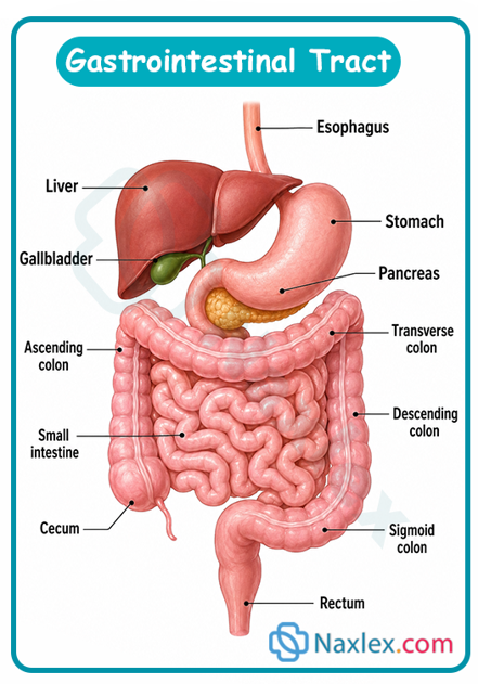
Functions Of The Gastrointestinal Tract
Core Physiological Processes
|
Function |
Primary Anatomical Site |
Mechanism / Key Enzymes |
Pediatric Considerations |
|
Ingestion & Propulsion |
Oral Cavity, Esophagus, Stomach |
Swallowing reflex, peristaltic wave contractions, segmentation |
Uncoordinated swallowing in premies; reduced LES tone leads to physiologic reflux. |
|
Chemical Digestion |
Stomach, Duodenum, Jejunum |
Hydrochloric acid, Pepsinogen converted to pepsin, Pancreatic Lipase, Amylase, Trypsin, Disaccharidases |
Amylase and Lipase deficient in early infancy; milk digestion depends on gastric/lingual enzymes. |
|
Nutrient Absorption |
Jejunum, Ileum |
Villi and microvilli (brush border) expand surface area; passive diffusion, active transport |
High mucosal surface area per body weight allows efficient milk nutrient extraction. |
|
Water & Electrolyte Absorption |
Colon (Large Intestine) |
Active sodium transport, passive water reabsorption |
High risk of fluid deficit during enterovirus/rotavirus infections due to rapid transit time. |
|
Immunological Defense |
Gut-Associated Lymphoid Tissue (GALT), Peyer's Patches |
Secretory IgA production, stomach acid barrier, tight cell junctions |
Immature IgA secretion and permeable mucosal junctions in early infancy. |
|
Excretion |
Rectum & Anal Canal |
Defecation reflex controlled by internal (involuntary) and external (voluntary) sphincters |
Voluntary sphincter control is not neurologically developed until 18–24 months of age. |
Assessment Of The Gastrointestinal Tract
Focused Pediatric Health History
- Current Symptoms: Onset, duration, character, and frequency of vomiting (bilious vs. non-bilious, projectile vs. regurgitation), diarrhea (watery, bloody, mucous, seed-like), abdominal pain (location, cramping, constant), constipation, or fever.
- Feeding & Diet: Breast milk vs. formula type, preparation, feeding volume/frequency, solid food introduction, recent changes in diet, fluid intake.
- Elimination Baseline: Baseline bowel pattern, stool color/consistency, diaper counts (urine output assessment: normal is 1–2 mL/kg/hr in infants, 1 mL/kg/hr in children).
- Growth Metrics: Percentiles for weight, length/height, and head circumference. Loss of weight or crossing downward percentiles indicates failure to thrive (FTT).
Systematic Physical Examination Sequence
Critical Nursing Rule: Always perform inspection, auscultation, percussion, and light-to-deep palpation in that exact order. Palpation or percussion performed before auscultation alters bowel sound frequency and quality.
Step 1: Inspection
- Contour & Shape: Flat, rounded, scaphoid (sunken, seen in diaphragmatic hernia), or distended (protuberant).
- Skin & Umbilicus: Color, venous patterns, umbilical hernia, erythema around umbilicus (omphalitis), surgical scars, or stomas.
- Abdominal Movement: Visible peristaltic waves (e.g., left-to-right gastric peristaltic waves seen in pyloric stenosis) or visible pulsations.
- Perianal Area: Inspection for diaper dermatitis, fissures, skin tags, rectal prolapse, or imperforate anus.
Step 2: Auscultation
- Bowel Sounds: Listen using the warm diaphragm of the stethoscope in all four quadrants (RLQ, RUQ, LUQ, LLQ), starting in the RLQ (ileocecal valve region).
- Normal (Normoactive): Irregular clicks and gurgles occurring 5 to 30 times per minute.
- Hyperactive: Loud, high-pitched, rapid gurgling (gastroenteritis, early intestinal obstruction).
- Hypoactive: Soft, infrequent sounds (post-operative ileus, peritonitis, severe hypokalemia).
- Absent: Complete silence after listening for a full 3 to 5 minutes in all quadrants (paralytic ileus, acute abdomen).
Step 3: Percussion
- Tympany: Dominant sound percussed over air-filled stomach and intestinal loops.
- Dullness: Heard over solid organs (liver, spleen), full bladder, fluid collections (ascites), or fecal masses.
Step 4: Palpation
- Comfort Measures: Flex the child's knees, warm hands, use distraction, save painful areas for last.
- Light Palpation (1 cm depth): Assess for superficial tenderness, muscle guarding, cutaneous hypersensitivity, and abdominal rigidity.
- Deep Palpation (2–4 cm depth): Assess for deep masses, organomegaly (liver, spleen), or localized pain.
- Key Pediatric Diagnostic Signs:
- Olive-shaped Mass: Firm, mobile mass in the right epigastrium/RUQ (pathognomonic for pyloric stenosis).
- Sausage-shaped Mass: Mass in the RUQ or epigastrium with an empty RLQ (Dance Sign) (pathognomonic for intussusception).
- McBurney’s Point Tenderness: Localized RLQ tenderness (indicative of appendicitis).
- Rebound Tenderness / Involuntary Guarding: Indicates peritoneal irritation/peritonitis.
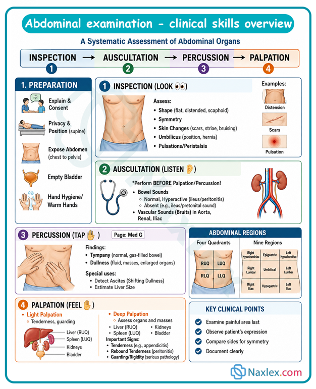
Common Gastrointestinal Laboratory And Diagnostic Tests
LABORATORY DIAGNOSTIC TESTS
|
Test Name |
Primary Purpose / Clinical Indications |
Normal Values / Key Findings |
Nursing Responsibilities & Interpretation |
|
Stool Culture & Ova/Parasites (O&P) |
Identify bacterial (Salmonella, Shigella, C. difficile) or parasitic pathogens. |
Negative for enteric pathogens. |
Collect stool avoiding urine contamination. Transport immediately to lab in transport media. |
|
Stool pH & Reducing Substances |
Detect carbohydrate malabsorption (e.g., lactose intolerance). |
Stool pH > 6.0; Reducing substances < 0.25%. |
pH < 5.5 or reducing substances ≥ 0.5% indicates carbohydrate malabsorption. |
|
Stool Guaiac / Fecal Occult Blood Test (FOBT) |
Detect hidden microscopic blood loss in GI tract. |
Negative (no blue color change). |
False positives caused by red meat or iron supplements. Blue color change indicates presence of blood. |
|
Serum Electrolytes & BUN/Creatinine |
Evaluate hydration status, metabolic acidosis/alkalosis from emesis/diarrhea. |
Normal Na: 135–145 mEq/L; K: 3.5–5.0 mEq/L; HCO3: 20–28 mEq/L. |
Hypokalemic, hypochloremic metabolic alkalosis seen in severe vomiting (pyloric stenosis). |
|
Serum Liver Function Tests (LFTs) |
Evaluate hepatic cell injury, cholestasis, biliary patency (ALT, AST, Total/Direct Bilirubin). |
Direct Bilirubin: < 0.3 mg/dL; Total Bilirubin: < 1.2 mg/dL (varies in neonates). |
Elevated Direct (Conjugated) Bilirubin indicates biliary obstruction or hepatitis (e.g., Biliary Atresia). |
|
Celiac Disease Serology (Tissue Transglutaminase IgA - tTG-IgA) |
Screen for Celiac disease (gluten-sensitive enteropathy). |
Negative antibody titers. |
Patient must be consuming a gluten-containing diet prior to testing to avoid false-negative results. |
DIAGNOSTIC IMAGING AND INVASIVE PROCEDURES
Plain Abdominal Radiograph (KUB / Flat Plate)
- Indications: Suspected bowel obstruction, intestinal perforation, foreign body ingestion, or severe constipation.
- Key Findings: Dilated bowel loops, air-fluid levels (obstruction), free air under the diaphragm (perforation), or Pneumatosis intestinalis (gas within the bowel wall, pathognomonic for necrotizing enterocolitis [NEC]).
Abdominal Ultrasound
- Indications: Gold standard diagnostic imaging for Hypertrophic Pyloric Stenosis, Intussusception, and Acute Appendicitis.
- Key Findings: Pyloric muscle thickness > 3 mm or length > 15 mm; "Target" or "Doughnut" sign in intussusception; non-compressible appendix > 6 mm in diameter.
Upper GI Fluoroscopic Series (Barium/Water-Soluble Swallow)
- Indications: Evaluate esophageal strictures, gastroesophageal reflux, tracheoesophageal fistula, or intestinal malrotation with volvulus.
- Nursing Care: NPO prior to procedure (age-dependent: 3–8 hours). Post-procedure: Encourage fluids to clear barium; educate parents that stools will be chalky white for 24–48 hours.
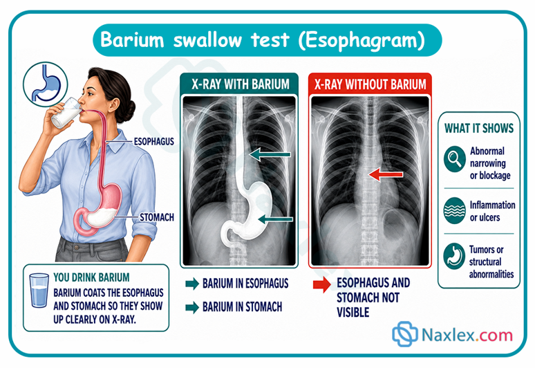
Therapeutic Contrast / Air Enema
Indications: Both diagnostic and therapeutic treatment for intussusception. - Mechanism: Air or radiopaque contrast is instilled into the rectum under pressure to reduce (un-invaginate) the telescoped bowel segment.
- Nursing Insight: If the child passes a normal, fully formed brown stool prior to the procedure, notify the provider immediately as this indicates spontaneous reduction of the intussusception.
-
24-Hour Esophageal pH Probe / Impedance Monitoring
- Indications: Gold standard for quantifying severe Gastroesophageal Reflux Disease (GERD).
- Procedure: A thin probe is inserted transnasally into the distal esophagus. Monitors pH drops below 4.0.
- Nursing Care: Keep a detailed diary of feeding, sleeping, positioning, and coughing symptoms during the 24-hour test period.
Summary
- The pediatric gastrointestinal (GI) system undergoes structural maturation, enzymatic development, and functional adaptation from infancy through adolescence.
- Recognizing the physiological and anatomical variations unique to pediatric clients is critical for pediatric nursing practice.
- Neonates and infants are characterized by a smaller gastric capacity, lower lower esophageal sphincter (LES) tone, hyper-permeable mucosal linings, and lower baseline levels of digestive enzymes such as pancreatic amylase and lipase.
- These characteristics explain common pediatric phenomena including physiological gastroesophageal reflux, limited starch digestion prior to 4–6 months, and an increased risk for macromolecular absorption and systemic infections.
- Furthermore, infants possess a higher percentage of total body water stored within the extracellular fluid (ECF) compartment coupled with a high basal metabolic rate, rendering them susceptible to rapid, life-threatening dehydration and electrolyte imbalances during acute episodes of vomiting or diarrhea.
- Anatomically, the GI tract is organized into upper and lower divisions, supported by key accessory organs including the liver, gallbladder, and pancreas.
- Understanding structural milestones, such as the completion of spinal cord myelination around 18–24 months enabling voluntary bowel control, helps nurses guide caregivers on developmental readiness for toilet training.
- Clinical physical assessment of the pediatric abdomen must adhere to a strict sequence: inspection, auscultation, percussion, and palpation, ensuring bowel sounds are not artificially altered.
- Pediatric GI disorders present with pathognomonic clinical signs, such as the olive-shaped epigastric mass in hypertrophic pyloric stenosis, sausage-shaped mass with currant-jelly stools in intussusception, and McBurney’s point tenderness in appendicitis.
- Nurses must be proficient in interpreting diagnostic tests ranging from stool pH and reducing substances to abdominal ultrasounds, pH probe studies, and therapeutic air/contrast enemas.
Naxlex
Videos
Login to View Video
Click here to loginTake Notes on Pediatric Anatomy And Physiology Of The Gastrointestinal Tract
This filled cannot be empty
Join Naxlex Nursing for nursing questions & guides! Sign Up Now