





 Video Library


Anatomy And Physiology Bio 168 Proctored Exam
Total Questions : 60
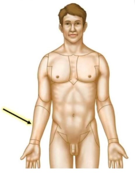
Showing 10 questions, Sign in for moreIdentify region indicated by arrow.
Explanation
Brief Discussion:
The forearm is the portion of the upper limb located between the elbow and the wrist, making it the correct region where the arrow points to this segment. It is distinct from the arm (brachium), which extends from the shoulder to the elbow, and from the hand, which begins distal to the wrist. Therefore, a structure located between the elbow and wrist is identified as the forearm rather than any adjacent upper-limb region.
Rationale:
The forearm is situated distal to the elbow joint and proximal to the wrist joint. It contains the radius and ulna, which are connected by the interosseous membrane and surrounded by anterior (flexor-pronator) and posterior (extensor-supinator) muscle compartments. Major neurovascular structures, including the radial and ulnar arteries and the median, ulnar, and radial nerves, traverse this region to supply the forearm and hand. Physiologically, the forearm provides the muscular force required for wrist and finger movements, as well as pronation and supination of the hand through coordinated movement of the radius around the ulna.
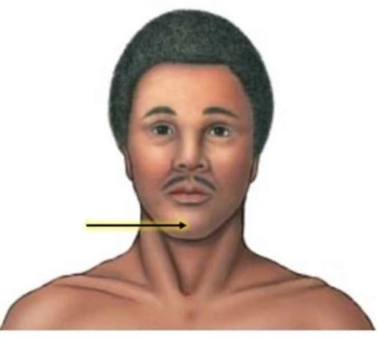
Identify region indicated by arrow.
Explanation
Brief Discussion:
The mentum is the anatomical term for the chin, forming the most inferior and anterior prominence of the face. It is correctly identified by the arrow pointing to the chin rather than the adjacent lower lip (labial region), cheek (buccal region), or neck (cervical region). Its distinct location at the anterior aspect of the mandible differentiates it from surrounding facial regions.
Rationale:
The mentum is located on the anterior midline of the face, overlying the mandibular symphysis, where the two halves of the mandible fuse during early development. It lies inferior to the lower lip, superior to the cervical region, and medial to the mandibular (jaw) regions. Structurally, it is supported by the body of the mandible and is covered by skin, subcutaneous tissue, and muscles involved in facial expression, particularly the mentalis muscle. Physiologically, the mentum contributes to facial contour and aesthetics, supports movements of the lower lip during speech and facial expression, and serves as an attachment site for muscles that elevate and protrude the lower lip.
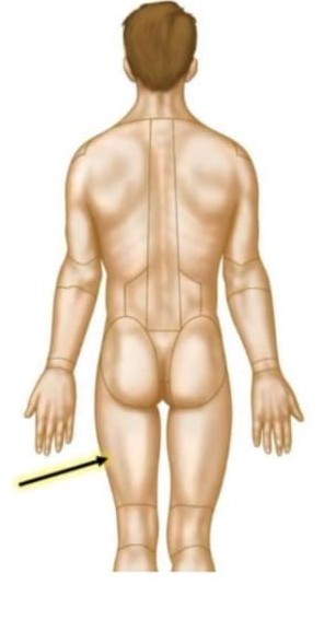
Identify region indicated by arrow.
Explanation
Brief Discussion:
The posterior thigh region is the portion of the lower limb located on the back of the thigh, extending from the gluteal region superiorly to the popliteal region posterior to the knee inferiorly. It is the correct identification when the arrow points to the back of the thigh rather than the anterior thigh (femoral region), buttock (gluteal region), or the posterior knee (popliteal region). Its location between these adjacent regions distinguishes it anatomically.
Rationale:
The posterior thigh region is situated on the posterior aspect of the thigh, overlying the femur and extending from the inferior border of the gluteal fold to the popliteal fossa. It is primarily occupied by the hamstring muscle group, comprising the biceps femoris, semitendinosus, and semimembranosus, which receive their principal innervation from the tibial division of the sciatic nerve. The sciatic nerve also courses through this region before dividing near the popliteal fossa into the tibial and common fibular nerves. The posterior thigh muscles are responsible for extension of the hip, flexion of the knee, and stabilization of the pelvis and lower limb during standing, walking, running, and other weight-bearing activities.
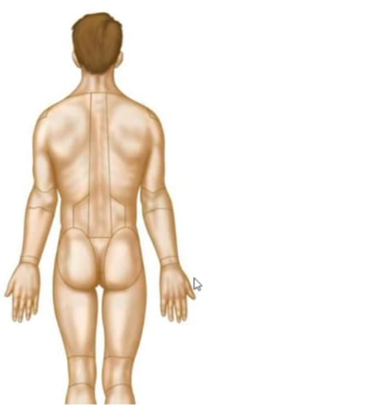
Fill in the blank: The popliteal region is
Explanation
Brief Discussion:
The popliteal region is proximal to the tarsal region because it is located closer to the trunk along the lower limb. The popliteal region is situated at the posterior aspect of the knee, whereas the tarsal region refers to the ankle. Since anatomical directional terms on the limbs are based on distance from the point of attachment to the trunk, the knee is proximal to the ankle, making "proximal" the correct term rather than distal.
Rationale:
The popliteal region is located on the posterior aspect of the knee joint, forming the depression known as the popliteal fossa. It contains important neurovascular structures, including the popliteal artery, popliteal vein, tibial nerve, and common fibular nerve. The tarsal region is located at the ankle and consists of the seven tarsal bones, which connect the leg to the foot and contribute to ankle stability and mobility. Physiologically, the popliteal region serves as a major conduit for blood vessels and nerves supplying the leg and foot while permitting knee flexion and extension. The tarsal region functions in weight-bearing, shock absorption, and movement of the foot during standing and ambulation. Because the popliteal region lies closer to the trunk than the tarsal region, it is anatomically described as proximal to the tarsal region.
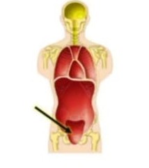
Identify the indicated body cavity.
Explanation
Brief Discussion:
The pelvic cavity is the inferior subdivision of the abdominopelvic cavity enclosed by the bony pelvis. It is the correct identification when the arrow points to the space within the pelvis rather than the abdominal cavity, which lies superior to it, or the vertebral cavity, which is located posteriorly within the vertebral column. Its position below the abdominal cavity and within the pelvic girdle distinguishes it from adjacent body cavities.
Rationale:
The pelvic cavity is located inferior to the abdominal cavity and is bounded by the pelvic inlet superiorly, the pelvic diaphragm inferiorly, and the bones of the pelvis laterally and anteriorly. It contains the urinary bladder, distal ureters, rectum, and internal reproductive organs, including the uterus, ovaries, and uterine tubes in females, or the prostate gland and seminal vesicles in males. Physiologically, the pelvic cavity protects and supports the pelvic viscera, provides passageways for major blood vessels, nerves, and lymphatics, and accommodates structures involved in urinary, reproductive, and distal gastrointestinal function. In females, it also expands to facilitate pregnancy and childbirth. Its anatomical location within the bony pelvis and its role in housing and protecting pelvic organs distinguish it from the adjacent abdominal cavity.
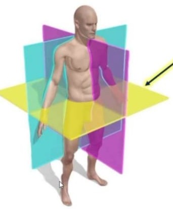
Fill in the blank.
A
Explanation
Brief Discussion:
The transverse plane is the anatomical plane that divides the body or an organ into superior (upper) and inferior (lower) portions. It is distinguished from the sagittal plane, which divides the body into right and left portions, and the frontal (coronal) plane, which separates the body into anterior and posterior portions. Therefore, only the transverse plane correctly creates upper and lower sections.
Rationale:
The transverse plane, also known as the horizontal or axial plane, passes perpendicular to the body's longitudinal axis. It can intersect the body or an organ at any level, producing superior and inferior segments. This plane is widely used in anatomy, diagnostic imaging (such as computed tomography [CT] and magnetic resonance imaging [MRI]), and surgical planning to visualize cross-sectional anatomy. Physiologically, the transverse plane does not alter body function but serves as an essential anatomical reference for accurately describing the location of structures, interpreting imaging studies, and communicating the level of injuries, lesions, or surgical procedures. Its orientation perpendicular to the body's vertical axis uniquely distinguishes it from the sagittal and coronal planes.
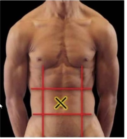
Identify the indicated abdominal region.
Explanation
Brief Discussion:
The umbilical region is the central region of the nine-region abdominal scheme and surrounds the umbilicus (navel). It is the correct identification when the arrow points to the middle portion of the abdomen rather than the epigastric region superiorly, the hypogastric (pubic) region inferiorly, or the lumbar regions laterally. Its central location makes it distinct from the surrounding abdominal regions.
Rationale:
The umbilical region is located in the center of the anterior abdominal wall, bounded superiorly by the epigastric region, inferiorly by the hypogastric (pubic) region, and laterally by the right and left lumbar regions. It commonly overlies portions of the small intestine (primarily the jejunum and ileum), the transverse colon, and the inferior part of the duodenum. Physiologically, the organs within this region are primarily involved in digestion, nutrient absorption, and the propulsion of intestinal contents. The umbilical region also serves as an important anatomical landmark for physical examination, abdominal pain localization, surgical planning, and the description of intra-abdominal pathology. Its central position surrounding the umbilicus distinguishes it from adjacent abdominal regions.
Fill in the blank.
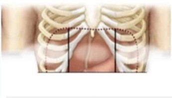
The epigastric region is
Explanation
Brief Discussion:
The epigastric region is medial to the right and left hypochondriac regions because it occupies the central upper portion of the abdomen. The hypochondriac regions lie on either side of the epigastric region beneath the costal cartilages. Therefore, the epigastric region is located closer to the body's midline than the hypochondriac regions, making "medial" the correct directional term rather than lateral.
Rationale:
The epigastric region is one of the nine anatomical regions of the abdomen and is situated in the superior central portion of the anterior abdominal wall. It is bordered laterally by the right and left hypochondriac regions and inferiorly by the umbilical region. This region overlies structures such as the stomach, left lobe of the liver, pancreas, proximal duodenum, and portions of the abdominal aorta. Physiologically, these organs are involved in digestion, secretion of digestive enzymes and hormones, nutrient processing, and blood circulation. The hypochondriac regions, in contrast, lie lateral to the epigastric region and contain structures such as the liver on the right and the spleen on the left. Because the epigastric region lies along the body's midline, it is anatomically described as medial to both hypochondriac regions.
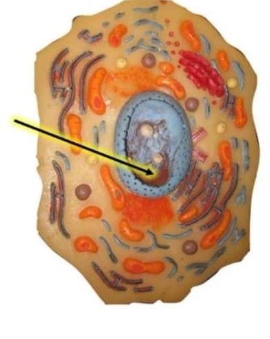
Identify the structure represented by the red "threads" within the nucleus.
Explanation
Brief Discussion:
Chromatin is the thread-like material found within the nucleus and is correctly identified in the diagram showing fine red "threads" dispersed through the nuclear interior. These threads are distinct from the nucleolus, which appears as a dense, rounded structure involved in ribosome production, and from the nuclear envelope, which forms the boundary surrounding the nucleus. Therefore, the thread-like appearance within the nucleus specifically represents chromatin.
Rationale:
Chromatin is a complex of deoxyribonucleic acid (DNA) and histone proteins located within the nucleus of eukaryotic cells. It is distributed throughout the nucleoplasm and exists in two functional forms: euchromatin, which is loosely packed and transcriptionally active, and heterochromatin, which is densely packed and largely transcriptionally inactive. Physiologically, chromatin packages the cell's genetic material into an organized structure that fits within the nucleus while regulating DNA replication, gene expression, DNA repair, and chromosome formation during cell division. Prior to mitosis and meiosis, chromatin condenses to form visible chromosomes, ensuring the accurate segregation of genetic material to daughter cells. Its characteristic thread-like appearance within the nucleus distinguishes it from other nuclear structures.
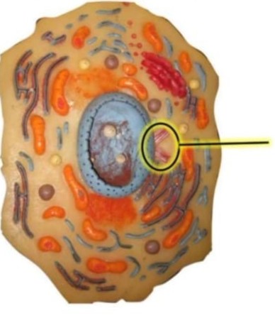
Identify the indicated organelle.
Explanation
Brief Discussion:
Centrioles are cylindrical organelles typically found as a paired structure within the centrosome near the nucleus. They are correctly identified in the diagram as two short, barrel-shaped cylinders arranged at right angles to one another. This appearance distinguishes them from adjacent organelles such as mitochondria, which are oval with internal cristae, or the Golgi apparatus, which consists of flattened membranous sacs.
Rationale:
Centrioles are non-membrane-bound organelles located in the centrosome of most animal cells, immediately adjacent to the nucleus. Each centriole is composed of nine triplets of microtubules arranged in a cylindrical pattern (9 × 3 arrangement). Physiologically, centrioles serve as microtubule-organizing centers, playing a critical role in the formation of the mitotic spindle during mitosis and meiosis to ensure accurate separation of chromosomes into daughter cells. They also give rise to basal bodies, which initiate the formation of cilia and flagella involved in cellular movement and fluid transport across epithelial surfaces. Their characteristic paired, perpendicular arrangement near the nucleus and their essential role in microtubule organization distinguish them from other cytoplasmic organelles.
Sign Up or Login to view all the 60 Questions on this Exam
Join over 100,000+ nursing students using Naxlex’s science-backend flashcards, practice tests and expert solutions to improve their grades and reach their goals.
Sign Up Now