
BIOL 252 Anatomy And Physiology II Module 2 Proctored Exam
Total Questions : 69
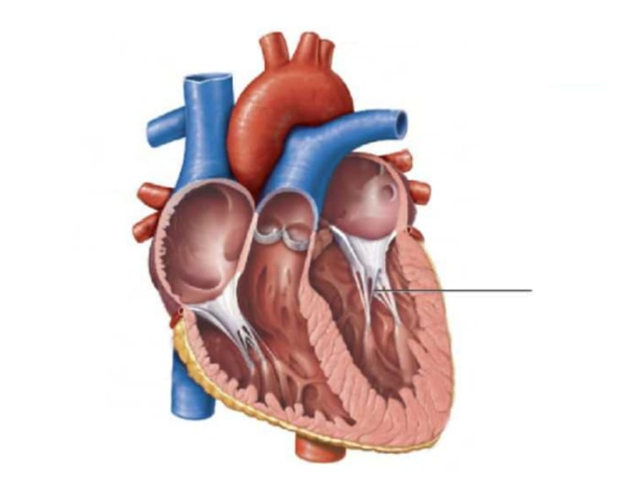
Showing 10 questions, Sign in for moreIdentify the structure pointed by the line
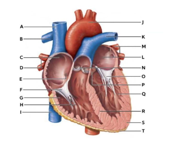
Identify the letter of the heart component that is being described in the statement.
Shallow depression that is a remnant of the foramen ovale.
The nurse plans to use role playing as a therapeutic measure. Which individual is most likely to benefit from this type of therapeutic intervention?
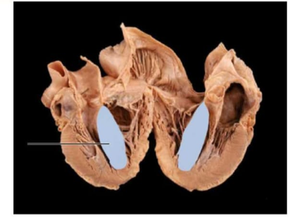
Identify the highlighted section using the drop down below.
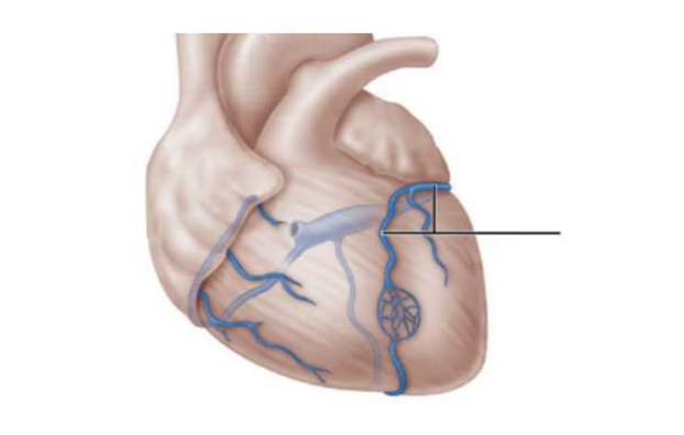
What structure of the electrical conduction system passes through the highlighted area?
Explanation
Correct answer:
- Interventricular septum
- Atrioventricular (AV) bundle / Bundle of His
The highlighted area represents the interventricular septum, the thick muscular wall that separates the right and left ventricles. It forms the medial wall of both ventricles and extends from the atrioventricular valves superiorly to the apex inferiorly. Its primary physiologic function is to prevent mixing of oxygenated blood in the left ventricle with deoxygenated blood in the right ventricle while also contributing to ventricular contraction. The electrical conduction structure that passes through this area is the atrioventricular (AV) bundle / Bundle of His. The Bundle of His passes from the atrioventricular node into the membranous portion of the interventricular septum. It then divides into the right and left bundle branches, which travel along the septum toward the apex to distribute electrical impulses to both ventricles. This allows coordinated ventricular depolarization and synchronized contraction.
Match the structure to its description.
|
Description |
Structure |
|
A cluster of cells located in the interatrial septum that delays the electrical signal before it passes to the ventricles. |
dropdown
|
|
The pacemaker of the heart, located in the right atrium, responsible for initiating the heartbeat. |
dropdown
|
|
The valve located between the right atrium and right ventricle. |
dropdown
|
|
The layer of the pericardium that covers the heart directly, also known as the epicardium. |
dropdown
|
|
Blood vessels carrying oxygenated blood from the lungs to the left atrium. |
dropdown
|
Explanation
Correct answer:
- A cluster of cells located in the interatrial septum that delays the electrical signal before it passes to the ventricles: Atrioventricular node
- The pacemaker of the heart, located in the right atrium, responsible for initiating the heartbeat: Sinoatrial node
- The valve located between the right atrium and right ventricle: Tricuspid valve
- The layer of the pericardium that covers the heart directly, also known as the epicardium: Visceral pericardium
- Blood vessels carrying oxygenated blood from the lungs to the left atrium: Pulmonary veins
• Atrioventricular node: The AV node is located in the interatrial septum and functions to delay the electrical impulse received from the SA node. This delay allows the atria to contract and complete ventricular filling before ventricular contraction. Its position and timing role are crucial for coordinated cardiac conduction.
• Sinoatrial node: The SA node, located in the right atrium near the superior vena cava, acts as the heart’s natural pacemaker. It generates electrical impulses that initiate each heartbeat, setting the rhythm for the entire heart. Proper SA node function is essential for synchronized atrial and ventricular activity.
• Tricuspid valve: The tricuspid valve sits between the right atrium and right ventricle, preventing backflow of blood during ventricular contraction. Its three leaflets open during diastole to allow atrial emptying into the ventricle. Proper valve function maintains unidirectional blood flow and cardiac efficiency.
• Visceral pericardium: Also called the epicardium, this layer of the pericardium lies directly on the heart surface. It provides protection, reduces friction during heartbeats, and contains blood vessels supplying the myocardium. Its anatomical position distinguishes it from the parietal pericardium.
• Pulmonary veins: Pulmonary veins carry oxygen-rich blood from the lungs to the left atrium. They are unique among veins as they transport oxygenated rather than deoxygenated blood. Their flow ensures that systemic circulation receives oxygenated blood.
Blood flows from the right coronary artery into the
The pulmonary veins carry deoxygenated blood from the lungs to the left atrium.
The blood vessel on the image is the
A patient with a defective tricuspid valve undergoes valve replacement surgery. After surgery, the patient should be monitored for proper opening of the valve during which part of the cardiac cycle?
Which letter represents ventricular depolarization?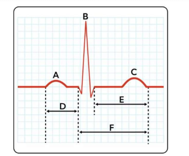
Sign Up or Login to view all the 69 Questions on this Exam
Join over 100,000+ nursing students using Naxlex’s science-backend flashcards, practice tests and expert solutions to improve their grades and reach their goals.
Sign Up Now
