
Biol 252 Anatomy And Physiology Module 3 Proctored Exam
Total Questions : 68
Showing 10 questions, Sign in for moreThe point at which the Korotkoff sounds disappear is recorded as the diastolic pressure.
Identify the following structures by letter and make sure to number your answer for grading.
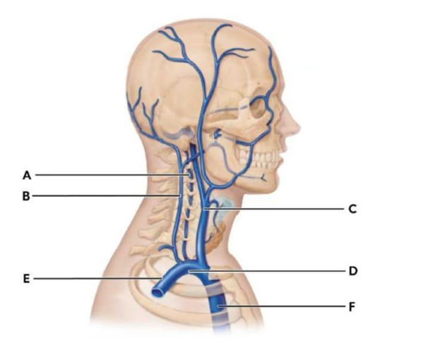
- Brachiocephalic vein dropdown
- Vertebral vein dropdown
- Internal jugular vein dropdown
- Superior vena cava dropdown
Explanation
A. E (Brachiocephalic vein): The brachiocephalic veins are large veins formed by the union of the internal jugular and subclavian veins on each side of the body. The left and right brachiocephalic veins converge to form the superior vena cava. They serve as major conduits for venous blood returning from the head, neck, upper limbs, and thorax to the heart.
B. A (Vertebral vein): The vertebral veins accompany the vertebral arteries within the transverse foramina of the cervical vertebrae. They drain the cervical spinal cord, vertebrae, deep neck muscles, and portions of the posterior brain. These veins empty into the brachiocephalic veins, providing a deep venous return pathway from the neck and posterior cranial structures.
C. C (Internal jugular vein): The internal jugular veins are the principal veins that drain blood from the brain, superficial face, and neck. They originate from the dural venous sinuses at the base of the skull and descend within the carotid sheath lateral to the internal carotid artery and common carotid artery. They join the subclavian veins to form the brachiocephalic veins.
D. F (Superior vena cava): The superior vena cava is a large, short vein that carries deoxygenated blood from the head, neck, upper limbs, and thoracic structures into the right atrium. It is formed by the convergence of the left and right brachiocephalic veins and lies in the superior mediastinum, anterior to the trachea and to the right of the ascending aorta.
Which veins drain blood from the brain into the brachiocephalic vein?
All of the following blood vessels bring blood to the brain except:
Identify the following blood vessels by matching them to the letter on the image.
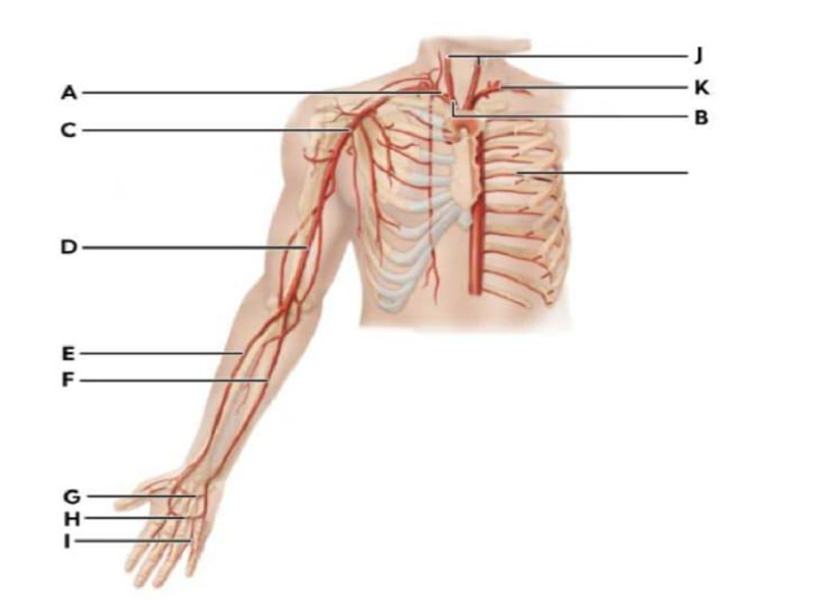
- Axillary artery dropdown
- Left Subclavian artery dropdown
- Ulnar artery dropdown
- Superficial palmar arch dropdown
Explanation
Axillary Artery: C
Left Subclavian Artery: K
Ulnar Artery: F
Superficial Palmar Arch: H
A. Axillary artery: The axillary artery, presented by letter C is the continuation of the subclavian artery. It courses through the axilla and becomes the brachial artery at the inferior border of the teres major muscle. It gives off key branches including the thoracoacromial, lateral thoracic, subscapular, and anterior and posterior circumflex humeral arteries. It supplies the shoulder joint, lateral thoracic wall, and upper limb.
B. Left Subclavian artery: The left subclavian artery presented as K arises from the arch of the aorta, posterior to the left common carotid artery. It travels laterally toward the upper limb, arching over the apex of the lung and passing between the anterior and middle scalene muscles. It supplies blood to the brain (via the vertebral artery), thoracic wall, and left upper extremity.
C. Ulnar artery: The ulnar artery as F is one of the two terminal branches of the brachial artery, arising in the cubital fossa opposite the neck of the radius. It travels along the medial (ulnar) side of the forearm. It primarily forms the superficial palmar arch and supplies the medial forearm, hand muscles, and digits, particularly the medial three and a half fingers.
D. Superficial palmar arch: The superficial palmar arch as H, is an arterial arcade in the palm primarily formed by the ulnar artery, with contribution from the superficial branch of the radial artery. From this arch arise the common palmar digital arteries, which divide into proper digital arteries supplying the fingers, ensuring collateral circulation within the hand.
Match the letter to its blood vessel:
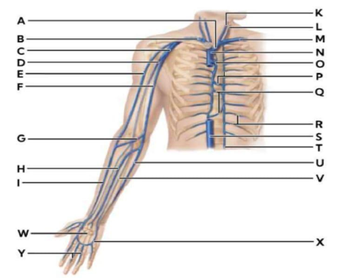
- T dropdown
- I dropdown
- M dropdown
- C dropdown
Explanation
A. T (Ascending lumbar vein): The ascending lumbar vein is a paired longitudinal vein located along the posterior abdominal wall, running vertically on either side of the vertebral column. It connects the common iliac veins inferiorly, it serves as an important collateral pathway between the inferior and superior vena cava and drains the lumbar region and posterior abdominal wall.
B. I (Cephalic Vein): The cephalic vein is a superficial vein of the upper limb that originates from the dorsal venous network of the hand. It ascends along the lateral (radial) side of the forearm and arm, traveling within the deltopectoral groove before piercing the clavipectoral fascia. It drains into the axillary vein.
C. M (Subclavian vein): The subclavian vein is the continuation of the axillary vein at the lateral border of the first rib. It runs medially beneath the clavicle and anterior to the subclavian artery and anterior scalene muscle. It joins with the internal jugular vein to form the brachiocephalic vein. It drains venous blood from the upper limb, as well as parts of the thoracic wall and neck.
D. C (Axillary vein): The axillary vein is formed by the union of the brachial veins and the basilic vein at the inferior border of the teres major muscle. It ascends through the axilla medial to the axillary artery and receives tributaries corresponding to branches of the axillary artery, including the cephalic vein. At the lateral border of the first rib, it becomes the subclavian vein.
Which pathway describes blood flow from the heart to the radial side of the forearm?
Which of the following is the primary function of the celiac trunk?
Identify the following blood vessels by matching them to the letter on the image.
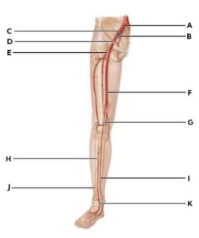
- Anterior tibial artery dropdown
- External iliac artery dropdown
- Popliteal artery dropdown
- Dorsalis pedis dropdown
Explanation
Anterior tibial artery: H
External iliac artery: A
Popliteal artery: G
Dorsalis pedis: K
A. Anterior tibial artery (H): The anterior tibial artery arises from the popliteal artery at the inferior border of the popliteus muscle. It passes through an opening in the interosseous membrane to enter the anterior compartment of the leg. It supplies the anterior leg musculature and continues distally as the dorsalis pedis artery at the level of the ankle joint.
B. External iliac artery (A): The external iliac artery is one of the terminal branches of the common iliac artery. It courses along the pelvic brim and passes deep to the inguinal ligament, where it becomes the femoral artery. It provides major blood supply to the lower limb and gives off important branches such as the inferior epigastric and deep circumflex iliac arteries.
C. Popliteal artery (G): The popliteal artery is the continuation of the femoral artery after it passes through the adductor hiatus. It lies deep within the popliteal fossa posterior to the knee joint and is closely related to the popliteal vein and tibial nerve. It supplies the knee joint and surrounding structures and bifurcates into the anterior and posterior tibial arteries.
D. Dorsalis pedis (K): The dorsalis pedis artery is the continuation of the anterior tibial artery distal to the ankle joint. It runs along the dorsum of the foot, lateral to the tendon of the extensor hallucis longus, and is commonly palpated to assess peripheral circulation. It contributes to the dorsal arterial arch and provides branches to the dorsal foot and toes.
Match the letter to its blood vessel:
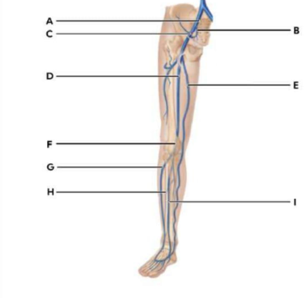
- H dropdown
- C dropdown
- F dropdown
- E dropdown
Explanation
H: Anterior tibial vein
C: Femoral vein
F: Popliteal vein
E: Great saphenous vein
A. H (Anterior tibial vein): The anterior tibial vein is a deep vein of the lower leg that accompanies the anterior tibial artery. It originates from the dorsal venous arch of the foot and ascends through the anterior compartment of the leg between the tibia and fibula. It drains blood from the dorsum of the foot and anterior leg musculature, and it joins the posterior tibial vein to form the popliteal vein.
B. C (Femoral vein): The femoral vein is a major deep vein of the thigh and is the continuation of the popliteal vein after passing through the adductor hiatus. It travels superiorly within the femoral triangle medial to the femoral artery and receives tributaries such as the great saphenous vein and deep femoral vein. It becomes the external iliac vein as it passes beneath the inguinal ligament.
C. F (Popliteal vein): The popliteal vein is located in the popliteal fossa posterior to the knee joint. It is formed by the union of the anterior and posterior tibial veins. It ascends through the posterior knee region, receives the small saphenous vein, and continues proximally to become the femoral vein after passing through the adductor hiatus.
D. E (Great saphenous vein): The great saphenous vein is the longest superficial vein in the body. It begins on the medial side of the dorsal venous arch of the foot, ascends anterior to the medial malleolus, and travels along the medial aspect of the leg and thigh. It drains into the femoral vein at the saphenofemoral junction.
Sign Up or Login to view all the 68 Questions on this Exam
Join over 100,000+ nursing students using Naxlex’s science-backend flashcards, practice tests and expert solutions to improve their grades and reach their goals.
Sign Up Now
