
BIOL 252 Anatomy And Physiology II Module 7 Proctored Exam
Total Questions : 72
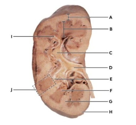
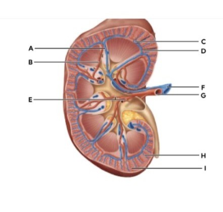
Showing 10 questions, Sign in for moreIdentify the fibrous capsule.
Identify the following structures by letter and make sure to number your answer for grading.
|
|
|
|
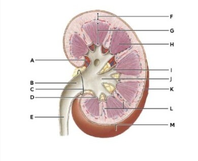
Minor calyx |
dropdown
|
|
Renal medulla |
dropdown
|
|
Renal cortex |
dropdown
|
Explanation
Minor calyx (I): The minor calyx is a small, cup-shaped structure that collects urine from the renal papilla at the tip of each pyramid. It is part of the drainage system, channeling urine into the major calyces and then into the renal pelvis. Look for the small funnel-like extensions directly attached to the renal papillae. These are distinctly smaller than the major calyces.
Renal medulla(H): The renal medulla is the inner region of the kidney, composed of renal pyramids. It plays a crucial role in concentrating urine through the countercurrent mechanism in the loops of Henle and collecting ducts. Appears as darker, triangular or striated regions deeper inside the kidney, between the cortex and the renal pelvis.
Renal cortex (F): The renal cortex is the outer functional tissue of the kidney, lying just beneath the fibrous capsule. It contains glomeruli and convoluted tubules, making it the primary site of blood filtration. Appears as the lighter-colored outer rim of the kidney, surrounding the medulla.
Identify the part of the organ that corresponds with the statement.
The innermost part of the kidney, containing the renal pyramids and involved in concentrating urine.
Explanation
Correct answer: B
Renal medulla: The renal medulla is the deepest (innermost) region of the kidney. It is composed of renal pyramids, which are cone-shaped structures containing loops of Henle and collecting ducts. These structures are essential for the countercurrent mechanism, which allows the kidney to concentrate urine by reabsorbing water and solutes. The medulla lies beneath the renal cortex and surrounds the renal pelvis, making it central to urine formation and concentration.
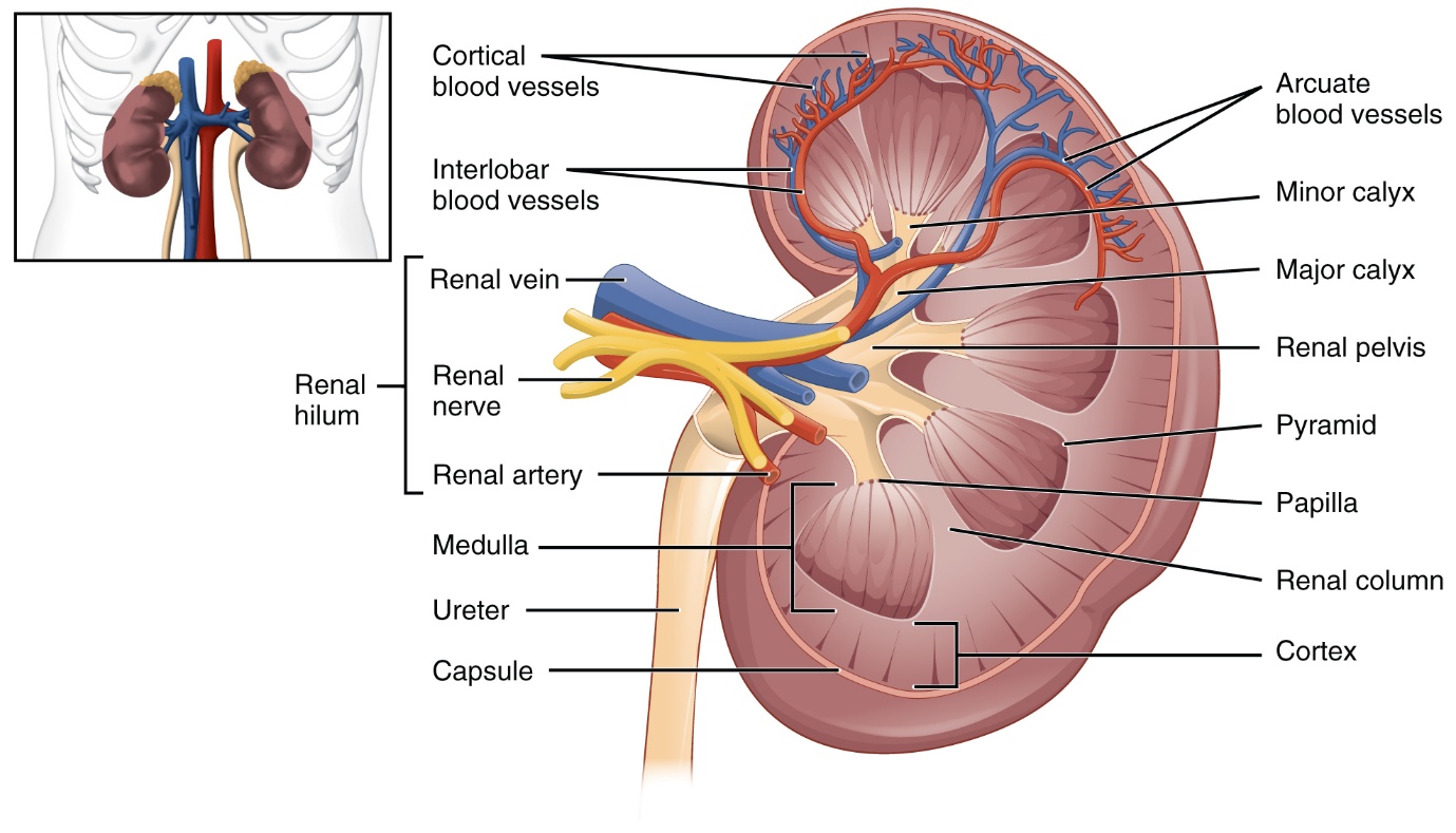
Identify the following structures.
|
|
|
|
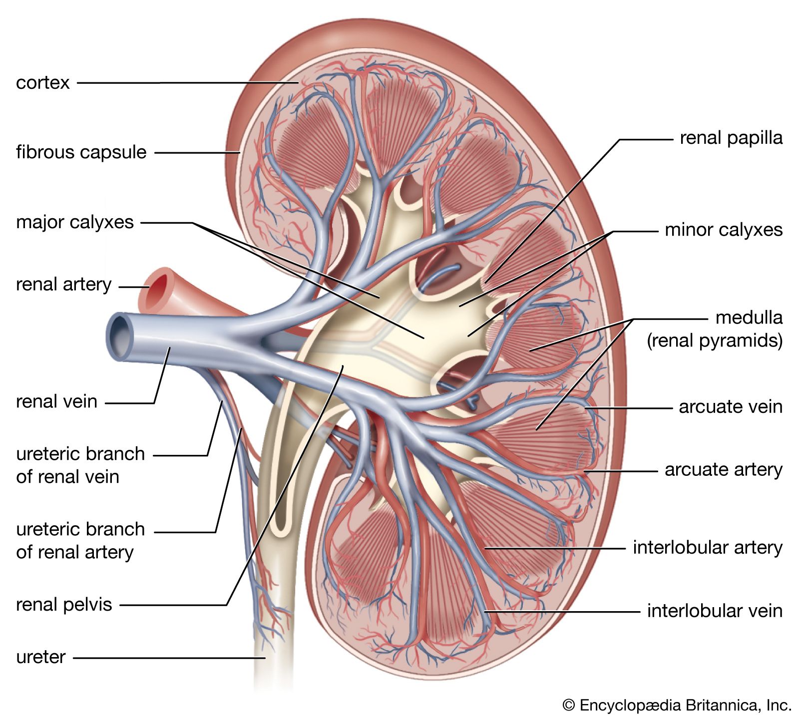
Renal artery |
dropdown
|
|
Interlobar vein |
dropdown
|
|
Cortical radiate vein |
dropdown
|
Explanation
Renal artery → Label G
The renal artery is the thick-walled vessel entering the kidney at the hilum. It carries oxygenated blood from the abdominal aorta into the kidney. In the diagram, G is positioned at the hilum and has the appearance of a robust arterial vessel.
Interlobar vein → Label A
Interlobar veins run between the renal pyramids, draining blood toward the renal vein. In the diagram, A is shown coursing between the medullary pyramids, consistent with the location of interlobar vessels.
Cortical radiate vein → Label I
Cortical radiate veins (interlobular veins) drain blood from the cortex into arcuate veins. In the diagram, I is located in the renal cortex, radiating outward toward the capsule, which matches the expected position of cortical radiate veins.
Which of the following has the blood vessels listed out of order?
What type of muscle is found in the muscularis layer of the ureters?
Identify the following structures.
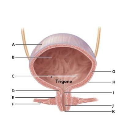
|
|
|
|
Internal urethral sphincter |
dropdown
|
|
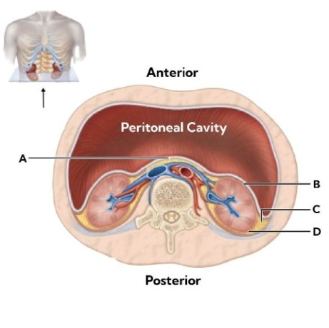
Peritoneum |
dropdown
|
|
External urethral sphincter |
dropdown
|
Explanation
Internal urethral sphincter (D): This is a smooth muscle sphincter located at the junction of the bladder and urethra. It is involuntary, controlled by the autonomic nervous system. Its role is to prevent urine leakage by keeping the urethra closed until micturition is initiated.Found at the bladder neck, just where the urethra begins.
Peritoneum (A): The peritoneum is the serous membrane lining the abdominal cavity. It covers the superior surface of the bladder when the bladder is full. Appears as the outermost layer draped over the top of the bladder in anatomical diagrams.
External urethral sphincter (E ): This is a skeletal muscle sphincter located further down the urethra. It is voluntary, controlled by the somatic nervous system. Its role is to allow conscious control over urination. Found in the urogenital diaphragm, surrounding the urethra outside the bladder.
What condition might a low specific gravity indicate?
In the kidney filtration experiment, the results suggest that cells, proteins, and large molecules were too large to diffuse through the semi-permeable membrane.
The kidneys are located on the ventral side of the body in the superior lumbar region.
Sign Up or Login to view all the 72 Questions on this Exam
Join over 100,000+ nursing students using Naxlex’s science-backend flashcards, practice tests and expert solutions to improve their grades and reach their goals.
Sign Up Now
