
Biol 252 Anatomy and Physiology II Module 5 Proctored Exam
Total Questions : 68
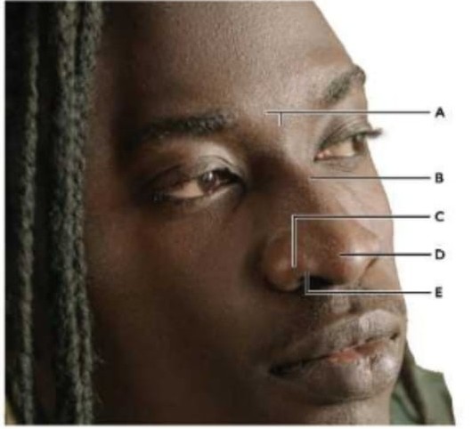
Showing 10 questions, Sign in for moreWhich letter is labelling the ala of the nose?
What is the role of oxygen in a cellular respiration?
What is NOT a function of the nose?
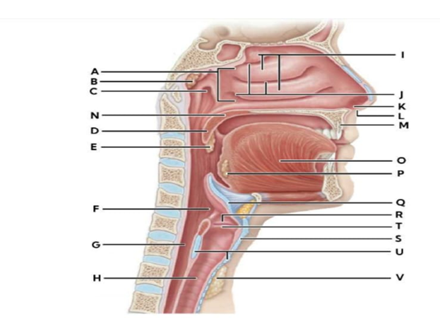
Identify the structures labelled by the following letters:
|
|
|
|
D M H T
|
dropdown
dropdown
dropdown
dropdown
|
Explanation
D: Uvula- The uvula is a small, conical projection of soft tissue hanging from the posterior edge of the soft palate. It helps close off the nasopharynx during swallowing to prevent food or liquid from entering the nasal cavity and plays a minor role in speech articulation.
M: Hard palate- The hard palate forms the anterior bony roof of the mouth and separates the oral cavity from the nasal cavity. It provides a rigid surface against which the tongue can press during chewing and speech and contributes to proper airflow and resonance during respiration.
H: Trachea- The trachea, or windpipe, is a tubular structure composed of C-shaped cartilaginous rings and smooth muscle that connects the larynx to the bronchi. It provides a rigid but flexible airway, ensuring continuous airflow to the lungs while allowing slight expansion during swallowing.
T: Vocal cord- The vocal cords (true vocal folds) are paired, elastic structures within the larynx that vibrate as air passes through them, producing sound. They also help protect the airway by closing during swallowing and regulating airflow during breathing.
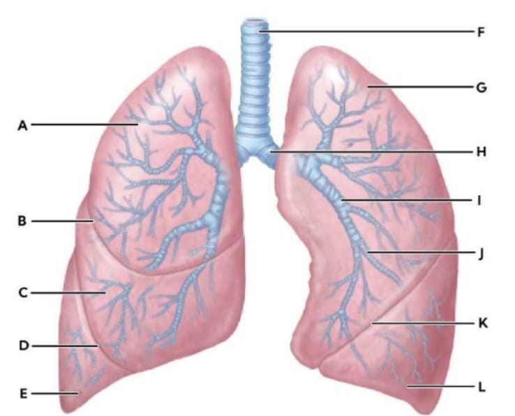
Identify each of the structures by selecting its corresponding letter.
- Bronchioles dropdown
- Left primary bronchus dropdown
- Primary bronchi dropdown
Explanation
A. Bronchioles: Bronchioles, labelled as C, are small-diameter airway passages that branch from the secondary (lobar) bronchi and lead to the alveolar ducts. They lack cartilage and are composed mainly of smooth muscle, which allows them to constrict or dilate to regulate airflow resistance and distribution within the lungs.
B. Left primary bronchus: The left primary bronchus, labelled as H, is a major airway that branches directly from the trachea and enters the left lung. It is wider, shorter, and more vertical on the right side than the left, but on the left, it angles more horizontally due to the heart’s position. It contains cartilage rings to maintain patency and directs airflow into the left lung.
C. Primary bronchi: Primary bronchi refer collectively to the right and left main bronchi that originate from the trachea. They are reinforced with cartilage and lined with ciliated epithelium and mucus-secreting cells to trap particles and propel mucus upward, protecting the lower respiratory tract from inhaled debris and pathogens.
Match the following letters to their structures.
I
K
B
Explanation
I: Lobar bronchus- The lobar (secondary) bronchi branch from the primary bronchi, with one serving each lobe of the lung—three on the right and two on the left. They are supported by cartilage and lined with ciliated epithelium, distributing inhaled air to specific lobes and aiding in filtration and mucus clearance.
K: Left oblique fissure- The left oblique fissure is a deep groove that separates the left lung’s superior and inferior lobes. It provides structural division for the lung lobes, helping compartmentalize airflow and facilitating independent expansion and contraction of each lobe during respiration.
B: Horizontal fissure- The horizontal (minor) fissure is found only in the right lung and separates the superior and middle lobes. Like the oblique fissure, it helps define lung lobes anatomically, directing airflow appropriately and allowing for efficient ventilation and surgical orientation.
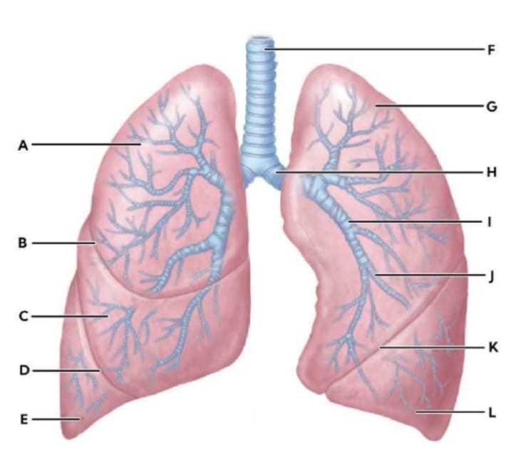
- Match the statements to their correct answers.
|
Structure |
Characteristic |
|
Space between the visceral and parietal pleura. |
dropdown
|
|
Secondary branches of the bronchi leading to lung lobes. |
dropdown
|
|
Smaller airways with no cartilage in their walls. |
dropdown
|
|
Smallest airways in the conducting zone. |
dropdown
|
|
Tertiary branches of the bronchi serving bronchopulmonary segments. |
dropdown
|
Explanation
Correct answer:
- Space between the visceral and parietal pleura: Pleural cavity
- Secondary branches of the bronchi leading to lung lobes: Lobar bronchi
- Smaller airways with no cartilage in their walls: Bronchioles
- Smallest airways in the conducting zone: Terminal bronchioles
- Tertiary branches of the bronchi serving bronchopulmonary segments: Segmental bronchi
Rationale for correct choices:
• Pleural cavity: The pleural cavity is the potential space located between the visceral pleura (covering the lungs) and the parietal pleura (lining the thoracic wall). It contains a thin layer of lubricating pleural fluid that reduces friction during respiration. This space also maintains negative pressure to help keep the lungs expanded.
• Lobar bronchi: Lobar bronchi are the secondary branches that arise from the primary bronchi. Each lobar bronchus supplies one lobe of the lung—three on the right and two on the left. They conduct air deeper into the lung and further divide into segmental bronchi.
• Bronchioles: Bronchioles are smaller conducting airways that branch from the segmental bronchi. Unlike bronchi, they lack cartilage in their walls and instead contain smooth muscle, allowing them to constrict or dilate. This makes them important in conditions such as asthma.
• Terminal bronchioles: Terminal bronchioles are the smallest airways within the conducting zone. They represent the final purely conducting structures before the respiratory zone begins. Beyond them are respiratory bronchioles, which participate in gas exchange.
• Segmental bronchi: Segmental bronchi are the tertiary branches of the bronchial tree. Each supplies a bronchopulmonary segment, which is a functionally independent unit of lung tissue. These segments are clinically significant because they can be surgically removed without affecting adjacent segments.
Rationale for incorrect options
• Bronchopulmonary segments: Bronchopulmonary segments are anatomical subdivisions of the lungs supplied by segmental bronchi. They are not airway structures themselves but regions of lung tissue. Therefore, they do not match any of the structural descriptions provided.
• Primary bronchi: Primary bronchi are the first branches of the trachea and enter each lung. They divide into lobar bronchi but do not directly supply lobes or segments as described in the statements. Thus, they do not correspond to the listed characteristics.
• Alveoli: Alveoli are microscopic air sacs located in the respiratory zone where gas exchange occurs. They are not part of the conducting airway system and do not function as bronchi or bronchioles. None of the statements describe gas exchange structures.
Which of the following structures does not belong grouped with the others?
The terminal bronchi enters the alveoli and become the alveolar ducts.
What separates the superior and middle lobes of the right lung?
Sign Up or Login to view all the 68 Questions on this Exam
Join over 100,000+ nursing students using Naxlex’s science-backend flashcards, practice tests and expert solutions to improve their grades and reach their goals.
Sign Up Now
