The emergency department nurse is assessing a patient who is in the compensatory stage of hypovolemic shock. Which manifestations does the nurse expect? Select all that apply.
Elevated heart rate.
Elevated respiratory rate.
Decreased pulse rate.
Decrease systolic blood pressure.
Decreased urine output.
Bilateral crackles in the lung bases.
Correct Answer : A,B,D,E
These manifestations occur as compensatory mechanisms in response to decreased blood volume and compromised tissue perfusion. The body attempts to compensate for the inadequate circulating volume by increasing heart rate (A) and respiratory rate (B) to enhance oxygen delivery.
D. The decreased systolic blood pressure (D) is a result of decreased cardiac output and vasoconstriction in an attempt to maintain perfusion to vital organs.
E. The decreased urine output (E) is a result of decreased renal perfusion due to decreased blood volume.
C. Decreased pulse rate in (option C) is incorrect because it is not typically seen in the compensatory stage of hypovolemic shock. The body tries to increase heart rate to maintain cardiac output and compensate for the decreased blood volume.
F. Bilateral crackles in (option F) is incorrect because the lung bases are more commonly associated with conditions such as pulmonary edema or fluid overload, rather than the compensatory stage of hypovolemic shock.
It's important to note that the manifestations of shock can vary depending on individual patient factors and the underlying cause of shock. Therefore, a comprehensive assessment and clinical judgment are necessary to fully evaluate the patient's condition.
Nursing Test Bank
Naxlex Comprehensive Predictor Exams
Related Questions
Correct Answer is B
Explanation
A. Auscultate for the presence of bilateral breath sounds.
It's an important check, but it is not the most reliable initial method because breath sounds can sometimes be misleading (for example, sounds may be heard in the stomach or transmitted incorrectly).
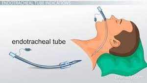
B. Use an end-tidal CO₂ monitor to check for placement in the trachea.
It's the correct answer. Continuous waveform capnography or end-tidal CO₂ detection is the most reliable and immediate method to confirm that the endotracheal tube is in the trachea and not in the esophagus. Presence of CO₂ indicates effective airway placement.
C. Observe the chest for symmetrical movement with ventilation.
Chest rise is helpful, but it is not specificboth esophageal and tracheal intubation may show chest movement.
D. Obtain a portable chest radiograph to check tube placement.
It's the gold standard for final confirmation, but it is not the initial bedside method because it takes time.
Correct Answer is B
Explanation
Mean arterial pressure (MAP) is a measure of the average pressure within the arteries during one cardiac cycle. It represents the perfusion pressure that drives blood flow to organs and tissues. MAP is calculated using the formula:
MAP = Diastolic blood pressure + 1/3 (Systolic blood pressure - Diastolic blood pressure)
Blood loss, particularly in cases of significant hemorrhage, leads to a decrease in blood volume. When blood volume decreases, there is less circulating blood available to generate pressure within the arterial system. This reduction in blood volume results in decreased MAP.
Therefore, in the case of massive blood loss after trauma, the student can correlate it with a lower blood volume, which in turn leads to a lower MAP. The decrease in blood volume reduces the perfusion pressure, compromising organ and tissue perfusion
A. It causes vasoconstriction and increased MAP in (option A) is incorrect because: While vasoconstriction can occur as a compensatory mechanism to maintain blood pressure, it does not necessarily lead to an increased MAP in the context of significant blood loss.
C. It raises cardiac output and MAP in (option C) is incorrect because Blood loss typically leads to a reduction in cardiac output due to decreased blood volume. Therefore, it does not raise cardiac output and MAP.
D. There is no direct correlation to MAP in (option D) is incorrect because: There is indeed a direct correlation between blood loss and MAP. As blood volume decreases, MAP decreases as well.
Therefore, the correct correlation between blood loss and MAP is that lower blood volume lowers MAP.
Whether you are a student looking to ace your exams or a practicing nurse seeking to enhance your expertise , our nursing education contents will empower you with the confidence and competence to make a difference in the lives of patients and become a respected leader in the healthcare field.
Visit Naxlex, invest in your future and unlock endless possibilities with our unparalleled nursing education contents today