
A 48-year-old female patient with septic shock presents with a BP of 70/46, pulse 136, respirations 32, temperature 104.0° F, and blood glucose of 246 mg/dl. The following have been ordered. Which order will the nurse accomplish first?
Draw an arterial blood gas (ABG).
Start insulin drip to maintain blood glucose at 150 mg/dl or lower.
Give a normal saline bolus IV of 30mL/kg.
Titrate norepinephrine (Levophed) to keep MAP greater than 65 mm Hg.
The Correct Answer is C
Septic shock is characterized by inadequate tissue perfusion and hypotension, which can lead to organ dysfunction and failure. The administration of intravenous fluids, such as a normal saline bolus, is the initial priority in the management of septic shock to restore intravascular volume and improve perfusion.
A. Draw an arterial blood gas (ABG) in (option A) is incorrect because: ABG may be ordered to assess the patient's acid-base status and oxygenation, but addressing hypotension and restoring perfusion through fluid administration takes priority.
B. Start insulin drip to maintain blood glucose at 150 mg/dl or lower in (option B) is incorrect because: Hyperglycaemia is commonly observed in critically ill patients, including those with septic shock. While controlling blood glucose is important, it is not the immediate priority compared to addressing hypotension and restoring intravascular volume.
D. Titrate norepinephrine (Levophed) to keep mean arterial pressure (MAP) greater than 65 mm Hg in (option D) is incorrect because: Norepinephrine is a vasopressor medication used to increase blood pressure and perfusion in septic shock. While it may be necessary for the management of septic shock, fluid resuscitation should be initiated first to optimize intravascular volume before starting vasopressors.
Therefore, the first order that the nurse should accomplish in this scenario is to give a normal saline bolus IV of 30 mL/kg to address the hypotension and restore intravascular volume.
Nursing Test Bank
Naxlex Comprehensive Predictor Exams
Related Questions
Correct Answer is C
Explanation
This method, known as the 6-second method, involves counting the number of QRS complexes in a 6-second interval on the electrocardiogram (ECG) strip and then multiplying that number by 10 to calculate the heart rate per minute. The advantage of this method is that it provides a relatively quick estimate of the heart rate.
A. Printing a 1-minute ECG strip and counting the number of QRS complexes in (option A) is incorrect because it can be time-consuming and may not be practical in situations where a quick estimate is needed.
B. Calculating the number of small squares between one QRS complex and the next and dividing into 1500 in (option B) is incorrect because it is a method used to calculate heart rate, known as the "1500 method," but it is not as quick as the 6-second method and requires more time and measurement precision.
D. Counting the number of large squares in the R-R interval and dividing by 300 is another method used to calculate heart rate, known as the "300 method," but it is also less quick and less accurate for assessing heart rate in patients with regular rhythms.
It's important to note that if the heart rhythm is irregular, these methods may not provide an accurate estimate of the heart rate, and a longer monitoring period or a different approach may be necessary.
Correct Answer is A
Explanation
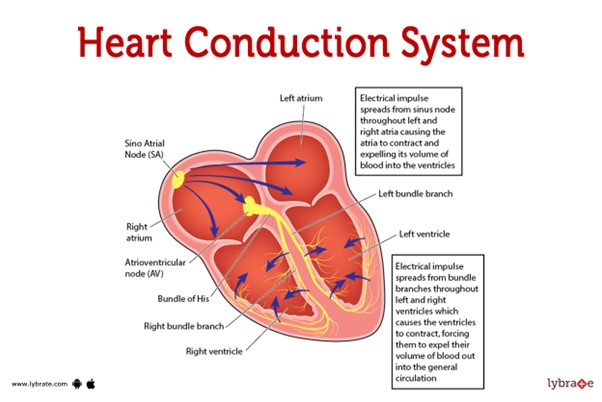 This pathway represents the normal sequence of electrical impulses that coordinate the contraction and relaxation of the heart chambers.
The electrical signal originates from the sinoatrial (SA) node, which is often referred to as the natural pacemaker of the heart. It is located in the right atrium and generates the electrical impulses that initiate each heartbeat. From the SA node, the electrical signal travels to the atrioventricular (AV) node, which is located at the junction between the atria and ventricles.
After passing through the AV node, the electrical impulse travels through the bundle of His (also known as the atrioventricular bundle) and divides into the right and left bundle branches. These branches continue the conduction pathway and deliver the electrical signal to the Purkinje fibers.
The Purkinje fibers spread the electrical impulse rapidly throughout the ventricles, stimulating the contraction of the ventricular muscle and allowing for efficient pumping of blood out of the heart.
Therefore, the correct sequence of the normal conduction pathway in the heart is:
A. SA node - AV node - bundle of His - bundle branches - Purkinje fibers.
Whether you are a student looking to ace your exams or a practicing nurse seeking to enhance your expertise , our nursing education contents will empower you with the confidence and competence to make a difference in the lives of patients and become a respected leader in the healthcare field.
Visit Naxlex, invest in your future and unlock endless possibilities with our unparalleled nursing education contents today