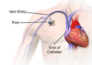
Following surgery, a patient's central venous pressure (CVP) monitor indicates high pressures. Which action will the nurse anticipate taking?
Increase the IV fluid infusion rate.
Administer IV diuretic medications.
Elevate the head of the patient's bed to 45 degrees.
Document the CVP and continue to monitor.
The Correct Answer is B
Central venous pressure (CVP) is a measurement of the pressure in the central veins, which reflects the blood volume and right-sided cardiac function. High CVP readings may indicate fluid overload or impaired cardiac function, and intervention is necessary to address the underlying cause.
Administering IV diuretic medications can help reduce fluid volume by increasing urine output and promoting fluid elimination. By removing excess fluid, the diuretic medications can help lower the CVP and alleviate the high pressures.
The other options mentioned are not the anticipated actions for addressing high CVP:
A. Increasing the IV fluid infusion rate in (option A) is incorrect because: If the CVP is already indicating high pressures, increasing the IV fluid infusion rate would further contribute to fluid overload and exacerbate the problem. This action would not be appropriate for high CVP readings.
C. Elevating the head of the patient's bed to 45 degrees in (option C) is incorrect because Positioning the patient with the head of the bed elevated is commonly done to prevent complications such as aspiration or improve respiratory function. While it may have other benefits, it does not directly address the high CVP.
D. Documenting the CVP and continuing to monitor in (option D) is incorrect because Documenting the CVP and continuing to monitor is important for ongoing assessment and evaluation. However, in the presence of high CVP readings, intervention is necessary to address the underlying issue rather than solely documenting and monitoring.
Therefore, when a patient's CVP monitor indicates high pressures following surgery, the nurse would anticipate administering IV diuretic medications to help reduce fluid volume and lower the CVP.

Nursing Test Bank
Naxlex Comprehensive Predictor Exams
Related Questions
Correct Answer is C
Explanation
Septic shock is characterized by inadequate tissue perfusion and hypotension, which can lead to organ dysfunction and failure. The administration of intravenous fluids, such as a normal saline bolus, is the initial priority in the management of septic shock to restore intravascular volume and improve perfusion.
A. Draw an arterial blood gas (ABG) in (option A) is incorrect because: ABG may be ordered to assess the patient's acid-base status and oxygenation, but addressing hypotension and restoring perfusion through fluid administration takes priority.
B. Start insulin drip to maintain blood glucose at 150 mg/dl or lower in (option B) is incorrect because: Hyperglycaemia is commonly observed in critically ill patients, including those with septic shock. While controlling blood glucose is important, it is not the immediate priority compared to addressing hypotension and restoring intravascular volume.
D. Titrate norepinephrine (Levophed) to keep mean arterial pressure (MAP) greater than 65 mm Hg in (option D) is incorrect because: Norepinephrine is a vasopressor medication used to increase blood pressure and perfusion in septic shock. While it may be necessary for the management of septic shock, fluid resuscitation should be initiated first to optimize intravascular volume before starting vasopressors.
Therefore, the first order that the nurse should accomplish in this scenario is to give a normal saline bolus IV of 30 mL/kg to address the hypotension and restore intravascular volume.
Correct Answer is C
Explanation
Septic shock is a life-threatening condition characterized by severe infection, systemic inflammation, and inadequate tissue perfusion. In this critical situation, one of the initial priorities is to restore intravascular volume and improve tissue perfusion. Initiation of an intravenous line allows for the administration of fluids and other necessary medications to support the patient's hemodynamic stability.
While the other interventions mentioned are also important components of septic shock management, the immediate priority is to address hypotension and tissue hypoperfusion through fluid resuscitation:
A. Obtaining wound and blood cultures in (option A) is incorrect because: Cultures are important to identify the source and causative organisms of the infection. However, fluid resuscitation should take priority over obtaining cultures, as it is necessary to stabilize the patient's hemodynamics.
B. Removing or controlling potentially infected sources in (option B) is incorrect because: Identifying and controlling the source of infection is crucial in septic shock management to prevent further progression. However, initiating fluid resuscitation is more time-sensitive and should be prioritized.
D. Drawing blood for hematology and chemistry studies in (option D) is incorrect because Laboratory studies are important for evaluating organ function and guiding treatment. However, the immediate focus should be on fluid resuscitation to address the underlying hypoperfusion and stabilize the patient's condition.
Therefore, the intervention considered a priority when treating a patient who presents with septic shock is the initiation of an intravenous line and fluid administration to restore intravascular volume and improve tissue perfusion.
Whether you are a student looking to ace your exams or a practicing nurse seeking to enhance your expertise , our nursing education contents will empower you with the confidence and competence to make a difference in the lives of patients and become a respected leader in the healthcare field.
Visit Naxlex, invest in your future and unlock endless possibilities with our unparalleled nursing education contents today
Report Wrong Answer on the Current Question
Do you disagree with the answer? If yes, what is your expected answer? Explain.
Kindly be descriptive with the issue you are facing.
