The ICU charge nurse will evaluate that teaching about hemodynamic monitoring for a new staff nurse has been effective when the new nurse does which of the following?
Position the limb with the catheter insertion site at the level of the transducer.
Positions the transducer level with the phlebostatic axis.
Ensures that the patient is lying with the head of the bed flat for all readings.
Balances and calibrates the hemodynamic monitoring equipment every hour.
The Correct Answer is B
Positioning the transducer level with the phlebostatic axis is a crucial step in accurate hemodynamic monitoring. The phlebostatic axis is an imaginary reference point located at the fourth intercostal space, mid-anterior/posterior chest. Placing the transducer at this level ensures that the pressure measurements obtained are reflective of the patient's true hemodynamic status.
A. Positioning the limb with the catheter insertion site at the level of the transducer in (option A) is incorrect because: While it is important to position the limb appropriately to avoid kinks or occlusions in the catheter tubing, this is not directly related to the accurate measurement of hemodynamic parameters.
C. Ensuring that the patient is lying with the head of the bed flat for all readings in (option C) is incorrect because The position of the patient's head does not directly impact the accuracy of hemodynamic monitoring unless it specifically relates to changes in preload or intracranial pressure monitoring.
D. Balancing and calibrating the hemodynamic monitoring equipment every hour in (option D) is incorrect because: While it is important to ensure that the monitoring equipment is calibrated and functioning properly, doing so every hour may not be necessary. Calibration frequency may vary based on institutional policies and patient stability.
Therefore, the correct action that demonstrates effective teaching about hemodynamic monitoring is positioning the transducer level with the phlebostatic axis.
Nursing Test Bank
Naxlex Comprehensive Predictor Exams
Related Questions
Correct Answer is A
Explanation
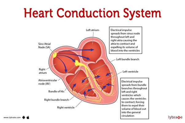 This pathway represents the normal sequence of electrical impulses that coordinate the contraction and relaxation of the heart chambers.
The electrical signal originates from the sinoatrial (SA) node, which is often referred to as the natural pacemaker of the heart. It is located in the right atrium and generates the electrical impulses that initiate each heartbeat. From the SA node, the electrical signal travels to the atrioventricular (AV) node, which is located at the junction between the atria and ventricles.
After passing through the AV node, the electrical impulse travels through the bundle of His (also known as the atrioventricular bundle) and divides into the right and left bundle branches. These branches continue the conduction pathway and deliver the electrical signal to the Purkinje fibers.
The Purkinje fibers spread the electrical impulse rapidly throughout the ventricles, stimulating the contraction of the ventricular muscle and allowing for efficient pumping of blood out of the heart.
Therefore, the correct sequence of the normal conduction pathway in the heart is:
A. SA node - AV node - bundle of His - bundle branches - Purkinje fibers.
Correct Answer is C
Explanation
In the initial 24 hours after burn injury, fluid resuscitation is a critical priority in the management of burn patients. Burn injuries can lead to significant fluid loss, both locally at the burn site and systemically due to increased capillary permeability. Fluid resuscitation aims to restore and maintain adequate intravascular volume, ensuring sufficient tissue perfusion and organ function.
The Parkland Formula is commonly used to guide fluid resuscitation in burn patients. It involves calculating the total volume of fluid needed in the first 24 hours, with a portion given in the initial hours after injury and the remainder given over the remaining hours.
A. Sterile dressing changes (option A) are incorrect because they are important in wound care management for burn patients to prevent infection. However, fluid resuscitation takes precedence within the first 24 hours.
B. Emotional support (option B) is incorrect because it is an essential aspect of burn care, as burn injuries can have a significant psychological impact. While emotional support is crucial for the patient's overall well-being, it may not be the highest priority within the first 24 hours compared to addressing the physiological needs of fluid resuscitation.
D. Range-of-motion exercises (option D) are incorrect because they are important for preventing contractures and maintaining joint mobility in burn patients. However, they are typically initiated after the initial fluid resuscitation phase and wound stabilization.
Therefore, the priority the nurse anticipates within the first 24 hours for a 31-year-old male patient with burn injuries is fluid resuscitation.
Whether you are a student looking to ace your exams or a practicing nurse seeking to enhance your expertise , our nursing education contents will empower you with the confidence and competence to make a difference in the lives of patients and become a respected leader in the healthcare field.
Visit Naxlex, invest in your future and unlock endless possibilities with our unparalleled nursing education contents today