Which of the following clinical manifestations of Disseminated intravascular coagulation (DIC)?
Decreased hematocrit, increased platelet counts, increased D-dimer
Decreased platelet counts, increased D-dimer, increased prothrombin time
Decreased Antithrombin III, increased platelet counts, increased fibrinogen
Decreased D-dimer, increased platelet counts, Increased hemoglobin
The Correct Answer is B
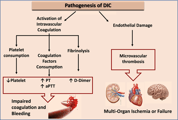
Disseminated Intravascular Coagulation (DIC) is a condition characterized by widespread activation of the coagulation system, leading to both excessive clot formation and consumption of clotting factors and platelets. This process can result in both bleeding and thrombosis.
The manifestations mentioned in option B are commonly seen in DIC:
Decreased platelet counts: DIC leads to platelet consumption and destruction, resulting in low platelet counts (thrombocytopenia).
Increased D-dimer: D-dimer is a fibrin degradation product, and its levels are increased DIC due to the breakdown of fibrin clots.
Increased prothrombin time (PT): DIC can lead to the depletion of clotting factors, resulting in prolonged prothrombin time, indicating impaired coagulation.
The other options mentioned do not represent the typical clinical manifestations of DIC:
A. Decreased hematocrit, increased platelet counts, and increased D-dimer in (option A) are incorrect because While platelet counts and D-dimer are increased in DIC, decreased hematocrit is not a characteristic finding.
C. Decreased Antithrombin III, increased platelet counts, and increased fibrinogen in (option C) is incorrect because: Decreased Antithrombin III can be seen in DIC, but increased platelet counts and fibrinogen levels are not specific to DIC.
D. Decreased D-dimer, increased platelet counts, and increased hemoglobin in (option D) is incorrect because Decreased D-dimer and increased hemoglobin are not typical findings in DIC, while increased platelet counts can be seen in some cases.
Nursing Test Bank
Naxlex Comprehensive Predictor Exams
Related Questions
Correct Answer is C
Explanation
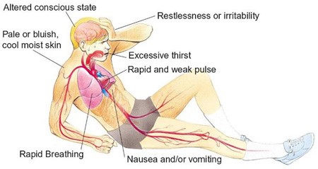
Hypovolemic shock is a life-threatening condition resulting from severe blood or fluid loss. The patient in this scenario exhibits signs of hypovolemic shock, such as low blood pressure, tachycardia, cool and clammy skin, and decreased urine output.
When assessing the prescription options, the nurse should consider the appropriateness of each intervention for hypovolemic shock. Plasmanate is a type of plasma protein fraction that is used for volume expansion in certain situations. However, in hypovolemic shock, the primary intervention is to restore intravascular volume promptly. Plasmanate alone may not be sufficient for rapid-volume resuscitation.
In hypovolemic shock, the initial management typically involves the administration of crystalloid solutions, such as Lactated Ringers or Normal Saline, to restore intravascular volume. Therefore, the prescription of Plasmanate as the primary intervention raises concerns and should be questioned by the nurse.
A. Dopamine (Intropin) 12 mcg/min in (option A) is incorrect because: Dopamine is a vasopressor medication used to increase blood pressure and cardiac output. It is a suitable option for hypovolemic shock to support blood pressure and tissue perfusion.
B. Dobutamine (Dobutrex) 5 mcg/kg/min in (option B) is incorrect because: Dobutamine is an inotropic medication that helps improve cardiac contractility and cardiac output. It can be beneficial in cases of hypovolemic shock with signs of poor cardiac function.
D. Bumetanide (Bumex) 1 mg IV in (option D) is incorrect because: Bumetanide is a loop diuretic used to promote diuresis. However, in the context of hypovolemic shock, diuretics are generally not the first-line treatment as they can further reduce intravascular volume and worsen the patient's condition.
It is essential for the nurse to consult with the healthcare provider regarding the prescription order of Plasmanate and consider alternative interventions for rapid volume resuscitation in hypovolemic shock.
Correct Answer is B
Explanation
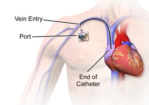
Central venous pressure (CVP) is a measurement of the pressure in the central veins, which reflects the blood volume and right-sided cardiac function. High CVP readings may indicate fluid overload or impaired cardiac function, and intervention is necessary to address the underlying cause.
Administering IV diuretic medications can help reduce fluid volume by increasing urine output and promoting fluid elimination. By removing excess fluid, the diuretic medications can help lower the CVP and alleviate the high pressures.
The other options mentioned are not the anticipated actions for addressing high CVP:
A. Increasing the IV fluid infusion rate in (option A) is incorrect because: If the CVP is already indicating high pressures, increasing the IV fluid infusion rate would further contribute to fluid overload and exacerbate the problem. This action would not be appropriate for high CVP readings.
C. Elevating the head of the patient's bed to 45 degrees in (option C) is incorrect because Positioning the patient with the head of the bed elevated is commonly done to prevent complications such as aspiration or improve respiratory function. While it may have other benefits, it does not directly address the high CVP.
D. Documenting the CVP and continuing to monitor in (option D) is incorrect because Documenting the CVP and continuing to monitor is important for ongoing assessment and evaluation. However, in the presence of high CVP readings, intervention is necessary to address the underlying issue rather than solely documenting and monitoring.
Therefore, when a patient's CVP monitor indicates high pressures following surgery, the nurse would anticipate administering IV diuretic medications to help reduce fluid volume and lower the CVP.
Whether you are a student looking to ace your exams or a practicing nurse seeking to enhance your expertise , our nursing education contents will empower you with the confidence and competence to make a difference in the lives of patients and become a respected leader in the healthcare field.
Visit Naxlex, invest in your future and unlock endless possibilities with our unparalleled nursing education contents today