Which of the following clinical manifestations is NOT considered part of Beck's triad (classic indications of cardiac tamponade)?
muffled heart tones
marked hypotension
distended jugular veins
widening pulse pressure
The Correct Answer is D
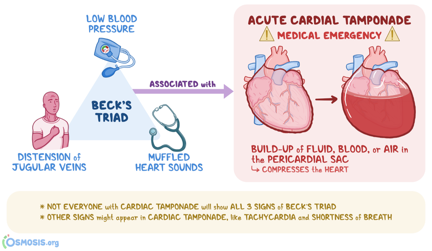
Beck's triad consists of three classic clinical manifestations that are suggestive of cardiac tamponade, which is the compression of the heart by accumulated fluid or blood within the pericardial sac. The three components of Beck's triad include:
A. Muffled heart tones in (option A) are incorrect because Cardiac tamponade can dampen or muffle heart sounds due to the presence of fluid or blood around the heart, which can impair sound transmission.
B. Marked hypotension in (option B) is incorrect because Cardiac tamponade can cause decreased cardiac output, leading to hypotension, which is characterized by low blood pressure.
C. Distended jugular veins in (option C) is incorrect because Elevated venous pressure resulting from impaired filling and elevated right-sided heart pressures can lead to jugular vein distension, which is commonly seen in cardiac tamponade.
However, widening pulse pressure (the difference between systolic and diastolic blood pressure) is not typically part of Beck's triad. Widening pulse pressure is associated with her conditions such as aortic regurgitation, hyperthyroidism, or conditions involving increased stroke volume, rather than cardiac tamponade specifically.
Nursing Test Bank
Naxlex Comprehensive Predictor Exams
Related Questions
Correct Answer is ["21"]
Explanation
flow rate for an infusion= (Volume in mL * Drop factor) / Time in minutes.
volume of the infusion bag is 250 mL, the drop factor is 10 gtts/mL, and the time is 2 hours, which is 120 minutes.
(250 mL * 10 gtts/mL) / 120 minutes = 2500 gtts / 120 minutes ≈ 20.83 gtts/minute. Therefore, the nurse should run the infusion at a rate of approximately 21 drops per minute to deliver 1 unit of packed red blood cells over the 2-hour period.
Correct Answer is C
Explanation
This method, known as the 6-second method, involves counting the number of QRS complexes in a 6-second interval on the electrocardiogram (ECG) strip and then multiplying that number by 10 to calculate the heart rate per minute. The advantage of this method is that it provides a relatively quick estimate of the heart rate.
A. Printing a 1-minute ECG strip and counting the number of QRS complexes in (option A) is incorrect because it can be time-consuming and may not be practical in situations where a quick estimate is needed.
B. Calculating the number of small squares between one QRS complex and the next and dividing into 1500 in (option B) is incorrect because it is a method used to calculate heart rate, known as the "1500 method," but it is not as quick as the 6-second method and requires more time and measurement precision.
D. Counting the number of large squares in the R-R interval and dividing by 300 is another method used to calculate heart rate, known as the "300 method," but it is also less quick and less accurate for assessing heart rate in patients with regular rhythms.
It's important to note that if the heart rhythm is irregular, these methods may not provide an accurate estimate of the heart rate, and a longer monitoring period or a different approach may be necessary.
Whether you are a student looking to ace your exams or a practicing nurse seeking to enhance your expertise , our nursing education contents will empower you with the confidence and competence to make a difference in the lives of patients and become a respected leader in the healthcare field.
Visit Naxlex, invest in your future and unlock endless possibilities with our unparalleled nursing education contents today