In an ECG pattern, the P wave is caused by:
Repolarization of atrial muscle fibers.
Depolarization of atrial muscle fibers.
Depolarization of ventricular muscle fibers.
Repolarization of ventricular muscle fibers.
The Correct Answer is B
This means that the electrical activity that causes the atria to contract starts from the sinoatrial node and spreads across the atria.
The P wave on the ECG reflects this atrial depolarization.
Choice A is wrong because repolarization of atrial muscle fibers is not visible on the ECG, as it occurs during the QRS complex when the ventricular depolarization masks it.
Choice C is wrong because the depolarization of ventricular muscle fibers is represented by the QRS complex on the ECG, not the P wave.
Choice D is wrong because the repolarization of ventricular muscle fibers is represented by the T wave on the ECG, not the P wave.
Normal ranges for the P wave are:
Duration: less than 0.12 seconds (less than 3 small squares)
Amplitude: less than 2.5 mm (0.25 mV) in the limb leads, less than 1.5 mm (0.15 mV) in the precordial leads
Axis: between 0° and +75°12
Nursing Test Bank
Naxlex Comprehensive Predictor Exams
Related Questions
Correct Answer is C
Explanation
Positive feedback mechanisms move conditions away from the normal state.
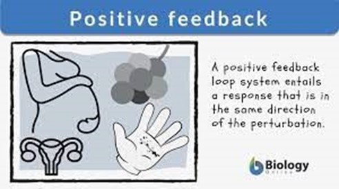
They amplify the original action and produce more of the same effect.
For example, blood clotting and childbirth are positive feedback mechanisms.
Choice A is wrong because positive feedback mechanisms usually produce unstable conditions.
They do not resist change but rather enhance it.
Choice B is wrong because positive feedback mechanisms do not cause long-term changes.
They are ultimately stopped by negative feedback loops once the process they were used for is complete.
Choice D is wrong because positive feedback mechanisms do not bring conditions back to the normal state.
That is the role of negative feedback mechanisms, which oppose the stimulus and restore homeostasis.
Correct Answer is A
Explanation
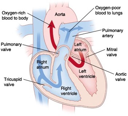
This is the correct sequence of parts through which blood moves from the vena cava to the lungs.
Choice B is wrong because it reverses the order of the right atrium and right ventricle. Blood flows from the right atrium to the right ventricle, not the other way around.
Choice C is wrong because it switches the positions of the tricuspid valve and the pulmonary valve.
Blood flows from the right atrium through the tricuspid valve to the right ventricle, and then through the pulmonary valve to the pulmonary artery.
Choice D is wrong because it also switches the positions of the tricuspid valve and the pulmonary valve, and reverses the order of the right atrium and right ventricle.
Blood flows from the right atrium through the tricuspid valve to the right ventricle, and then through the pulmonary valve to the pulmonary artery.
The normal range of blood pressure in the vena cava is about 0 to 5 mmHg, while in the pulmonary artery, it is about 15 to 25 mmHg.
The normal range of oxygen saturation in the vena cava is about 60% to 80%, while in the pulmonary vein, it is about 95% to 100%.
Whether you are a student looking to ace your exams or a practicing nurse seeking to enhance your expertise , our nursing education contents will empower you with the confidence and competence to make a difference in the lives of patients and become a respected leader in the healthcare field.
Visit Naxlex, invest in your future and unlock endless possibilities with our unparalleled nursing education contents today