Capillary walls consist of a single layer of epithelial cells, and they exchange substances in the blood for substances in the tissue fluid surrounding body cells.
True
False
The Correct Answer is A
Capillary walls consist of a single layer of epithelial cells, and they exchange substances in the blood for substances in the tissue fluid surrounding body cells.
This single layer of cells is called the endothelium and it forms the barrier between the blood and the interstitial fluid.
The endothelium can be either continuous or fenestrated, depending on the tissue type and function.
The capillaries are very thin and allow red blood cells to flow through them single file.
The capillaries also have a layer of a glycoprotein called the glycocalyx that covers their luminal surface.
Choice B. False is wrong because it contradicts the definition and structure of capillaries.
Capillaries are not made of multiple layers of cells, nor do they prevent the exchange of substances between the blood and the tissue fluid.
Nursing Test Bank
Naxlex Comprehensive Predictor Exams
Related Questions
Correct Answer is A
Explanation
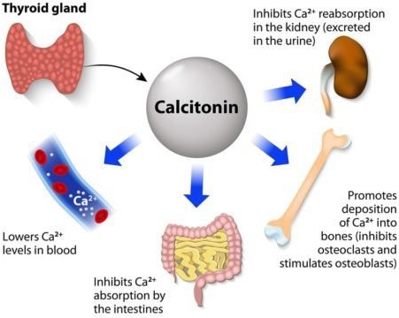
Calcitonin is a hormone that protects against excessive blood calcium levels by inhibiting bone turnover and decreasing reabsorption.
It is produced by the thyroid gland and acts on both osteoclasts and osteoblasts.
Choice B is wrong because parathyroid hormone (PTH) stimulates both resorption and formation of bone, and controls the level of calcium in the blood.
Choice C is wrong because thyroxine is a thyroid hormone that is required for skeletal maturation and influences adult bone maintenance but does not directly affect calcium deposition into bone.
Choice D is wrong because insulin is a hormone that regulates both bone formation and bone resorption but does not specifically stimulate calcium deposition into bone.
Correct Answer is B
Explanation
Innate defenses counter specific disease-causing agents, whereas adaptive defenses include mechanical and chemical barriers.
Choice A is wrong because it confuses the two types of defenses.
Innate defenses are nonspecific and include physical barriers such as the skin, molecules that are toxic to invaders, and phagocytic cells that ingest invaders.
Adaptive defenses are specific and are activated by the innate immune system.
They involve the production of antibodies and specialized cells that recognize and eliminate specific pathogens.
Normal ranges are not applicable in this question as it is about the definitions of innate and adaptive defenses.
Whether you are a student looking to ace your exams or a practicing nurse seeking to enhance your expertise , our nursing education contents will empower you with the confidence and competence to make a difference in the lives of patients and become a respected leader in the healthcare field.
Visit Naxlex, invest in your future and unlock endless possibilities with our unparalleled nursing education contents today