
Where is the location of the popliteal artery within the body?
Around the thymus.
Around the liver, spleen, and upper gastrointestinal tract.
Behind the knee.
The colon and rectum.
The Correct Answer is C
The popliteal artery is a continuation of the femoral artery that runs through the back of the leg and the knee.
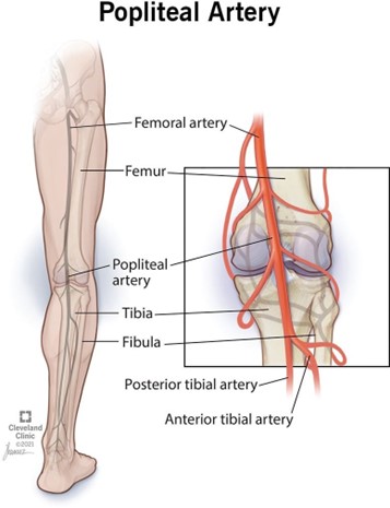
It is located in the popliteal fossa, a hollow space behind the knee. The popliteal pulse can be felt behind the knee.
Choice A is wrong because the thymus is a gland in the chest, not in the leg.
Choice B is wrong because the liver, spleen, and upper gastrointestinal tract are organs in the abdomen, not in the leg.
Choice D is wrong because the colon and rectum are parts of the large intestine, not the leg.
Nursing Test Bank
Naxlex Comprehensive Predictor Exams
Related Questions
Correct Answer is A
Explanation
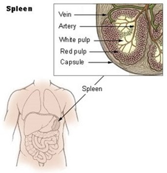
It is a major component of the lymphatic system and contains T and B lymphocytes.
Choice B is wrong because the spleen does not produce T lymphocytes, but rather stores them.
T lymphocytes are produced in the thymus.
Choice C is wrong because the spleen does not filter lymph, but rather blood. It traps bloodborne microbes and produces an immune response to them.
Choice D is wrong because the spleen consists of one lobe and is located in the upper left abdomen below the diaphragm.
The description in choice D matches the thymus, not the spleen.
The normal size of the spleen in adults is about 12 cm long, 8 cm broad, and 3-4 cm thick, weighing about 200 g.
The normal range of splenic index (the product of length, width, and thickness) is 120-480 cm.
Correct Answer is B
Explanation
This means that the electrical activity that causes the atria to contract starts from the sinoatrial node and spreads across the atria.
The P wave on the ECG reflects this atrial depolarization.
Choice A is wrong because repolarization of atrial muscle fibers is not visible on the ECG, as it occurs during the QRS complex when the ventricular depolarization masks it.
Choice C is wrong because the depolarization of ventricular muscle fibers is represented by the QRS complex on the ECG, not the P wave.
Choice D is wrong because the repolarization of ventricular muscle fibers is represented by the T wave on the ECG, not the P wave.
Normal ranges for the P wave are:
Duration: less than 0.12 seconds (less than 3 small squares)
Amplitude: less than 2.5 mm (0.25 mV) in the limb leads, less than 1.5 mm (0.15 mV) in the precordial leads
Axis: between 0° and +75°12
Whether you are a student looking to ace your exams or a practicing nurse seeking to enhance your expertise , our nursing education contents will empower you with the confidence and competence to make a difference in the lives of patients and become a respected leader in the healthcare field.
Visit Naxlex, invest in your future and unlock endless possibilities with our unparalleled nursing education contents today