The hormone that stimulates calcium deposition into bone is:
Calcitonin.
Parathyroid hormone.
Thyroxine.
Insulin.
The Correct Answer is A
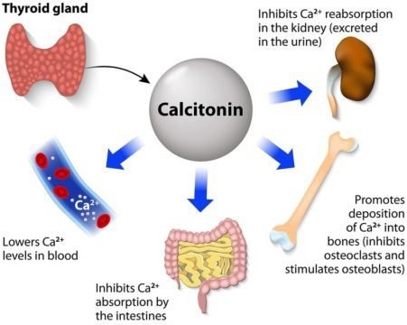
Calcitonin is a hormone that protects against excessive blood calcium levels by inhibiting bone turnover and decreasing reabsorption.
It is produced by the thyroid gland and acts on both osteoclasts and osteoblasts.
Choice B is wrong because parathyroid hormone (PTH) stimulates both resorption and formation of bone, and controls the level of calcium in the blood.
Choice C is wrong because thyroxine is a thyroid hormone that is required for skeletal maturation and influences adult bone maintenance but does not directly affect calcium deposition into bone.
Choice D is wrong because insulin is a hormone that regulates both bone formation and bone resorption but does not specifically stimulate calcium deposition into bone.
Nursing Test Bank
Naxlex Comprehensive Predictor Exams
Related Questions
Correct Answer is A
Explanation
Calcitonin is a hormone that protects against excessive blood calcium levels by inhibiting bone turnover and decreasing reabsorption.
It is produced by the thyroid gland and acts on both osteoclasts and osteoblasts.
Choice B is wrong because parathyroid hormone (PTH) stimulates both resorption and formation of bone, and controls the level of calcium in the blood.
Choice C is wrong because thyroxine is a thyroid hormone that is required for skeletal maturation and influences adult bone maintenance but does not directly affect calcium deposition into bone.
Choice D is wrong because insulin is a hormone that regulates both bone formation and bone resorption but does not specifically stimulate calcium deposition into bone.
Correct Answer is B
Explanation
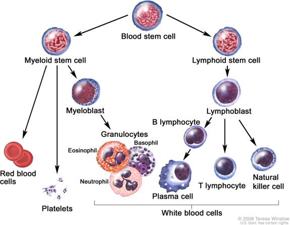
B cells are a type of lymphocyte that originate from the bone marrow and are involved in humoral immunity.
When they encounter a foreign substance (antigen), they differentiate into plasma cells, which secrete antibodies.
Antibodies are proteins that bind to the antigen and neutralize it.
Choice A is wrong because megakaryocytes are large cells that produce platelets, not antibodies.
Choice C is wrong because antibodies are not cells, but products of plasma cells.
Choice D is wrong because T cells are another type of lymphocyte that originate from the thymus and are involved in cell-mediated immunity, not antibody production.
Whether you are a student looking to ace your exams or a practicing nurse seeking to enhance your expertise , our nursing education contents will empower you with the confidence and competence to make a difference in the lives of patients and become a respected leader in the healthcare field.
Visit Naxlex, invest in your future and unlock endless possibilities with our unparalleled nursing education contents today