After an infection, many dead and fragmented bacterial cells must be filtered from the body.
Which of the following cells will clear out the cell debris?
Lymphocytes.
Cytokines.
Mast cells.
Macrophages.
The Correct Answer is D
Macrophages are the main cells that clear out the cell debris by phagocytosis, a process that involves recognition, engulfment, and degradation of the disposable particles.
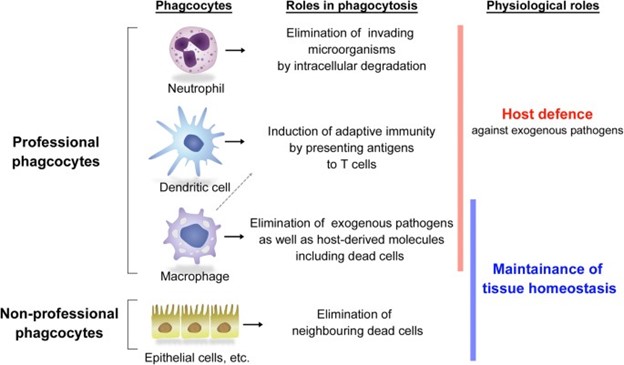
Macrophages are professional phagocytes that can be found in various tissues and organs, where they perform efferocytosis, the removal of dead and dying cells.
Choice A is wrong because lymphocytes are not phagocytes, but rather immune cells that mediate adaptive immunity by producing antibodies or killing infected cells.
Choice B is wrong because cytokines are not cells, but rather soluble molecules that regulate inflammation and immunity by acting as signals between cells.
Choice C is wrong because mast cells are not primarily involved in clearing cell debris, but rather in allergic reactions and innate immunity by releasing histamine and other mediators.
Nursing Test Bank
Naxlex Comprehensive Predictor Exams
Related Questions
Correct Answer is B
Explanation
Kidneys are not part of the lymphatic system.
The lymphatic system is a network of vessels and organs that drain excess fluid from the tissues, transport fats and immune cells, and protect the body from infections.
Kidneys are part of the urinary system, which filters blood, regulates fluid and electrolyte balance, and produces urine.
Choice A is wrong because kidneys do not have a direct connection to the lymphatic system.
Although kidneys have lymphatic vessels in their cortex, they do not originate from the lymphatic system.
Kidneys receive blood from the renal arteries and return it to the renal veins.
The lymphatic vessels in the kidney cortex drain interstitial fluid and immune cells from the kidney tissue to the regional lymph nodes.
Some additional sentences are:
Choice B is right because kidneys are part of the urinary system, not the lymphatic system.
The urinary system and the lymphatic system have different functions and structures in the body.
Normal ranges for kidney function tests include blood urea nitrogen (BUN) of 7 to 20 mg/dL, serum creatinine of 0.6 to 1.2 mg/dL, and glomerular filtration rate (GFR) of more than 90 mL/min/1.73 m.
Normal ranges for lymphatic system tests include white blood cell (WBC) count of 4,000 to 11,000 cells per microliter, lymphocyte count of 1,000 to 4,800 cells per microliter, and immunoglobulin levels of IgG (700 to 1,600 mg/dL), IgA (70 to 400 mg/dL), IgM (40 to 230
mg/dL), IgE (0 to 100 IU/mL), and IgD (0.5 to 5 mg/dL).
Correct Answer is B
Explanation
Innate defenses counter specific disease-causing agents, whereas adaptive defenses include mechanical and chemical barriers.
Choice A is wrong because it confuses the two types of defenses.
Innate defenses are nonspecific and include physical barriers such as the skin, molecules that are toxic to invaders, and phagocytic cells that ingest invaders.
Adaptive defenses are specific and are activated by the innate immune system.
They involve the production of antibodies and specialized cells that recognize and eliminate specific pathogens.
Normal ranges are not applicable in this question as it is about the definitions of innate and adaptive defenses.
Whether you are a student looking to ace your exams or a practicing nurse seeking to enhance your expertise , our nursing education contents will empower you with the confidence and competence to make a difference in the lives of patients and become a respected leader in the healthcare field.
Visit Naxlex, invest in your future and unlock endless possibilities with our unparalleled nursing education contents today