The right atrium receives blood directly from:
The superior vena cava, inferior vena cava, and coronary sinus.
The superior vena cava and inferior vena cava only.
The superior vena cava, inferior vena cava, and pulmonary veins.
The pulmonary veins.
The Correct Answer is A
The right atrium receives blood directly from the superior vena cava, inferior vena cava, and coronary sinus.
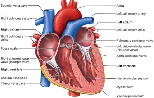
The superior vena cava and inferior vena cava bring deoxygenated blood from the upper and lower body, respectively.
The coronary sinus brings blood from the heart muscle.
Choice B is wrong because it excludes the coronary sinus, which also empties into the right atrium.
Choice C is wrong because it includes the pulmonary veins, which carry oxygenated blood from the lungs to the left atrium, not the right atrium.
Choice D is wrong because it only includes the pulmonary veins, which are not connected to the right atrium at all.
Nursing Test Bank
Naxlex Comprehensive Predictor Exams
Related Questions
Correct Answer is B
Explanation
Monocytes are a type of agranulocytes, which are white blood cells that lack visible granules in their cytoplasm.
Agranulocytes also include lymphocytes, which are involved in adaptive immunity.
Choice A is wrong because basophils are a type of granulocytes, which are white blood cells that have granules in their cytoplasm.
Granulocytes also include neutrophils and eosinophils, which are involved in innate immunity.
Choice C is wrong because neutrophils are also a type of granulocyte.
Neutrophils are the most abundant white blood cells and are responsible for phagocytizing bacteria and fungi.
Choice D is wrong because eosinophils are also a type of granulocytes. Eosinophils are involved in allergic reactions and parasitic infections.
Normal ranges for white blood cells vary depending on age, gender, and health status, but generally, they are between 4,000 and 11,000 cells per microliter of blood.
Correct Answer is C
Explanation
Basophils usually account for the smallest percentage of leukocytes in a blood sample. Basophils are a type of white blood cell that is involved in allergic reactions and inflammation.
Choice A is wrong because eosinophils are not the least common type of leukocyte.
Eosinophils are another type of white blood cell that is involved in allergic responses and parasitic infections.
They typically make up about 1-6% of the total leukocyte count.
Choice B is wrong because monocytes are not the least common type of leukocyte.
Monocytes are a type of white blood cell that can differentiate into macrophages and dendritic cells, which are important for phagocytosis and antigen presentation.
They typically make up about 2-10% of the total leukocyte count.
Choice D is wrong because neutrophils are not the least common type of leukocyte.
Neutrophils are a type of white blood cell that are the first responders to bacterial infections and tissue damage.
They typically make up about 55-70% of the total leukocyte count.
Whether you are a student looking to ace your exams or a practicing nurse seeking to enhance your expertise , our nursing education contents will empower you with the confidence and competence to make a difference in the lives of patients and become a respected leader in the healthcare field.
Visit Naxlex, invest in your future and unlock endless possibilities with our unparalleled nursing education contents today