The antibodies produced in allergic reactions:
Belong to the immunoglobulin E group.
Are called allergens.
Belong to the immunoglobulin G group.
Are secreted by mast cells.
The Correct Answer is A
The antibodies produced in allergic reactions belong to the immunoglobulin E group.
These antibodies bind to an allergen, triggering a release of histamine, which causes allergy symptoms.
They also help to fight parasitic infections.
Choice B is wrong because allergens are not antibodies, but substances that cause an allergic reaction.
Choice C is wrong because the antibodies that belong to the immunoglobulin G group are not involved in allergic reactions, but in fighting bacterial and viral infections.
Choice D is wrong because mast cells are not antibodies, but cells that release histamine and other chemicals when activated by IgE antibodies.
Nursing Test Bank
Naxlex Comprehensive Predictor Exams
Related Questions
Correct Answer is B
Explanation
The correct answer is choice B. True.
As a result of the allergen-antibody reaction, mast cells release histamine.
This is because when mast cells are exposed to an allergen that binds to IgE antibodies attached to their FcɛRI receptors, they release histamine and other inflammatory mediators.
Histamine causes vasodilation, increased capillary permeability, mucus secretion, and smooth muscle contraction, which result in allergic symptoms such as itching, swelling, sneezing, and wheezing.
Choice A is wrong because it is the opposite of the correct answer.
Mast cells do release histamine in response to allergen-antibody reactions. Some additional sentences are:
Normal ranges of histamine levels vary depending on the tissue and the method of measurement, but they are usually low in healthy individuals and elevated in allergic conditions.
Histamine can be degraded by enzymes such as diamine oxidase and histamine N-methyltransferase, or inhibited by drugs such as antihistamines.
Correct Answer is B
Explanation
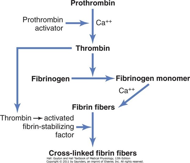
This is because fibrinogen is a soluble protein in the blood plasma that is converted to insoluble fibrin strands by the enzyme thrombin during blood clotting. Fibrin forms a mesh-like network that traps platelets and other blood cells to form a clot.
Choice A is wrong because thrombin is not converted to prothrombin, but rather prothrombin is converted to thrombin by another enzyme called prothrombinase.
Choice C is wrong because vitamin K is not converted to prothrombin, but rather vitamin K is required for the synthesis of prothrombin and other clotting factors in the liver.
Choice D is wrong because fibrin is not converted to fibrinogen, but rather fibrinogen is converted to fibrin as explained above.
Normal ranges of fibrinogen in the blood are 200 to 400 mg/dL.
Normal ranges of prothrombin time (a measure of how long it takes blood to clot) are 11 to
13.5 seconds.
Whether you are a student looking to ace your exams or a practicing nurse seeking to enhance your expertise , our nursing education contents will empower you with the confidence and competence to make a difference in the lives of patients and become a respected leader in the healthcare field.
Visit Naxlex, invest in your future and unlock endless possibilities with our unparalleled nursing education contents today