Cardiac output is calculated by multiplying the stroke volume by the systolic blood pressure.
True
False
The Correct Answer is B
Cardiac output is calculated by multiplying the stroke volume by the heart rate, not by the systolic blood pressure.
Stroke volume is the amount of blood circulated by the heart with each beat. Heart rate is the number of beats per minute.
Systolic blood pressure is the pressure in the arteries when the heart contracts. Choice A is wrong because it confuses systolic blood pressure with heart rate.
Systolic blood pressure is not directly related to cardiac output, although it can be affected by it.
1: Cardiac Output- Definition, Factors Affecting, Cardiac Index - BYJU’S 2: Cardiac Output (Fick’s Formula) - MDCalc 3: Calculating how much blood is pumped by the heart - Cellular respiration and transport - Edexcel - GCSE Biology (Single Science) Revision - Edexcel - BBC Bitesize 4: Cardiac output - Structure and function of the heart - Higher Human Biology Revision - BBC Bitesize : Blood Pressure: What Is Normal? How To Measure Blood Pressure (healthline.com)
Nursing Test Bank
Naxlex Comprehensive Predictor Exams
Related Questions
Correct Answer is A
Explanation
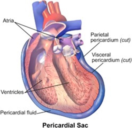
The fibrous pericardium is the loose-fitting sac around the heart that protects it and anchors it to surrounding structures.

Choice B is wrong because the epicardium is the outer layer of the heart wall, also called the visceral pericardium, and it is not a sac.
Choice C is wrong because the endocardium is the inner layer of the heart wall that forms the lining of all heart chambers, and it is not a sac.
Choice D is wrong because the visceral pericardium is another name for the epicardium, and it is not a loose-fitting sac.
Correct Answer is B
Explanation

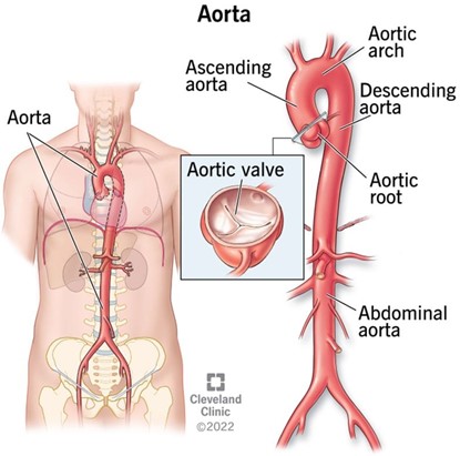
The aorta is the largest artery in the human body, as well as the main artery in the circulatory system.
It originates from the left ventricle of the heart and extends down to the abdomen, where it splits into two smaller arteries (the common iliac arteries).
The aorta distributes oxygenated blood to all parts of the body through the systemic circulation.
Choice A. Carotid is wrong because the carotid artery is not the largest artery in the body, but one of the main arteries that pumps blood from the heart to the brain and the rest of the head.
It has a diameter of 4.3 mm-7.7 mm and a blood flow of 350-550 milliliters per minute.
Choice C. Celiac is wrong because the celiac artery is not the largest artery in the body, but a major branch of the abdominal aorta that supplies oxygenated blood to the liver, stomach, spleen, pancreas, and duodenum.
Choice D. Femoral is wrong because the femoral artery is not the largest artery in the body, but the largest artery found in the leg region.
It runs down the inner thigh and carries out the important role of supplying blood to the lower body.
It has a diameter of 6.6 mm and a blood flow of 284 milliliters per minute.
Whether you are a student looking to ace your exams or a practicing nurse seeking to enhance your expertise , our nursing education contents will empower you with the confidence and competence to make a difference in the lives of patients and become a respected leader in the healthcare field.
Visit Naxlex, invest in your future and unlock endless possibilities with our unparalleled nursing education contents today
Report Wrong Answer on the Current Question
Do you disagree with the answer? If yes, what is your expected answer? Explain.
Kindly be descriptive with the issue you are facing.
