Positive feedback mechanisms:
Usually produce stable conditions.
Cause long-term changes.
Move conditions away from the normal state.
Bring conditions back to the normal state.
The Correct Answer is C
Positive feedback mechanisms move conditions away from the normal state.
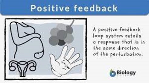
They amplify the original action and produce more of the same effect.
For example, blood clotting and childbirth are positive feedback mechanisms.
Choice A is wrong because positive feedback mechanisms usually produce unstable conditions.
They do not resist change but rather enhance it.
Choice B is wrong because positive feedback mechanisms do not cause long-term changes.
They are ultimately stopped by negative feedback loops once the process they were used for is complete.
Choice D is wrong because positive feedback mechanisms do not bring conditions back to the normal state.
That is the role of negative feedback mechanisms, which oppose the stimulus and restore homeostasis.
Nursing Test Bank
Naxlex Comprehensive Predictor Exams
Related Questions
Correct Answer is A
Explanation
The celiac artery supplies blood to the liver, spleen, and stomach.
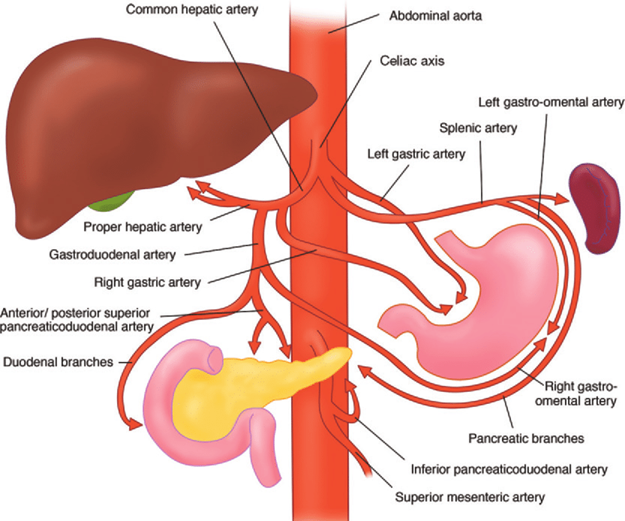
It is one of the three major branches of the abdominal aorta, along with the superior mesenteric artery and the inferior mesenteric artery.
The celiac artery divides into three branches: the left gastric artery, the splenic artery, and the common hepatic artery.
Choice B is wrong because the brachiocephalic artery is a large vessel that arises from the aortic arch and supplies blood to the right side of the head and neck and the right arm.
It has no connection to the liver or spleen.
Choice C is wrong because the renal arteries are paired vessels that arise from the abdominal aorta and supply blood to the kidneys.
They are located below the superior mesenteric artery and above the inferior mesenteric artery.
Choice D is wrong because the tibial arteries are branches of the popliteal artery that supply blood to the lower leg and foot.
They are located in the posterior and anterior compartments of the leg.
The normal range of blood pressure in the celiac artery is about 100-120 mmHg systolic and 60-80 mmHg diastolic.
The normal range of blood flow in the celiac artery is about 200-300 ml/min. The normal diameter of the celiac artery is about 5-7 mm.
Correct Answer is A
Explanation
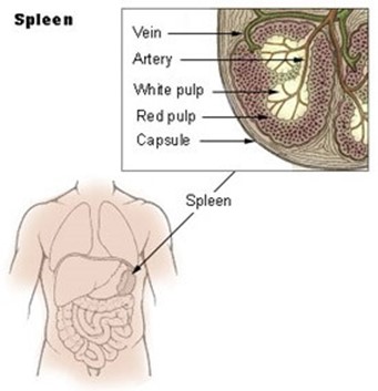
It is a major component of the lymphatic system and contains T and B lymphocytes.
Choice B is wrong because the spleen does not produce T lymphocytes, but rather stores them.
T lymphocytes are produced in the thymus.
Choice C is wrong because the spleen does not filter lymph, but rather blood. It traps bloodborne microbes and produces an immune response to them.
Choice D is wrong because the spleen consists of one lobe and is located in the upper left abdomen below the diaphragm.
The description in choice D matches the thymus, not the spleen.
The normal size of the spleen in adults is about 12 cm long, 8 cm broad, and 3-4 cm thick, weighing about 200 g.
The normal range of splenic index (the product of length, width, and thickness) is 120-480 cm.
Whether you are a student looking to ace your exams or a practicing nurse seeking to enhance your expertise , our nursing education contents will empower you with the confidence and competence to make a difference in the lives of patients and become a respected leader in the healthcare field.
Visit Naxlex, invest in your future and unlock endless possibilities with our unparalleled nursing education contents today