A nurse is planning care for a child who has epiglottitis. Which of the following actions should the nurse plan to take?
Obtain a throat culture.
Visualize the epiglottis using a tongue depressor.
Provide moist air to reduce the inflammation of the epiglottis
Initiate airborne precautions.
The Correct Answer is C
A. Obtain a throat culture.
This option is not appropriate as a primary nursing action in the acute management of epiglottitis. While obtaining a throat culture may be necessary for diagnostic purposes, it is not a priority in the immediate care of a child with suspected epiglottitis. The focus should be on ensuring airway patency and providing emergency treatment.
B. Visualize the epiglottis using a tongue depressor.
This option is contraindicated in the acute management of epiglottitis. Direct visualization of the epiglottis using a tongue depressor or other instruments can provoke spasm of the epiglottis and worsen airway obstruction. Attempting to visualize the epiglottis should be avoided until the child's airway has been secured in a controlled environment, such as in the operating room under anesthesia.
C. Provide moist air to reduce the inflammation of the epiglottis.
This option is appropriate. Providing moist air, such as humidified oxygen or a cool mist, can help soothe the inflamed tissues of the epiglottis and upper airway. Moist air may help alleviate discomfort and reduce inflammation, although it will not directly address the risk of airway obstruction. It is often used as supportive therapy in conjunction with other interventions.
D. Initiate airborne precautions.
This option is not necessary for the care of a child with epiglottitis. Epiglottitis is not typically transmitted through airborne droplets. The priority in the management of epiglottitis is ensuring a patent airway and providing appropriate treatment to reduce inflammation and prevent complications.
Nursing Test Bank
Naxlex Comprehensive Predictor Exams
Related Questions
Correct Answer is C
Explanation
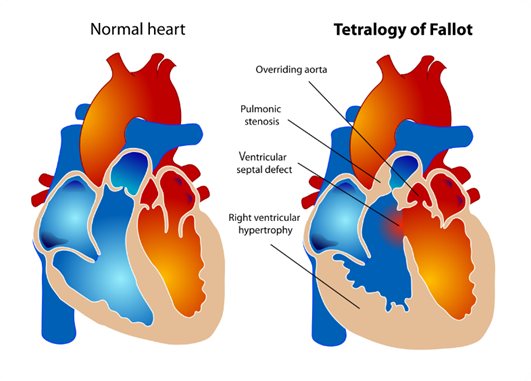
A. Overriding aorta: In Tetralogy of Fallot, the aorta is positioned over the ventricular septal defect (VSD), rather than solely over the left ventricle as it would be in a normal heart. This is called overriding aorta, which allows blood from both the right and left ventricles to enter the aorta.
B. Pulmonary stenosis: This is a critical component of Tetralogy of Fallot. Pulmonary stenosis refers to narrowing of the pulmonary valve or the area just below it, which restricts blood flow from the right ventricle to the pulmonary artery. This results in decreased blood flow to the lungs for oxygenation.
C. Left ventricular hypertrophy: This choice is not typically associated with Tetralogy of Fallot. Left ventricular hypertrophy refers to an enlargement or thickening of the muscular wall of the left ventricle of the heart. It is often seen in conditions where the left ventricle has to work harder to pump blood, such as in hypertension or aortic stenosis, but it is not a characteristic feature of Tetralogy of Fallot.
D. Ventricular septal defect: This defect is one of the four components of Tetralogy of Fallot. A ventricular septal defect (VSD) is a hole in the septum, the muscular wall that separates the left and right ventricles of the heart. In Tetralogy of Fallot, the VSD allows oxygen-poor blood from the right ventricle to flow directly into the left ventricle and out to the body.

Correct Answer is ["B","D"]
Explanation
A. Cranberry juice
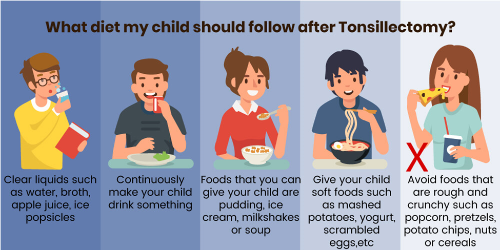
Cranberry juice may be too acidic and could irritate the surgical site. It's best to avoid acidic beverages immediately following a tonsillectomy.
B. Ice-cream
This option is suitable. Ice-cream is cold and soothing, and it can help numb the throat, providing relief from discomfort after a tonsillectomy. However, it's essential to ensure that the ice-cream is not too cold to avoid causing discomfort.
C. Hot tea
Hot tea is not recommended immediately following a tonsillectomy. Hot liquids can irritate the surgical site and may cause discomfort. It's best to avoid hot beverages until the throat has had time to heal.
D. Italian ice
Italian ice is a frozen dessert similar to a slushy, and it can be a suitable option after a tonsillectomy. Like ice-cream, Italian ice is cold and can help numb the throat, providing relief from discomfort.

Whether you are a student looking to ace your exams or a practicing nurse seeking to enhance your expertise , our nursing education contents will empower you with the confidence and competence to make a difference in the lives of patients and become a respected leader in the healthcare field.
Visit Naxlex, invest in your future and unlock endless possibilities with our unparalleled nursing education contents today
Report Wrong Answer on the Current Question
Do you disagree with the answer? If yes, what is your expected answer? Explain.
Kindly be descriptive with the issue you are facing.
