A nurse is collecting data from an infant who has coarctation of the aorta. Which of the following manifestations should the nurse expect?
Machine-like murmur
Severe cyanosis
Decreased blood pressure in the legs
Pulmonary edema
The Correct Answer is C
A. Machine-like murmur:
A machine-like murmur typically refers to a continuous murmur, which can be heard throughout systole and diastole. While machine-like murmurs can be associated with certain cardiac conditions, such as patent ductus arteriosus (PDA), they are not typically heard in coarctation of the aorta. In coarctation of the aorta, a systolic ejection murmur may be heard over the upper left sternal border due to turbulent blood flow across the narrowed aortic segment.
B. Severe cyanosis:
Cyanosis refers to a bluish discoloration of the skin and mucous membranes due to decreased oxygenation of the blood. While cyanosis can occur in various congenital heart defects, such as tetralogy of Fallot, it is not a characteristic manifestation of coarctation of the aorta. Coarctation of the aorta typically results in decreased blood flow to the lower extremities rather than mixing of oxygenated and deoxygenated blood.
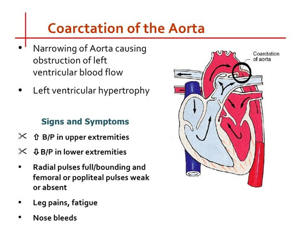
C. Decreased blood pressure in the legs:
This is the correct choice. Coarctation of the aorta is characterized by narrowing of the aorta, which leads to decreased blood flow to the lower extremities. Consequently, blood pressure measurements in the legs may be lower compared to those in the arms. This finding is often a key indicator of coarctation of the aorta.
D. Pulmonary edema:
Pulmonary edema refers to the accumulation of fluid in the lungs and is typically associated with conditions such as heart failure or fluid overload. While some congenital heart defects may lead to heart failure and subsequent pulmonary edema, coarctation of the aorta does not directly cause pulmonary edema. Instead, it primarily affects blood flow to the lower extremities due to the narrowing of the aorta.
Nursing Test Bank
Naxlex Comprehensive Predictor Exams
Related Questions
Correct Answer is B
Explanation
A. "When I use this technique the medication will not run out of the ear."
This explanation is not entirely accurate. While pulling the auricle down and back may help prevent ear drops from immediately dripping out of the ear, the primary purpose of this technique is to straighten the ear canal, facilitating the passage of the medication into the inner ear region for optimal effectiveness. The prevention of medication runoff is a secondary benefit.
B. “This opens the ear canal, allowing medication to reach the inner ear region.”
This explanation is correct. Pulling the auricle down and back helps to straighten the ear canal, making it easier for the ear drops to enter the ear canal and reach the inner ear where they can effectively treat the condition. This is the main purpose of using this technique.
C. “This is the safest and easiest way to administer this medication.”
While pulling the auricle down and back is a commonly used technique for administering ear drops, describing it as the safest and easiest way may not fully capture its purpose. Safety and ease of administration are important considerations, but the primary rationale for this technique is to facilitate the delivery of medication to the inner ear.
D. “When I use the technique, your child experiences less pain.”
This explanation is inaccurate. Pulling the auricle down and back may not directly reduce pain. The main purpose of this technique is to ensure that the medication reaches the inner ear region for effective treatment. While discomfort during administration may be minimized with proper technique, the primary focus is on medication delivery rather than pain reduction.
Correct Answer is B
Explanation
Infusion rate (mL/hour) = Total volume (mL) / Total time (hours)
Given:
Child's weight: 10 kg
Ordered volume: 40 mL/kg
Total time: 4 hours
First, calculate the total volume of Lactated Ringer's solution needed:
Total volume = 40 mL/kg × 10 kg = 400 mL
Then, divide the total volume by the total time to find the infusion rate:
Infusion rate = 400 mL / 4 hours = 100 mL/hour
Whether you are a student looking to ace your exams or a practicing nurse seeking to enhance your expertise , our nursing education contents will empower you with the confidence and competence to make a difference in the lives of patients and become a respected leader in the healthcare field.
Visit Naxlex, invest in your future and unlock endless possibilities with our unparalleled nursing education contents today