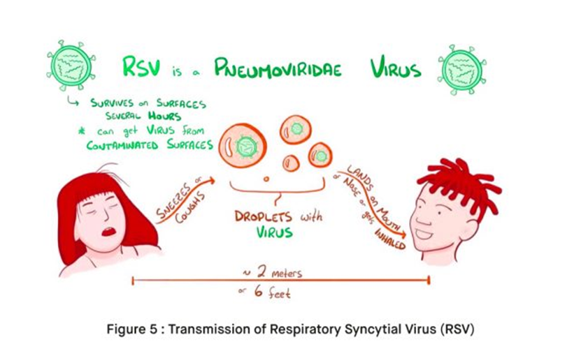
A nurse is assisting with the admission of an infant who has respiratory syncytial virus (RSV), which of the following rooms should the nurse assign the infant?
A room with a toddler who has pneumonia
A private room with reverse isolation
A private room with contact/droplet precautions
A room with an infant who has croup
The Correct Answer is C
A. A room with a toddler who has pneumonia.
This option is not ideal because both RSV and pneumonia are respiratory infections that can spread to other patients. Placing these two patients together could increase the risk of cross-infection.
B. A private room with reverse isolation.
Reverse isolation is typically used to protect immunocompromised patients from acquiring infections from others. However, in the case of RSV, reverse isolation is not necessary because RSV primarily affects infants and young children who are generally not immunocompromised. Therefore, this option is not appropriate for an infant with RSV.
C. A private room with contact/droplet precautions.
This option is the most appropriate. RSV is primarily spread through respiratory droplets and direct contact with respiratory secretions. Placing the infant in a private room with contact/droplet precautions helps to minimize the risk of transmission to other patients. Healthcare workers and visitors entering the room should adhere to appropriate precautions, including wearing personal protective equipment (PPE) such as masks, gloves, and gowns.
D. A room with an infant who has croup.
Placing an infant with RSV in the same room as an infant with croup is not ideal because both conditions involve respiratory symptoms and may increase the risk of cross-infection.
Nursing Test Bank
Naxlex Comprehensive Predictor Exams
Related Questions
Correct Answer is C
Explanation
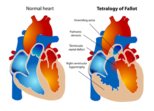
A. Overriding aorta: In Tetralogy of Fallot, the aorta is positioned over the ventricular septal defect (VSD), rather than solely over the left ventricle as it would be in a normal heart. This is called overriding aorta, which allows blood from both the right and left ventricles to enter the aorta.
B. Pulmonary stenosis: This is a critical component of Tetralogy of Fallot. Pulmonary stenosis refers to narrowing of the pulmonary valve or the area just below it, which restricts blood flow from the right ventricle to the pulmonary artery. This results in decreased blood flow to the lungs for oxygenation.
C. Left ventricular hypertrophy: This choice is not typically associated with Tetralogy of Fallot. Left ventricular hypertrophy refers to an enlargement or thickening of the muscular wall of the left ventricle of the heart. It is often seen in conditions where the left ventricle has to work harder to pump blood, such as in hypertension or aortic stenosis, but it is not a characteristic feature of Tetralogy of Fallot.
D. Ventricular septal defect: This defect is one of the four components of Tetralogy of Fallot. A ventricular septal defect (VSD) is a hole in the septum, the muscular wall that separates the left and right ventricles of the heart. In Tetralogy of Fallot, the VSD allows oxygen-poor blood from the right ventricle to flow directly into the left ventricle and out to the body.
Correct Answer is C
Explanation
A. Coloring book with crayons:
Coloring activities with crayons are typically more suitable for older children who have developed fine motor skills and hand-eye coordination. At 10 months old, infants are still in the early stages of motor development and may not have the dexterity to hold and manipulate crayons effectively. Additionally, infants at this age are more likely to put objects in their mouths, which poses a choking hazard with crayons.
B. Large-piece puzzles:
Puzzles with large pieces can be beneficial for older children's cognitive development by promoting problem-solving skills and hand-eye coordination. However, at 10 months old, infants are still developing their motor skills and may not have the ability to manipulate puzzle pieces effectively. Puzzles with small pieces can also pose a choking hazard for infants.
C. Crib gym:
A crib gym is a suitable toy for a 10-month-old infant as it provides opportunities for visual stimulation, reaching, grasping, and hand-eye coordination development. Crib gyms typically consist of hanging toys or objects that the infant can bat at or grasp while lying in their crib or playpen. This type of toy encourages exploration and interaction while ensuring safety within the confines of the crib.
D. Put-in take-out toy:
Put-in take-out toys involve placing objects into a container and then removing them, which can be engaging for infants. However, while this type of toy may provide some entertainment for a 10-month-old, it may not offer as much visual and tactile stimulation as a crib gym. Additionally, some put-in take-out toys may have smaller parts that pose a choking hazard for infants, so careful supervision is necessary.
Whether you are a student looking to ace your exams or a practicing nurse seeking to enhance your expertise , our nursing education contents will empower you with the confidence and competence to make a difference in the lives of patients and become a respected leader in the healthcare field.
Visit Naxlex, invest in your future and unlock endless possibilities with our unparalleled nursing education contents today