
A client at 20 weeks gestation reports discomfort after amniocentesis procedure to the nurse. Which of the following assessment findings warrants priority action by the nurse?
Braxton Hicks contraction
Amniotic fluid contains fetal urine
Prolonged vaginal bleeding
Lab results reveal abnormal chromosome cells
The Correct Answer is C
Explanation:
A. Braxton Hicks contraction
Braxton Hicks contractions are often referred to as "practice" contractions and are common during pregnancy. They are typically irregular and do not cause cervical changes. While discomfort after an amniocentesis procedure can sometimes trigger Braxton Hicks contractions, they are not usually a cause for immediate concern unless they become frequent, intense, or are accompanied by other signs of preterm labor, such as regular contractions, lower back pain, pelvic pressure, or changes in vaginal discharge.
B. Amniotic fluid contains fetal urine
The presence of fetal urine in the amniotic fluid is a normal and expected process during pregnancy. Fetal urine production contributes to the volume and composition of amniotic fluid, helping maintain the environment in the uterus and supporting fetal development. This finding is not directly related to the client's discomfort after an amniocentesis procedure and is not a cause for immediate concern unless there are other complications or abnormalities related to the amniotic fluid composition.
C. Prolonged vaginal bleeding
Prolonged vaginal bleeding after an amniocentesis procedure is a significant finding that requires immediate attention. While some spotting or mild bleeding can occur after the procedure, prolonged or significant bleeding may indicate complications such as uterine injury, placental abruption, or other issues that need urgent assessment and intervention by healthcare providers.
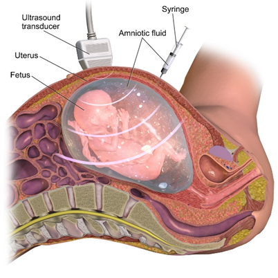
D. Lab results reveal abnormal chromosome cells
Abnormal chromosome cells found in the amniotic fluid sample obtained during amniocentesis may indicate genetic abnormalities or conditions in the fetus. While this finding is significant and requires follow-up and further evaluation, it is not an immediate concern in terms of the client's discomfort or the need for priority action by the nurse unless it is associated with other urgent clinical signs or symptoms.
Nursing Test Bank
Naxlex Comprehensive Predictor Exams
Related Questions
Correct Answer is A
Explanation
Explanation:
A. Excessive uterine enlargement
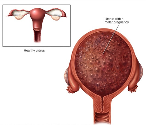
This choice is correct because a hydatidiform mole can cause rapid and excessive growth of the uterus due to the abnormal proliferation of placental tissue. This can lead to the uterus being larger than expected for the gestational age.
B. Profuse, clear vaginal discharge
This choice is not typically associated with a hydatidiform mole. While vaginal discharge can occur during pregnancy, a profuse and clear discharge is not specifically characteristic of a hydatidiform mole. Other causes, such as normal vaginal secretions or infections, could lead to such discharge.
C. Rapid decline in human chorionic gonadotropin (hCG) levels
This choice is not typical of a hydatidiform mole. In fact, one of the hallmarks of a molar pregnancy is an abnormally high level of hCG. The hCG levels may continue to rise instead of declining rapidly.
D. Irregular fetal heart rate
This choice is not associated with a hydatidiform mole because a molar pregnancy does not involve a viable fetus with a heartbeat. Instead, it is characterized by the abnormal growth of placental tissue, which can cause symptoms related to uterine enlargement and complications such as bleeding or preeclampsia, but not an irregular fetal heart rate.
Correct Answer is B
Explanation
Explanation:
A. Slightly below the umbilicus:
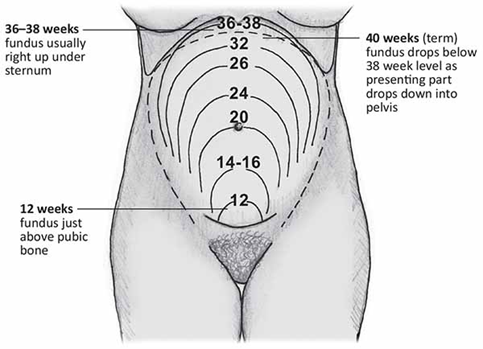
Around 16-20 weeks of gestation, the fundal height is typically palpated slightly below the umbilicus. This corresponds to the level of the fundus before it starts rising significantly above the umbilicus as the pregnancy progresses.
B. 3 cm above the umbilicus:
At 22 weeks of gestation, the fundal height is expected to be approximately 3 cm above the umbilicus. This is a common estimation for the fundal height at this stage of pregnancy, as the uterus continues to grow and the fundus ascends in a predictable manner.
C. 3 cm below the umbilicus:
Palpating the fundus 3 cm below the umbilicus would typically correspond to an earlier gestational age, such as around 16-18 weeks. As pregnancy advances, the fundal height increases, and by 22 weeks, it should be well above the level of the umbilicus.
D. Slightly above the umbilicus:
This location is closer to where the fundal height is expected to be at later gestational ages, such as 24-26 weeks. However, at 22 weeks of gestation, the fundal height should not be palpated slightly above the umbilicus but rather around 3 cm above it.
Whether you are a student looking to ace your exams or a practicing nurse seeking to enhance your expertise , our nursing education contents will empower you with the confidence and competence to make a difference in the lives of patients and become a respected leader in the healthcare field.
Visit Naxlex, invest in your future and unlock endless possibilities with our unparalleled nursing education contents today