Which of the following assessment findings should the nurse report to the practitioner? (Select all that apply)
Use of accessory muscles
Nail bed greater than 160 degrees
Circumoral cyanosis
Pursed lip breathing
Anteroposterior-to-transverse diameter of 1:1
Correct Answer : A,B,C,D,E
A. Use of accessory muscles
Explanation: Using accessory muscles during breathing indicates increased effort to breathe, which can be a sign of respiratory distress. It suggests that the client is having difficulty breathing and is using additional muscles to aid in the process. This finding should be reported to the practitioner for further evaluation.
B. Nail bed greater than 160 degrees
Explanation: A nail bed angle greater than 160 degrees, also known as clubbing, is an abnormal finding and can be associated with chronic respiratory or cardiovascular conditions. It may indicate insufficient oxygenation and should be reported to the practitioner for evaluation.
C. Circumoral cyanosis
Explanation: Circumoral cyanosis, which is a bluish discoloration around the mouth, indicates inadequate oxygenation. It can be a sign of respiratory or cardiac problems and should be reported to the practitioner for further assessment and intervention.
D. Pursed lip breathing
Explanation: Pursed lip breathing is a technique often used by individuals with respiratory difficulties to improve oxygen exchange. However, if it's observed in a person who does not normally use this technique, it could indicate respiratory distress and should be reported to the practitioner for evaluation.
E. Anteroposterior-to-transverse diameter of 1:1
Explanation: An anteroposterior-to-transverse diameter of 1:1 (also known as barrel chest) is an abnormal finding often associated with chronic obstructive pulmonary disease (COPD). It suggests overinflation of the lungs and can impair effective breathing. This finding should be reported to the practitioner for further evaluation.
Nursing Test Bank
Naxlex Comprehensive Predictor Exams
Related Questions
Correct Answer is ["B","C","D","E"]
Explanation
A. Age: While age itself is not modifiable, it is included in the list because aging increases the risk of developing cardiovascular disease. However, individuals cannot change their age, so it is not a modifiable risk factor.
B. Smoking: Smoking is a significant risk factor for cardiovascular disease. It damages the heart and blood vessels and can lead to atherosclerosis (narrowing and hardening of the arteries), which can result in heart attacks and strokes.
C. Hypertension: High blood pressure is a leading cause of cardiovascular disease. It can damage the arteries over time, making them more susceptible to atherosclerosis.
D. Diabetes: Diabetes, especially if poorly controlled, increases the risk of cardiovascular disease. High blood sugar levels can damage the blood vessels and the heart.
E. High cholesterol: Elevated levels of cholesterol, especially low-density lipoprotein (LDL) cholesterol, can lead to the buildup of plaque in the arteries, increasing the risk of atherosclerosis and cardiovascular events.
Correct Answer is A
Explanation
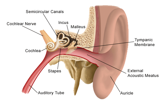
A. Auricle (Pinna):
The auricle, also known as the pinna, is the visible external part of the ear. It consists of movable cartilage and skin. When administering eardrops, pulling the auricle up and back helps to straighten the ear canal, allowing the drops to enter the ear effectively.
B. Mastoid Process:
The mastoid process is a bony prominence located behind the ear. It is not a part of the outer ear structure involved in administering eardrops.
C. Outer Meatus:
The outer meatus, also known as the external acoustic meatus or ear canal, is the tube-like structure leading from the auricle to the eardrum. It is the passage through which eardrops are administered. Pulling the auricle up and back helps to straighten the outer meatus for the proper administration of eardrops.
D. Concha:
The concha refers to the bowl-shaped depression next to the ear canal. While it is a part of the outer ear, pulling the concha is not a technique used for administering eardrops. The auricle, specifically, is manipulated to facilitate the process.

Whether you are a student looking to ace your exams or a practicing nurse seeking to enhance your expertise , our nursing education contents will empower you with the confidence and competence to make a difference in the lives of patients and become a respected leader in the healthcare field.
Visit Naxlex, invest in your future and unlock endless possibilities with our unparalleled nursing education contents today
Report Wrong Answer on the Current Question
Do you disagree with the answer? If yes, what is your expected answer? Explain.
Kindly be descriptive with the issue you are facing.
