The nurse recognizes that the following statement is true regarding the internal structures of the breast: The breast is made up of:
Glandular tissue, which supports the breast by attaching to the chest wall.
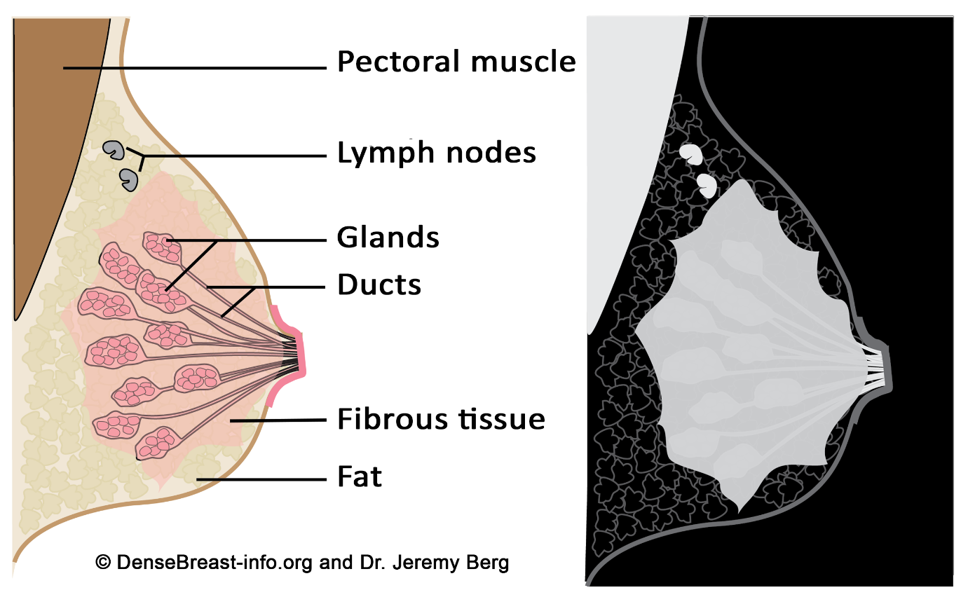
Fibrous, glandular, and adipose tissues
Primarily muscle with very little fibrous tissue.
Primarily milk ducts, known as lactiferous ducts.
The Correct Answer is B
A. Glandular tissue, which supports the breast by attaching to the chest wall: Glandular tissue is indeed a part of the breast structure, but it is not responsible for supporting the breast by attaching to the chest wall. It's the Cooper's ligaments, which are fibrous connective tissue, that provide structural support.
B. Fibrous, glandular, and adipose tissues: This statement is correct. The breast is composed of glandular tissue (responsible for milk production), fibrous tissue (including Cooper's ligaments for support), and adipose tissue (fat).
C. Primarily muscle with very little fibrous tissue: The breast contains very little muscle tissue. The main supportive structure is fibrous tissue, not muscle.
D. Primarily milk ducts, known as lactiferous ducts: Milk ducts are part of the glandular tissue and are responsible for carrying milk. However, the breast is not primarily made up of milk ducts; it consists of a combination of glandular, fibrous, and adipose tissues.
Nursing Test Bank
Naxlex Comprehensive Predictor Exams
Related Questions
Correct Answer is D
Explanation
A. Clear and equal breath sounds bilaterally
Explanation: Clear and equal breath sounds bilaterally indicate normal lung sounds, suggesting proper air exchange in both lungs. This is a normal finding and does not require immediate reporting.
B. Oxygen saturation of 98% on room air
Explanation: An oxygen saturation level of 98% on room air indicates adequate oxygenation of the blood. This is a normal and healthy oxygen saturation level and does not require immediate reporting.
C. Cough producing clear, thin sputum
Explanation: A cough producing clear, thin sputum is indicative of a non-infected or non-inflammatory condition in the respiratory system. Clear and thin sputum is often normal, especially in the absence of other symptoms. It does not require immediate reporting unless the client has other concerning symptoms.
D. Visible use of accessory muscles during inhalation
Explanation: Visible use of accessory muscles, such as neck or intercostal muscles, during inhalation suggests that the client is working hard to breathe. This could indicate respiratory distress, potentially due to conditions like asthma, COPD (Chronic Obstructive Pulmonary Disease), or other severe lung problems. It's a concerning sign and should be reported to the healthcare practitioner promptly for further evaluation and intervention.
Correct Answer is B
Explanation
A. AV node - SA node - bundle of His - bundle branches:
This sequence is incorrect. The SA node (sinoatrial node) initiates the electrical impulse in the heart, followed by the AV node (atrioventricular node), bundle of His, and then the bundle branches.
B. SA node - AV node - bundle of His - bundle branches:
This sequence is correct. The electrical stimulus of the cardiac cycle starts at the SA node, which is the natural pacemaker of the heart. From the SA node, the impulse travels to the AV node, then to the bundle of His, and finally to the bundle branches, which distribute the impulse to the ventricles, causing them to contract.
C. Bundle of His - AV node - SA node - Erb's Point:
This sequence is incorrect. Erb's Point is a point on the chest where heart sounds S2 and S3 can be heard most distinctly.
D. AV node - SA node - bundle of His - Erb's Point:
This sequence is incorrect. The AV node comes after the SA node in the electrical conduction system of the heart. Erb's Point is not a part of the normal cardiac conduction pathway; it is a location for auscultation on the chest.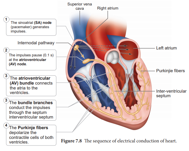
Whether you are a student looking to ace your exams or a practicing nurse seeking to enhance your expertise , our nursing education contents will empower you with the confidence and competence to make a difference in the lives of patients and become a respected leader in the healthcare field.
Visit Naxlex, invest in your future and unlock endless possibilities with our unparalleled nursing education contents today