Which of the following are agranulocytes?
Basophils.
Monocytes.
Neutrophils.
Eosinophils.
The Correct Answer is B
Monocytes are a type of agranulocytes, which are white blood cells that lack visible granules in their cytoplasm.
Agranulocytes also include lymphocytes, which are involved in adaptive immunity.
Choice A is wrong because basophils are a type of granulocytes, which are white blood cells that have granules in their cytoplasm.
Granulocytes also include neutrophils and eosinophils, which are involved in innate immunity.
Choice C is wrong because neutrophils are also a type of granulocyte.
Neutrophils are the most abundant white blood cells and are responsible for phagocytizing bacteria and fungi.
Choice D is wrong because eosinophils are also a type of granulocytes. Eosinophils are involved in allergic reactions and parasitic infections.
Normal ranges for white blood cells vary depending on age, gender, and health status, but generally, they are between 4,000 and 11,000 cells per microliter of blood.
Nursing Test Bank
Naxlex Comprehensive Predictor Exams
Related Questions
Correct Answer is D
Explanation
Atrial natriuretic peptide (ANP) is a hormone secreted by the heart when the atria are stretched by high blood pressure or volume.
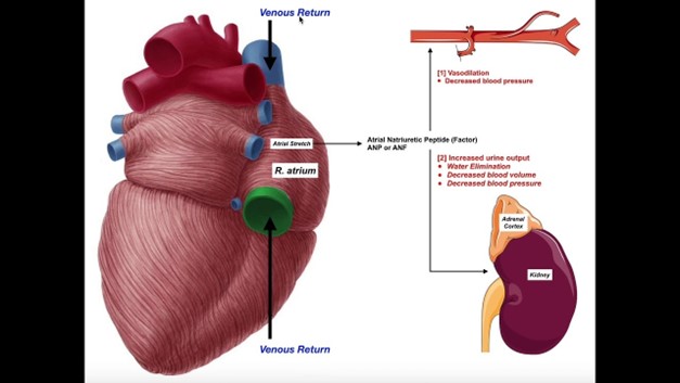
ANP has multiple effects, such as increasing urine and salt excretion, lowering blood pressure, and opposing the renin-angiotensin-aldosterone system.
Therefore, ANP inhibits the release of renin and aldosterone, which are hormones that increase blood pressure and sodium retention.
Choice A is wrong because ANP is not released from the adrenal cortex but from the cardiac atria.
ANP does not stimulate atrial hormones but rather inhibits them.
Choice B is wrong because ANP is not stimulated to release when blood volume decreases, but when it increases.
ANP acts to reduce blood volume by promoting diuresis and natriuresis.
Choice C is wrong because ANP does not raise blood pressure, but lowers it. ANP acts as a vasodilator and reduces peripheral resistance.
Correct Answer is A
Explanation
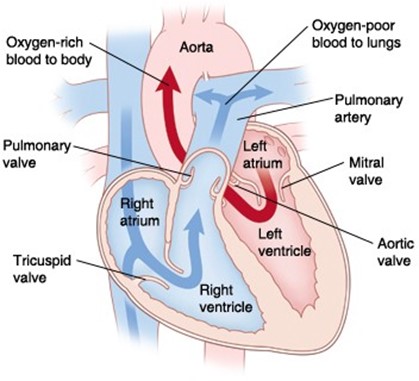
This is the correct sequence of parts through which blood moves from the vena cava to the lungs.
Choice B is wrong because it reverses the order of the right atrium and right ventricle. Blood flows from the right atrium to the right ventricle, not the other way around.
Choice C is wrong because it switches the positions of the tricuspid valve and the pulmonary valve.
Blood flows from the right atrium through the tricuspid valve to the right ventricle, and then through the pulmonary valve to the pulmonary artery.
Choice D is wrong because it also switches the positions of the tricuspid valve and the pulmonary valve, and reverses the order of the right atrium and right ventricle.
Blood flows from the right atrium through the tricuspid valve to the right ventricle, and then through the pulmonary valve to the pulmonary artery.
The normal range of blood pressure in the vena cava is about 0 to 5 mmHg, while in the pulmonary artery, it is about 15 to 25 mmHg.
The normal range of oxygen saturation in the vena cava is about 60% to 80%, while in the pulmonary vein, it is about 95% to 100%.
Whether you are a student looking to ace your exams or a practicing nurse seeking to enhance your expertise , our nursing education contents will empower you with the confidence and competence to make a difference in the lives of patients and become a respected leader in the healthcare field.
Visit Naxlex, invest in your future and unlock endless possibilities with our unparalleled nursing education contents today