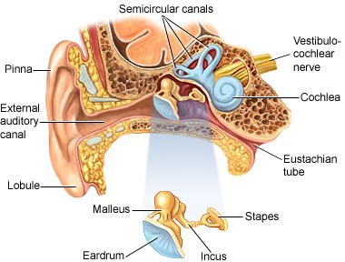
Which anatomical structure houses the malleus, incus and stapes?
Skull
Lungs
Ear
Mouth
The Correct Answer is C
The malleus, incus, and stapes are three small bones, collectively known as the ossicles, located in the middle ear. These bones work together to transmit sound waves from the eardrum to the inner ear, where they are converted into nerve impulses that are then sent to the brain. The malleus is atached to the eardrum, the incus is in between the malleus and the stapes, and the stapes is connected to the inner ear. Together, they form a chain that amplifies the sound waves and transmits them efficiently to the inner ear.
Nursing Test Bank
Naxlex Comprehensive Predictor Exams
Related Questions
Correct Answer is B
Explanation
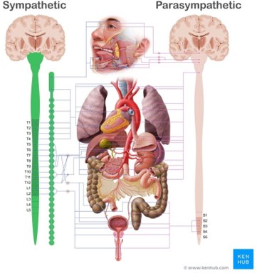
When the sympathetic nervous system is stimulated, it activates the "fight or flight" response, which prepares the body for physical activity or stressful situations. This response includes several physiological changes, such as:
Increased heart rate and cardiac output: The sympathetic nervous system releases adrenaline and noradrenaline, which increase heart rate and cardiac output to provide more oxygen and nutrients to the muscles.
Decreased uterine activity: The sympathetic nervous system inhibits uterine contractions to prevent
premature labor.
Decreased pancreatic activity: The sympathetic nervous system inhibits insulin secretion and promotes glucagon secretion to increase blood glucose levels.
Decreased gastrointestinal activity: The sympathetic nervous system inhibits digestive functions to divert blood flow to the muscles.
Correct Answer is D
Explanation
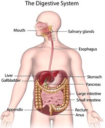
Digestion begins in the oral cavity, also known as the mouth, where food is ingested and broken down into smaller pieces through mechanical digestion by the teeth and chemical digestion by enzymes in saliva, such as amylase. Once the food is sufficiently broken down, it forms a bolus and is then swallowed, passing through the esophagus and into the stomach. In the stomach, the food is further broken down by stomach acid and enzymes before passing into the small intestine for the absorption of nutrients.
 |
Whether you are a student looking to ace your exams or a practicing nurse seeking to enhance your expertise , our nursing education contents will empower you with the confidence and competence to make a difference in the lives of patients and become a respected leader in the healthcare field.
Visit Naxlex, invest in your future and unlock endless possibilities with our unparalleled nursing education contents today