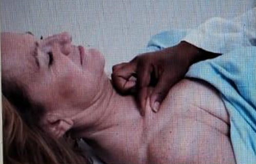
The nurse performs the action shown in this image during the assessment of a client. What is the nurse assessing?
Intercostal spaces
Lymph nodes
Skin Turgor
Carotid Pulse
The Correct Answer is C
A) Intercostal spaces:
Assessing intercostal spaces typically involves palpating or inspecting the area between the ribs to check for abnormalities such as retractions or tenderness, usually conducted with the client sitting or standing.
B) Lymph nodes:
Palpation of lymph nodes, such as in the cervical region, involves using fingertips to gently feel for enlarged or tender nodes. This examination focuses on areas like the neck, underarms, and groin.
C) Skin Turgor:
The image depicts a nurse pinching the skin, likely on the chest or forearm, which is a common method to assess skin turgor. Skin turgor evaluation helps determine hydration status; if the skin remains tented and returns slowly to its original position, it indicates dehydration.
D) Carotid Pulse:
Assessing the carotid pulse involves palpating the carotid artery along the side of the neck to evaluate the strength and rhythm of the pulse. This is typically done using the pads of the fingers, not by pinching the skin.
Nursing Test Bank
Naxlex Comprehensive Predictor Exams
Related Questions
Correct Answer is A
Explanation
a) The patient is unable to see in half of the visual field (same visual field) in each eye:
Homonymous hemianopsia is a condition where there is a loss of vision in the same side of the visual field in both eyes. This occurs due to damage to the visual pathways after the optic chiasm, often from a stroke or brain injury, resulting in the loss of either the right or left visual field in both eyes.
b) The patient can see from one eye but not through the other one:
This description fits a condition called monocular blindness, which is typically caused by damage to the optic nerve before it reaches the optic chiasm. Homonymous hemianopsia involves both eyes and specific visual fields, not complete loss of vision in one eye.
c) The patient is unable to see in half of the visual field (opposite visual field) in each eye:
This option describes bitemporal hemianopsia, which results in loss of vision in the outer (temporal) fields of both eyes and is often due to damage at the optic chiasm. Homonymous hemianopsia involves the same side of the visual field in both eyes, not the opposite visual fields.
d) No visual impairment:
Homonymous hemianopsia is characterized by significant visual impairment, specifically the loss of half the visual field in both eyes on the same side. Therefore, it is incorrect to say there is no visual impairment with this condition.
Correct Answer is C
Explanation
(a) Diarrhea: Diarrhea is an abnormal gastrointestinal response characterized by frequent, loose, or watery stools. It can be caused by infections, medications, or underlying gastrointestinal disorders. Pallor, or paleness of the skin, typically does not directly lead to diarrhea unless there are specific underlying conditions affecting both circulation and gastrointestinal function.
(b) Diaphoresis: Diaphoresis refers to excessive sweating, which can occur due to sympathetic nervous system activation, fever, or anxiety. While diaphoresis may be associated with conditions causing increased sympathetic activity, it is not directly related to pallor, which indicates reduced blood flow to the skin.
(c) Fainting: Pallor is often a sign of decreased blood flow to the skin, indicating potential hypoperfusion. If severe, this reduced circulation can lead to fainting (syncope) due to inadequate blood supply to the brain. Therefore, after noting pallor, the nurse should be prepared to manage the client for potential fainting episodes by ensuring safety and providing appropriate interventions.
(d) Vomiting: Vomiting is the forceful expulsion of stomach contents through the mouth and can be caused by various factors such as gastrointestinal irritation, infection, or systemic illnesses. Pallor does not directly cause vomiting, although severe systemic conditions affecting circulation could potentially lead to nausea and vomiting as part of a broader clinical picture.
Whether you are a student looking to ace your exams or a practicing nurse seeking to enhance your expertise , our nursing education contents will empower you with the confidence and competence to make a difference in the lives of patients and become a respected leader in the healthcare field.
Visit Naxlex, invest in your future and unlock endless possibilities with our unparalleled nursing education contents today