The nurse is testing a client's visual accommodation. Which of the following should the nurse recognize as an assessment finding from visual accommodation?
The pupils constrict when the examiner's index finger slowly moves toward the client's nose.
The client involuntary blinks in the presence of bright light directed over the pupils during the eye exam.
The client's peripheral vision becomes sharper when the examiner shines a light over the pupils.
The pupils dilate when the examiner's index finger slowly moves toward the client's nose.
The Correct Answer is A
A. The pupils constrict when the examiner's index finger slowly moves toward the client's nose.
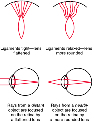
This statement is correct. Visual accommodation is the process by which the eye's lens changes shape to focus on objects at varying distances. When an object moves closer to the eyes, the pupils constrict to adjust and focus on the near object, preventing double vision.
B. The client involuntarily blinks in the presence of bright light directed over the pupils during the eye exam.
This statement describes the pupillary light reflex, not visual accommodation. The pupillary light reflex is the response of the pupils to light exposure.
C. The client's peripheral vision becomes sharper when the examiner shines a light over the pupils.
This statement is not accurate. Peripheral vision sharpness is not related to visual accommodation. Visual accommodation mainly involves adjusting focus for objects at varying distances.
D. The pupils dilate when the examiner's index finger slowly moves toward the client's nose.
This statement is incorrect. Pupils should constrict, not dilate, when focusing on a near object (as in visual accommodation). Dilation occurs in low-light conditions or in response to sympathetic stimulation.
Nursing Test Bank
Naxlex Comprehensive Predictor Exams
Related Questions
Correct Answer is B
Explanation
A. Wheezing: Wheezing is a continuous, high-pitched whistling sound usually heard during expiration. It is often associated with narrowed airways, such as in asthma or chronic obstructive pulmonary disease (COPD). Wheezing occurs due to the turbulent airflow through narrowed bronchi or bronchioles and is not typically associated with pleuritis.
B. Friction rub: Pleuritis, or inflammation of the pleura, can cause a friction rub. This sound occurs when the inflamed pleural layers rub against each other during breathing. It's a grating or rubbing sound heard on auscultation and is a hallmark sign of pleuritis.
C. Stridor: Stridor is a high-pitched, harsh sound heard during inspiration and sometimes expiration. It is often a sign of upper airway obstruction, such as in croup or anaphylaxis. Stridor results from turbulent airflow through a partially obstructed or narrowed larynx or trachea.
D. Crackles: Crackles, also known as rales, are brief, discontinuous, popping sounds heard on inspiration. They can be fine or coarse and are often associated with conditions that cause fluid or secretions in the alveoli or small airways, such as pneumonia or heart failure. Crackles are not typically associated with pleuritis.
Correct Answer is A
Explanation
A. When part of the lung is obstructed or collapsed: This statement is accurate. Unequal chest expansion can occur when part of the lung is obstructed or collapsed, preventing the affected area from expanding normally during inhalation.
B. When bulging of the intercostal spaces is present: This statement is not accurate. Unequal chest expansion typically refers to decreased expansion on one side, not bulging of intercostal spaces.
C. In an obese patient: This statement is not accurate. Obesity can affect breathing patterns and lung function, but it is not the primary cause of unequal chest expansion.
D. When accessory muscles are used to augment respiratory effort: This statement is not accurate. The use of accessory muscles to augment respiratory effort can be a sign of respiratory distress, but it doesn't directly cause unequal chest expansion. Unequal expansion is more indicative of specific lung conditions or issues with lung mechanics.
Whether you are a student looking to ace your exams or a practicing nurse seeking to enhance your expertise , our nursing education contents will empower you with the confidence and competence to make a difference in the lives of patients and become a respected leader in the healthcare field.
Visit Naxlex, invest in your future and unlock endless possibilities with our unparalleled nursing education contents today