The nurse is auscultating the lungs of a sleeping client and hears short, popping, crackling breath sounds that stop after a few breaths. The nurse recognizes that these breath sounds are:
Atelectatic crackles that do not have a pathologic cause.
Vesicular breath sounds.
Fine wheezes.
Fine crackles and may be a sign of pneumonia.
The Correct Answer is A
A. Atelectatic crackles that do not have a pathologic cause:
Atelectatic crackles are short, popping, crackling sounds heard during auscultation. They occur in individuals who are in a supine position and disappear after a few breaths. These crackles are not indicative of any pathological condition; they are common when the lungs are not fully aerated, especially when a person is lying down.
B. Vesicular breath sounds:
Vesicular breath sounds are normal lung sounds heard over the peripheral lung areas. They are soft, low-pitched, and continuous throughout inspiration and part of expiration. Vesicular breath sounds are the typical sounds heard during routine breathing and are not associated with crackling or popping noises.
C. Fine wheezes:
Wheezes are high-pitched whistling sounds heard during expiration. They occur due to narrowed airways and are commonly associated with conditions like asthma or bronchoconstriction. Fine wheezes suggest a partial obstruction in the smaller airways, causing turbulent airflow, leading to the characteristic sound.
D. Fine crackles and may be a sign of pneumonia:
Fine crackles are high-pitched, discontinuous, crackling sounds heard during inspiration. They can occur due to the sudden opening of small airways, and their presence may indicate fluid in the lungs or lung inflammation. Fine crackles are often associated with conditions such as pneumonia, heart failure, or interstitial lung diseases.
Nursing Test Bank
Naxlex Comprehensive Predictor Exams
Related Questions
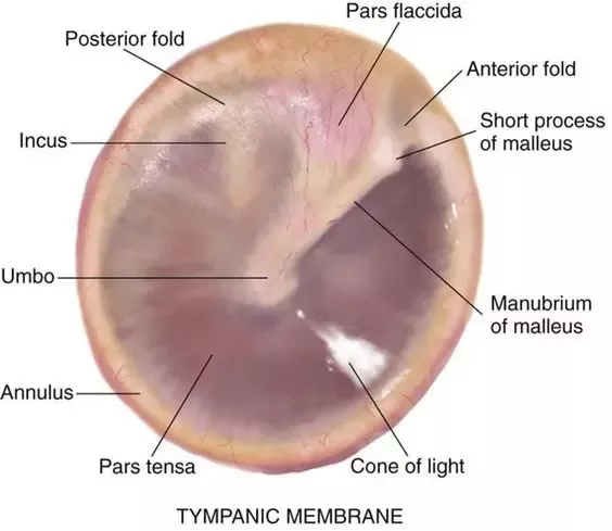
Correct Answer is D
Explanation
A. A shiny, pearly white color tympanic membrane: This is a normal finding. A healthy tympanic membrane often appears shiny and pearly white.
B. The presence of cerumen: This is a normal finding. Cerumen, or earwax, is a natural substance that helps protect the ear canal.
C. The presence of a cone of light: This is a normal finding. The cone of light is a reflection of the otoscope light on the tympanic membrane and is a normal variation.
D. A yellow or amber color to the tympanic membrane: This is considered an abnormal finding. A yellow or amber coloration of the tympanic membrane can indicate the presence of fluid or infection behind the eardrum, which may be a sign of otitis media or other ear conditions.
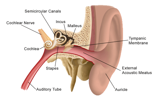
Correct Answer is A
Explanation
A. Auricle (Pinna):
The auricle, also known as the pinna, is the visible external part of the ear. It consists of movable cartilage and skin. When administering eardrops, pulling the auricle up and back helps to straighten the ear canal, allowing the drops to enter the ear effectively.
B. Mastoid Process:
The mastoid process is a bony prominence located behind the ear. It is not a part of the outer ear structure involved in administering eardrops.
C. Outer Meatus:
The outer meatus, also known as the external acoustic meatus or ear canal, is the tube-like structure leading from the auricle to the eardrum. It is the passage through which eardrops are administered. Pulling the auricle up and back helps to straighten the outer meatus for the proper administration of eardrops.
D. Concha:
The concha refers to the bowl-shaped depression next to the ear canal. While it is a part of the outer ear, pulling the concha is not a technique used for administering eardrops. The auricle, specifically, is manipulated to facilitate the process.
Whether you are a student looking to ace your exams or a practicing nurse seeking to enhance your expertise , our nursing education contents will empower you with the confidence and competence to make a difference in the lives of patients and become a respected leader in the healthcare field.
Visit Naxlex, invest in your future and unlock endless possibilities with our unparalleled nursing education contents today