A nurse is preparing a 9-year-old child for an IV catheter insertion. Which of the following actions should the nurse take first?
Allow the child to see and touch IV tubing and supplies.
Explain to the child's parents what role they will have during the procedure.
Describe the procedure using visual aids.
Ask the child what he knows about the procedure.
The Correct Answer is D
A. Allow the child to see and touch IV tubing and supplies.
Allowing the child to see and touch the IV tubing and supplies can help familiarize them with the equipment and reduce anxiety. However, there may be a more appropriate action to take first.
B. Explain to the child's parents what role they will have during the procedure.
While it's important to involve the child's parents and inform them of their role during the procedure, the priority should be to prepare the child for the insertion itself.
C. Describe the procedure using visual aids.
Using visual aids can be helpful in explaining the procedure to the child and providing a clear understanding of what will happen. However, there may be a more appropriate action to take first.
D. Ask the child what he knows about the procedure.
This is the correct answer. Asking the child what they already know about the procedure allows the nurse to assess their understanding and address any misconceptions or concerns they may have. It also helps the nurse tailor their explanation to the child's level of understanding and provide information that is relevant and meaningful to them.
Nursing Test Bank
Naxlex Comprehensive Predictor Exams
Related Questions
Correct Answer is ["A","D"]
Explanation
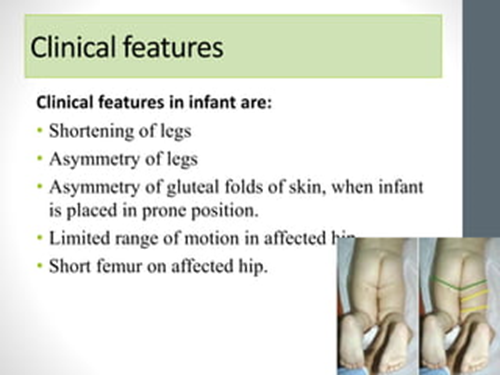
A. Inwardly turned foot on the affected side.
This finding is consistent with DDH. In infants with DDH, the affected leg may appear shortened and rotated inwardly due to hip instability or dislocation.
B. Lengthened thigh on the affected side.
This finding is not typically associated with DDH. In fact, the affected thigh may appear shortened rather than lengthened due to abnormal positioning of the hip joint.
C. Absent plantar reflexes.
Absent plantar reflexes are not directly related to DDH. Plantar reflexes assess the function of the spinal nerves in the lower extremities and are not typically affected by hip dysplasia.
D. Asymmetric thigh folds.
This finding is consistent with DDH. Asymmetric thigh folds, where one thigh appears fuller or has more skin folds compared to the other, can be indicative of hip dysplasia. The skin folds may be more prominent on the unaffected side due to the displacement of the femoral head on the affected side.

Correct Answer is C
Explanation
A. Machine-like murmur:
A machine-like murmur typically refers to a continuous murmur, which can be heard throughout systole and diastole. While machine-like murmurs can be associated with certain cardiac conditions, such as patent ductus arteriosus (PDA), they are not typically heard in coarctation of the aorta. In coarctation of the aorta, a systolic ejection murmur may be heard over the upper left sternal border due to turbulent blood flow across the narrowed aortic segment.
B. Severe cyanosis:
Cyanosis refers to a bluish discoloration of the skin and mucous membranes due to decreased oxygenation of the blood. While cyanosis can occur in various congenital heart defects, such as tetralogy of Fallot, it is not a characteristic manifestation of coarctation of the aorta. Coarctation of the aorta typically results in decreased blood flow to the lower extremities rather than mixing of oxygenated and deoxygenated blood.
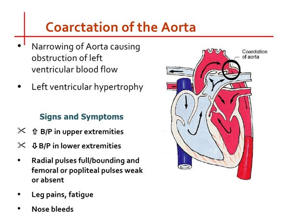
C. Decreased blood pressure in the legs:
This is the correct choice. Coarctation of the aorta is characterized by narrowing of the aorta, which leads to decreased blood flow to the lower extremities. Consequently, blood pressure measurements in the legs may be lower compared to those in the arms. This finding is often a key indicator of coarctation of the aorta.
D. Pulmonary edema:
Pulmonary edema refers to the accumulation of fluid in the lungs and is typically associated with conditions such as heart failure or fluid overload. While some congenital heart defects may lead to heart failure and subsequent pulmonary edema, coarctation of the aorta does not directly cause pulmonary edema. Instead, it primarily affects blood flow to the lower extremities due to the narrowing of the aorta.

Whether you are a student looking to ace your exams or a practicing nurse seeking to enhance your expertise , our nursing education contents will empower you with the confidence and competence to make a difference in the lives of patients and become a respected leader in the healthcare field.
Visit Naxlex, invest in your future and unlock endless possibilities with our unparalleled nursing education contents today
Report Wrong Answer on the Current Question
Do you disagree with the answer? If yes, what is your expected answer? Explain.
Kindly be descriptive with the issue you are facing.
