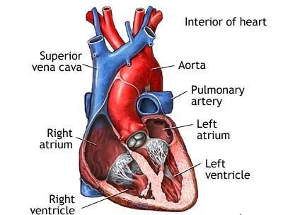
Which cardiac chamber has the thinnest wall and why?
The right and left atria because they are low pressure chambers that serve as storage units and conduits for blood.
The right and left atria because they are not involved directly in the preloaded, contractility or afterload of the heart
The left ventricle because the mean pressure of blood coming into the ventricle is from the lung, which has a low pressure
The right ventricle because it pumps blood into the pulmonary capillaries, which have a lower pressure compared with the systemic circulation
The Correct Answer is A

The walls of the atria are thin because they do not generate as much pressure as the ventricles, as their main function is to receive blood from the veins and pump it into the ventricles. The ventricles have thicker walls because they are responsible for generating the force necessary to pump blood out of the heart and into the systemic or pulmonary circulation.
Nursing Test Bank
Naxlex Comprehensive Predictor Exams
Related Questions
Correct Answer is B
Explanation
The correct answer is B. Systolic pressure less than 120 mmHg and diastolic pressure less than 80 mmHg is considered normal blood pressure in adults according to current guidelines. A systolic pressure between 140-150 mmHg (option A) would be classified as stage 1 hypertension, while a systolic pressure greater than 140 mmHg and diastolic pressure of 100 mmHg (option D) would be classified as stage 2 hypertension. A systolic pressure less than 100 mmHg regardless of diastolic pressure (option C) would be considered low blood pressure.
Correct Answer is B
Explanation
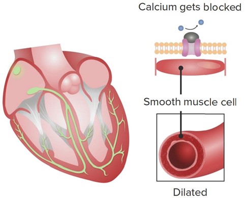
Calcium channel blockers (CCBs) are a class of medications that block the influx of calcium ions into cardiac and smooth muscle cells, leading to relaxation of these muscles and dilation of blood vessels.
In the heart, CCBs primarily affect the L-type calcium channels in the cardiac myocytes, which are responsible for the influx of calcium ions during the plateau phase of the cardiac action potential. By blocking these channels, CCBs decrease the amount of calcium that enters the cardiac myocytes, which in turn reduces the strength of cardiac contractions (i.e. contractility). 
This reduction in contractility can be beneficial in certain conditions where the heart is working too hard or experiencing insufficient blood flow, such as in hypertension, angina, or some forms of arrhythmia. By reducing the workload of the heart, CCBs can help to lower blood pressure, decrease oxygen demand, and improve blood flow to the heart.
While CCBs can also have effects on the rate and rhythm of cardiac contractions, these effects are generally less pronounced than the reduction in contractility. Some CCBs, such as verapamil and diltiazem, can slow the heart rate by blocking the L-type calcium channels in the sinoatrial (SA) and atrioventricular (AV) nodes, while others, such as nifedipine, have little effect on heart rate.
Whether you are a student looking to ace your exams or a practicing nurse seeking to enhance your expertise , our nursing education contents will empower you with the confidence and competence to make a difference in the lives of patients and become a respected leader in the healthcare field.
Visit Naxlex, invest in your future and unlock endless possibilities with our unparalleled nursing education contents today
Report Wrong Answer on the Current Question
Do you disagree with the answer? If yes, what is your expected answer? Explain.
Kindly be descriptive with the issue you are facing.
