
When performing an assessment, the nurse observes for bilateral equality. After performing a neurological assessment, which of the following will the nurse document when assessment findings indicate that there is left facial droop?
Inability to perform within normal limits
Symmetrical findings
Asymmetrical findings
Bilateral strength present
The Correct Answer is C
Choice A Reason: This choice is incorrect. Inability to perform within normal limits is a vague and general term that does not describe the specific finding of left facial droop. The nurse should document the exact observation and compare it to the expected or normal range.
Choice B Reason: This choice is incorrect. Symmetrical findings mean that both sides of the body or face are equal or similar in appearance or function. Left facial droop indicates that one side of the face is lower or weaker than the other, which is not symmetrical.
Choice C Reason: This is the correct choice. Asymmetrical findings mean that both sides of the body or face are unequal or different in appearance or function. Left facial droop indicates that one side of the face is lower or weaker than the other, which is asymmetrical.
Choice D Reason: This choice is incorrect. Bilateral strength present means that both sides of the body or face have normal or adequate muscle power or force. Left facial droop indicates that one side of the face has reduced or impaired muscle power or force, which is not bilateral strength present.
Nursing Test Bank
Naxlex Comprehensive Predictor Exams
Related Questions
Correct Answer is B
Explanation
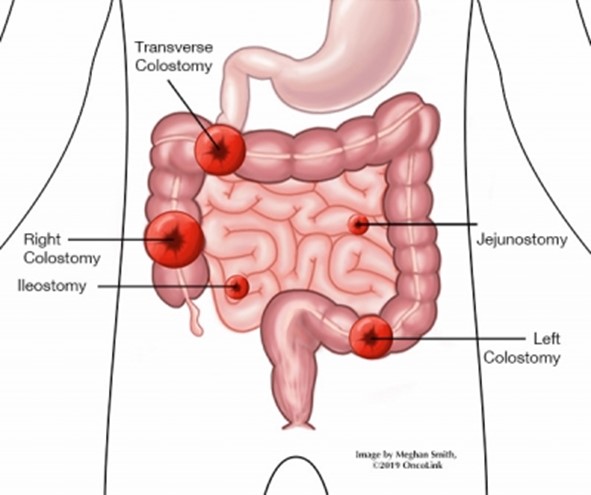
Choice A Reason: Soft pasty stool is normal for a transverse colostomy, as the stool has not reached the sigmoid colon where most of the water is absorbed.
Choice B Reason: This is the correct answer because purple discoloration of the stoma indicates ischemia or necrosis, which can lead to infection, perforation, or sepsis. It requires urgent intervention.
Choice C Reason: Stoma is beefy red is a normal finding for a healthy stoma, as it indicates adequate blood supply and healing.
Choice D Reason: There is skin excoriation around the stoma is a common complication of a colostomy, as the stool can irritate the skin. It can be managed with proper skin care and appliance fitting.
Correct Answer is ["A","B","D"]
Explanation
Choice A Reason: This is a correct choice. Standing next to the client when speaking is an action that the nurse should plan to take, as it helps the client hear better and see the nurse's facial expressions and lip movements. The nurse should also speak clearly and slowly, use simple words and sentences, and avoid covering their mouth.
Choice B Reason: This is a correct choice. Guiding the client away from background noise is an action that the nurse should plan to take, as it reduces distractions and interference with hearing. The nurse should also choose a well-lit and quiet place for communication and turn off any unnecessary devices or appliances.
Choice C Reason: This is an incorrect choice. Providing a copy of the instructions printed in Braille is not an action that the nurse should plan to take, as it is not helpful for clients with hearing loss. Braille is a system of raised dots that represents letters and numbers for people who are blind or visually impaired. The nurse should provide a copy of the instructions printed in large font or use pictures or diagrams to supplement verbal information.
Choice D Reason: This is a correct choice. Repeating any phrases that the client misunderstands is an action that the nurse should plan to take, as it ensures comprehension and clarification of important information. The nurse should also ask open-ended questions, encourage feedback, and summarize key points at the end of the conversation.
Whether you are a student looking to ace your exams or a practicing nurse seeking to enhance your expertise , our nursing education contents will empower you with the confidence and competence to make a difference in the lives of patients and become a respected leader in the healthcare field.
Visit Naxlex, invest in your future and unlock endless possibilities with our unparalleled nursing education contents today