The nurse is completing an assessment on a client suspected of having a transient ischemic attack. Which of the following techniques should the nurse use to assess the client's carotid arteries?
Simultaneously palpating both arteries to compare amplitude.
Listening with the diaphragm of the stethoscope to assess for bruits.
instructing the patient to take slow deep breaths during auscultation.
Palpating the artery at the base of the neck of the neck.
The Correct Answer is B
A. Simultaneously palpating both arteries to compare amplitude: While comparing amplitudes is important, using the diaphragm of the stethoscope to listen for bruits (abnormal whooshing sounds indicating turbulent blood flow) is a more specific and accurate method for assessing the carotid arteries for potential vascular issues.
B. Listening with the diaphragm of the stethoscope to assess for bruits: This technique allows the nurse to detect abnormal sounds (bruits) that could indicate partial blockages or stenosis in the carotid arteries, suggesting a risk of stroke or transient ischemic attack.
C. Instructing the patient to take slow deep breaths during auscultation: Deep breaths are more relevant during lung auscultation. Carotid artery assessment focuses on detecting abnormal sounds and assessing blood flow rather than respiratory patterns.
D. Palpating the artery at the base of the neck: Palpation alone does not provide enough information about potential blockages or abnormalities in the carotid arteries. Listening with a stethoscope allows for a more detailed assessment of blood flow and the presence of bruits. f the nurse hears a bruit during auscultation, they should not palpate the carotid artery. A bruit suggests partial obstruction (carotid stenosis), and compressing the artery further could worsen blood flow.
Nursing Test Bank
Naxlex Comprehensive Predictor Exams
Related Questions
Correct Answer is A
Explanation
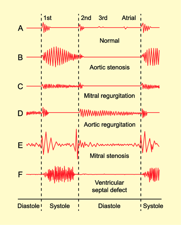
A. The third heart sound (S3):
The third heart sound (S3) is an abnormal heart sound that occurs during early diastole, immediately after S2 (the second heart sound). It is caused by the rapid filling of the ventricles and is often associated with conditions like heart failure. In heart failure, the ventricles become stiff, causing vibrations that produce the S3 sound.
B. A friction rub:
A friction rub is a high-pitched, scratchy sound heard during both systole and diastole. It is caused by the rubbing together of inflamed pericardial layers (pericarditis) and is usually heard best at the left lower sternal border. Friction rubs can indicate pericardial inflammation and are often heard in conditions such as pericarditis or after a myocardial infarction.
C. The fourth heart sound (S4):
The fourth heart sound (S4) occurs late in diastole, just before S1, and is caused by atrial contraction. It is associated with increased resistance to ventricular filling, often due to conditions like hypertension or aortic stenosis. The S4 sound is heard as a low-pitched "atrial gallop."
D. A split second heart sound S2:
The second heart sound (S2) represents the closure of the aortic and pulmonic valves. Normally, S2 has two components: A2 (aortic valve closure) and P2 (pulmonic valve closure). A split S2 occurs when A2 and P2 do not close simultaneously. A physiological split S2 is common during inspiration and occurs due to delayed closure of the pulmonic valve. An abnormal or fixed split S2 can indicate underlying heart conditions such as atrial septal defect (ASD) or right bundle branch block (RBBB).
Correct Answer is C
Explanation
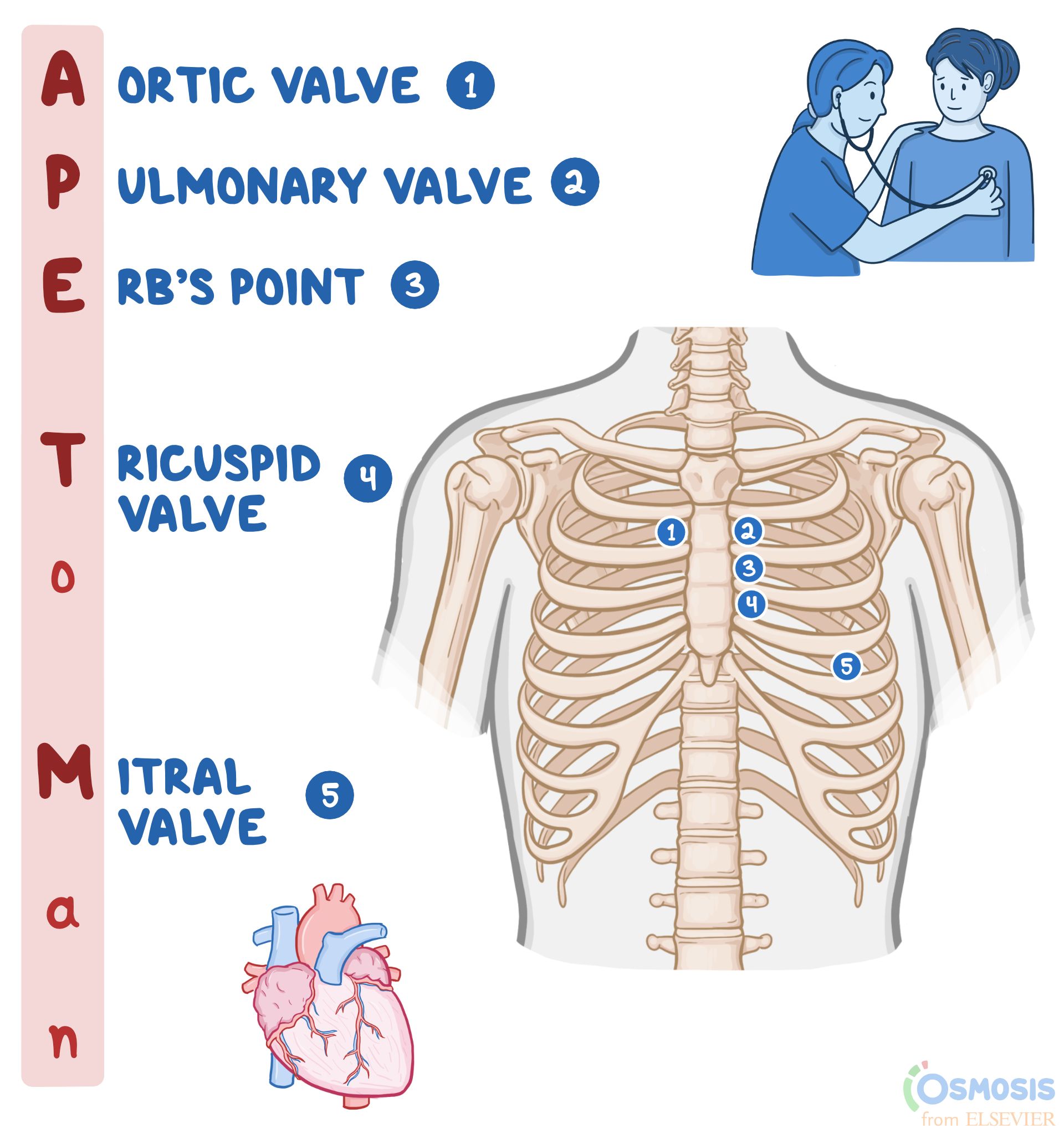
A. Fifth intercostal space, left of the midclavicular line: This placement is used to auscultate the mitral valve, which is best heard at the apex of the heart. The mitral valve sounds are typically heard around the fifth intercostal space, midclavicular line.
B. Left lower sternal border: This placement is used to auscultate the tricuspid valve, which is best heard at the lower left sternal border.
C. Second left intercostal space: This is the correct placement for auscultating the pulmonic valve. The pulmonic valve sounds are best heard at the second left intercostal space, which is close to the upper left sternal border.
D. Second right intercostal space: This placement is used to auscultate the aortic valve, which is best heard at the second right intercostal space, close to the upper right sternal border.
Whether you are a student looking to ace your exams or a practicing nurse seeking to enhance your expertise , our nursing education contents will empower you with the confidence and competence to make a difference in the lives of patients and become a respected leader in the healthcare field.
Visit Naxlex, invest in your future and unlock endless possibilities with our unparalleled nursing education contents today