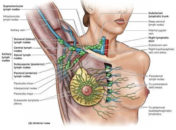
The lymph nodes of the axillary region receive lymph mainly from the:
Upper limb and mammary gland.
Abdominal viscera.
Scalp and face.
Thoracic viscera.
The Correct Answer is A
The lymph nodes of the axillary region receive lymph mainly from the upper limb and mammary gland.
This is because the axillary lymph nodes are located in the armpit area and drain the lymph vessels from the lateral quadrants of the breast and the arm.
Choice B is wrong because the abdominal viscera are drained by the celiac, superior mesenteric, and inferior mesenteric lymph nodes.
Choice C is wrong because the scalp and face are drained by the cervical lymph nodes.
Choice D is wrong because the thoracic viscera are drained by the bronchopulmonary, tracheobronchial, parasternal, and posterior mediastinal lymph nodes.
Nursing Test Bank
Naxlex Comprehensive Predictor Exams
Related Questions
Correct Answer is D
Explanation
Atrial natriuretic peptide (ANP) is a hormone secreted by the heart when the atria are stretched by high blood pressure or volume.
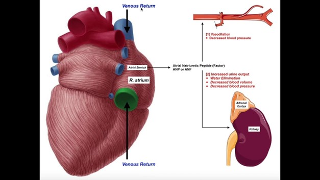
ANP has multiple effects, such as increasing urine and salt excretion, lowering blood pressure, and opposing the renin-angiotensin-aldosterone system.
Therefore, ANP inhibits the release of renin and aldosterone, which are hormones that increase blood pressure and sodium retention.
Choice A is wrong because ANP is not released from the adrenal cortex but from the cardiac atria.
ANP does not stimulate atrial hormones but rather inhibits them.
Choice B is wrong because ANP is not stimulated to release when blood volume decreases, but when it increases.
ANP acts to reduce blood volume by promoting diuresis and natriuresis.
Choice C is wrong because ANP does not raise blood pressure, but lowers it. ANP acts as a vasodilator and reduces peripheral resistance.
Correct Answer is A
Explanation
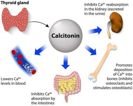
Calcitonin is a hormone that protects against excessive blood calcium levels by inhibiting bone turnover and decreasing reabsorption.
It is produced by the thyroid gland and acts on both osteoclasts and osteoblasts.
Choice B is wrong because parathyroid hormone (PTH) stimulates both resorption and formation of bone, and controls the level of calcium in the blood.
Choice C is wrong because thyroxine is a thyroid hormone that is required for skeletal maturation and influences adult bone maintenance but does not directly affect calcium deposition into bone.
Choice D is wrong because insulin is a hormone that regulates both bone formation and bone resorption but does not specifically stimulate calcium deposition into bone.
Whether you are a student looking to ace your exams or a practicing nurse seeking to enhance your expertise , our nursing education contents will empower you with the confidence and competence to make a difference in the lives of patients and become a respected leader in the healthcare field.
Visit Naxlex, invest in your future and unlock endless possibilities with our unparalleled nursing education contents today