
A nurse admits a client to the emergency department who reports nausea and vomiting that worsens when he lies down. Antacids do not help. The provider suspects acute pancreatitis. Which of the following laboratory test results should the nurse expect to see?
Increased serum amylase
Increased serum calcium
Decreased WBC
Decreased serum lipase
The Correct Answer is A
Choice A Reason:
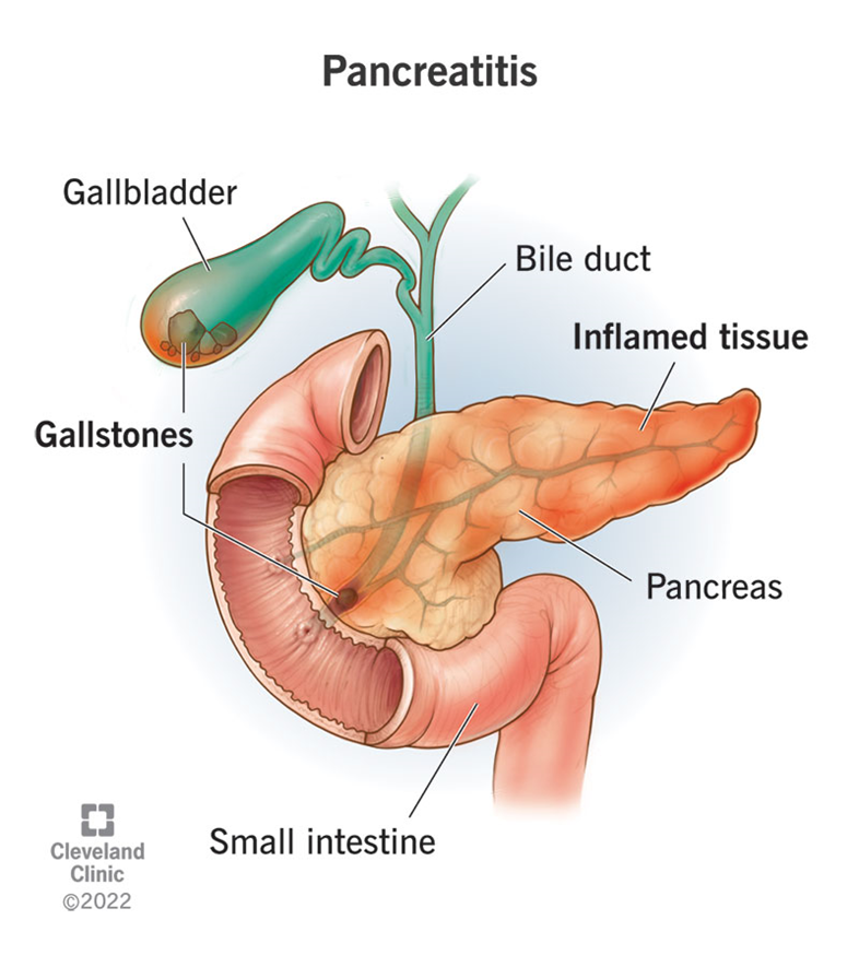
Increased serum amylase is a key indicator of acute pancreatitis. Amylase is an enzyme produced by the pancreas to help digest carbohydrates. In cases of acute pancreatitis, the pancreas becomes inflamed, leading to the release of amylase into the bloodstream. Elevated levels of serum amylase, typically more than three times the upper limit of normal, are a strong indication of acute pancreatitis. This enzyme level usually rises within a few hours of the onset of pancreatitis and can remain elevated for several days.
Choice B Reason:
Increased serum calcium is not typically associated with acute pancreatitis. In fact, acute pancreatitis can often lead to hypocalcemia (low calcium levels) due to fat saponification in the pancreas, where calcium binds with fatty acids. Therefore, an increase in serum calcium would not be expected in a patient with acute pancreatitis. Monitoring calcium levels is important, but an increase is not a diagnostic marker for this condition.
Choice C Reason:
Decreased WBC (white blood cell count) is not a characteristic finding in acute pancreatitis. On the contrary, acute pancreatitis often leads to an elevated WBC count due to the inflammatory response in the body. Leukocytosis (increased WBC) is a common finding in many inflammatory and infectious conditions, including acute pancreatitis. Therefore, a decreased WBC count would not be expected and does not support the diagnosis of acute pancreatitis.
Choice D Reason:
Decreased serum lipase is incorrect. Similar to amylase, lipase is another enzyme produced by the pancreas, which helps in the digestion of fats. In acute pancreatitis, serum lipase levels also increase significantly, often more than three times the upper limit of normal. Lipase levels tend to rise slightly later than amylase but remain elevated for a longer period, making it a useful marker for diagnosing acute pancreatitis. Therefore, decreased serum lipase would not be expected in this condition.
Nursing Test Bank
Naxlex Comprehensive Predictor Exams
Related Questions
Correct Answer is ["A","B","E"]
Explanation
Choice A: Nephropathy
Diabetic nephropathy is a common microvascular complication of diabetes mellitus. It is characterized by damage to the small blood vessels in the kidneys, leading to progressive kidney disease. The condition is often identified by the presence of protein in the urine (proteinuria) and can progress to end-stage renal disease if not managed properly. The primary mechanism involves hyperglycemia-induced damage to the glomeruli, the filtering units of the kidneys. This damage results in increased permeability and eventual scarring, impairing kidney function. Effective management of blood glucose levels and blood pressure is crucial in preventing or slowing the progression of diabetic nephropathy.
Choice B: Neuropathy
Diabetic neuropathy refers to nerve damage caused by chronic high blood sugar levels. It is another significant microvascular complication of diabetes. This condition can affect various types of nerves, including sensory, motor, and autonomic nerves. Symptoms may include pain, tingling, numbness, and loss of sensation, particularly in the extremities. Diabetic neuropathy can lead to severe complications such as foot ulcers and infections, which may necessitate amputation. The pathophysiology involves hyperglycemia-induced oxidative stress and inflammation, leading to nerve damage. Tight glycemic control and regular monitoring are essential in managing diabetic neuropathy.
Choice E: Retinopathy
Diabetic retinopathy is a leading cause of blindness among adults with diabetes. This microvascular complication involves damage to the small blood vessels in the retina, the light-sensitive tissue at the back of the eye. There are two main types: non-proliferative and proliferative retinopathy. Non-proliferative retinopathy is characterized by microaneurysms, hemorrhages, and exudates, while proliferative retinopathy involves the growth of new, fragile blood vessels that can bleed and cause retinal detachment. The primary cause is prolonged hyperglycemia, which damages the retinal blood vessels. Regular eye examinations and good glycemic control are vital in preventing and managing diabetic retinopathy.
Choice C: Peripheral Vascular Disease
Peripheral vascular disease (PVD) is not classified as a microvascular complication but rather a macrovascular one. It involves the narrowing or blockage of the blood vessels outside the heart and brain, primarily affecting the arteries in the legs. PVD is associated with atherosclerosis, where plaque builds up in the arterial walls, leading to reduced blood flow. Symptoms include leg pain, cramping, and ulcers. While PVD is a significant concern for individuals with diabetes, it is not considered a microvascular complication.
Choice D: Cerebral Vascular Disease
Cerebral vascular disease, which includes conditions such as stroke and transient ischemic attacks (TIAs), is also a macrovascular complication rather than a microvascular one. It involves the blood vessels supplying the brain and is primarily caused by atherosclerosis and hypertension. Diabetes increases the risk of cerebral vascular disease due to its association with other risk factors like high blood pressure and dyslipidemia. However, it is not classified as a microvascular complication.
Correct Answer is ["31"]
Explanation
Let’s calculate the IV infusion rate step by step.
Step 1: Determine the total volume to be infused.
The total volume ordered is 1,000 mL.
Step 2: Determine the total time for the infusion.
The total time is 8 hours.
Step 3: Calculate the infusion rate in mL per hour.
Total volume (1,000 mL) ÷ Total time (8 hours) = 125 mL per hour.
Result: 125
Step 4: Determine the drop factor.
The IV tubing delivers 15 drops per milliliter.
Step 5: Calculate the infusion rate in drops per minute.
Infusion rate (125 mL per hour) × Drop factor (15 drops per mL) = 1,875 drops per hour.
Result: 1,875
Step 6: Convert the infusion rate to drops per minute.
Total drops per hour (1,875 drops) ÷ 60 minutes = 31.25 drops per minute.
Result: 31.25
Step 7: Round the result to the nearest whole number if necessary.
31.25 rounded to the nearest whole number is 31.
The nurse should run the IV infusion at a rate of 31 drops per minute.
Whether you are a student looking to ace your exams or a practicing nurse seeking to enhance your expertise , our nursing education contents will empower you with the confidence and competence to make a difference in the lives of patients and become a respected leader in the healthcare field.
Visit Naxlex, invest in your future and unlock endless possibilities with our unparalleled nursing education contents today